Cracking the AP Biology Exam
11
Animal Structure and Function
III. THE CIRCULATORY SYSTEM
Most organisms need to carry out two tasks: (1) supply their bodies with nutrients and oxygen, and (2) get rid of wastes. Many simple aquatic organisms have no trouble moving materials across their membranes since their metabolic needs are met by diffusion. Larger organisms, on the other hand, particularly terrestrial organisms, can’t depend on diffusion. They therefore need special circulatory systems to accomplish internal transport.
There are two types of circulatory systems: an open circulatory system and a closed circulatory system. In an open circulatory system, blood is carried by open-ended blood vessels that spill blood into the body cavity. In arthropods, for example, blood vessels from the heart open into internal cavities known as sinuses. Other organisms have closed circulatory systems. That is, blood flows continuously through a network of blood vessels. Earthworms and some mollusks have a closed circulatory system, as do vertebrates.
THE HUMAN CIRCULATORY SYSTEM
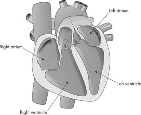
The heart is divided into four chambers, two on the left and two on the right. The four chambers of the heart are the right atrium, the right ventricle, the left atrium, and the left ventricle. Let’s take a look at a picture of the heart:
The heart pumps blood in a continuous circuit. Since blood makes a circuit in the body, it doesn’t matter where we begin to trace the flow of blood. For our purposes, we’ll begin at the point in the circulatory system where the blood leaves the heart and enters the body: the left ventricle. When blood leaves the left ventricle it will make a tour of the body. We call this systemic circulation.
Systemic Circulation
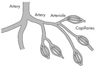
Blood leaves the heart through the aortic semilunar valve and enters a large blood vessel called the aorta. The aorta is the largest artery in the body. The aorta then branches out into smaller vessels called arteries.
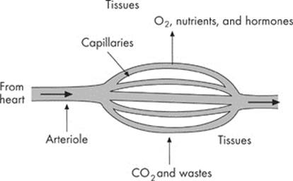
Arteries always carry blood away from the heart. Just remember “A” stands for “away” from the heart. They’re able to carry the blood because arteries are thick-walled, elastic vessels. The arteries become even smaller vessels called arterioles, and then the smallest vessels calledcapillaries.
There are thousands of capillaries. In fact, some estimate that the capillary routes in your bloodstream are as long as 100 kilometers! These vessels are so tiny that red blood cells must “squeeze” through them in single file. Capillaries intermingle with the tissues and exchange nutrients, gases, and wastes. Oxygen and nutrients leave the capillaries and enter the tissues; carbon dioxide and wastes leave the tissues and enter the capillaries.
Before we take a look at the next stage of circulation, let’s recap the pathway of blood through the body.
1. Blood leaves the heart’s left ventricle via the aorta.
2. It travels through the arteries to the arterioles, and eventually to the capillaries.
3. Gas and nutrient/waste exchange occurs between the blood and the tissues through the capillary walls.
Back to the Heart
After exchanging gases and nutrients with the cells, blood has very little oxygen left. Most of its oxygen was donated to the cells through the capillary walls. Since the blood is now depleted of oxygen, it is said to be deoxygenated. To get a fresh supply of oxygen the blood now needs to go to the lungs.
But the blood doesn’t go directly to the lungs. It must first go back to the heart. As the blood returns to the heart, the vessels get bigger and bigger.
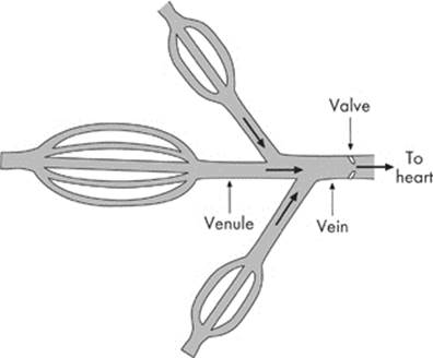
From the capillaries, blood travels through vessels called venules and then through larger vessels called veins. Veins always carry blood toward the heart. Veins are thin-walled vessels with valves that prevent the backward flow of blood.
Blood eventually enters the heart’s right atrium via two veins known as the superior vena cava and the inferior vena cava.
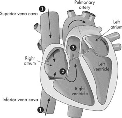
Blood now moves through the heart. Blood travels from the right atrium to the right ventricle through the right atrioventricular valve (or tricuspid). From the right ventricle, blood will go out again into the body, but this time toward the lungs. This is called pulmonary circulation.
The Pulmonary System
Blood leaves the right ventricle through the pulmonary semilunar valve and enters a large artery known as the pulmonary artery. Remember what we said about arteries? Blood vessels that leave the heart are always called arteries.
There’s one major feature you must remember about the blood in the pulmonary system. Whereas in systemic circulation the blood was rich with oxygen, the pulmonary artery is carrying deoxygenated blood. The pulmonary artery branches into the right and left pulmonary arteries which lead, respectively, to the right and left lungs. These arteries become smaller arterioles and then once again capillaries.
We just said that these vessels carry deoxygenated blood. In the lungs, the blood will pick up oxygen and dump carbon dioxide. Sound familiar? It should. It’s just like the gas exchange we discussed in the respiratory system. In the lungs, the blood fills with oxygen, or becomesoxygenated. The blood returns to the heart via the pulmonary veins and enters the left atrium.
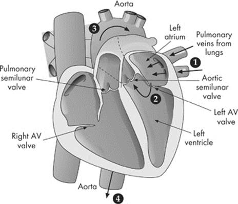
Blood then moves to the left ventricle through the left atrioventricular valve (or bicuspid or mitral valve). Now we’ve completed our tour of the heart. Let’s recap the events in pulmonary circulation.
1. Deoxygenated blood leaves the right ventricle via the pulmonary artery.
2. The pulmonary artery branches into the right and left pulmonary arteries, carrying the blood to the lungs.
3. Blood travels from the arteries to the arterioles, and eventually to the capillaries.
4. Gas exchange occurs between the capillaries and alveoli in the lungs.
5. Once the blood is oxygenated, it returns to the heart through the pulmonary veins.
Thermoregulation
Human homeostasis is characterized by thermoregulation, or the maintenance of a fairly stable body temperature regardless of external conditions. Animals that regulate their internal temperature are known as endotherms. Frequently referred to as “warm-blooded,” endotherms have arteries and veins arranged in a way that enables them to conserve heat through a process known as counter current exchange. The arteries carrying warm blood from the core to the outside are right next to the veins carrying cold blood in the opposite direction. Heat from the arterial blood warms the cold venous blood returning to the heart. Ectotherms, or “cold-blooded” animals, on the other hand, gain and lose heat by way of the environment.
HEART CYCLE
Your heart contracts and relaxes automatically, about 72 times a minute. A special conduction system makes sure that your heart beats rhythmically. The beat begins in tissues in the right atrium called the sinoatrial (SA) node (“the pacemaker”). The impulse then spreads through both atria and conducts directly to the atrioventricular (AV) node. From the AV node, the action potential spreads to the bundle of His and then to the Purkinje fibers in the walls of both ventricles. This generates a strong contraction. The part of the cycle in which contraction occurs is called systole, and the part in which relaxation occurs is called diastole.
THE CONTENTS OF BLOOD
Now let’s take a look at blood itself. Blood consists of two things:
- Plasma
- Cells and cell fragments suspended in the fluid
Blood carries three types of cells: red blood cells (also called erythrocytes), white blood cells (also called leukocytes), and platelets. Red blood cells are the oxygen-carrying cells in the body. They contain hemoglobin, the protein that actually carries the oxygen (and carbon dioxide) throughout the body. Mature red blood cells lack a nucleus. White blood cells fight infection by protecting the body against foreign organisms.
Platelets are cell fragments that are involved in blood clotting. When a blood vessel is damaged, platelets stick to the collagen fibers of the vessel wall. The damaged cells and platelets release substances that activate clotting factors and a series of reactions occur. First, a prothrombin activator converts prothrombin (a plasma protein) to thrombin. Then thrombin converts fibrinogen to fibrin threads, which strengthen the clot.
Here’s something you should remember for the test:
All of the blood cells are made in the bone marrow. The bone marrow is located in the center of the bones.
Blood Types
There are four blood groups: A, B, AB, and O. Blood types are pretty important and are based on the type of antigen(s) found on red blood cells. If a patient is given the wrong type of blood in a transfusion, it could be fatal! Why? Because red blood cells in the blood will clump if they are exposed to the wrong blood type. For example, if you’ve got blood type A (i.e., your red cells carry the A antigen) and you receive a blood transfusion of blood type B, your blood will clump. That’s because your blood contains antibodies, an immune substance that will bind and destroy the foreign blood.
What is important to remember about the different blood types is that type O blood is the universal donor and that type AB is the universal recipient. This means that anyone can receive a blood transfusion of type O blood, while those with type AB blood (which is very rare among Americans—only about 4 percent of the population) can receive any kind of blood without risk. Rh factors are also antigens found on red blood cells. Persons with these antigens are Rh+ and those without them are Rh–.
CIRCULATORY SYSTEM QUIZ
Directions: Each of the questions or incomplete statements below is followed by five suggested answers or completions. Select the answer that is best in each case. Answers can be found here.
1. Which of the following blood components is responsible for blood coagulation?
(A) Erythrocytes
(B) Leukocytes
(C) Platelets
(D) Lymphocytes
(E) Plasma
2. The blood type that is the universal recipient is
(A) O
(B) A
(C) AB
(D) B
(E) Rh
3. All of the following are associated with the distribution of CO2 in the bloodstream EXCEPT
(A) water
(B) hemoglobin
(C) plasma
(D) red blood cells
(E) platelets
Directions: Each group of questions consists of five lettered headings followed by a list of numbered phrases or sentences. For each numbered phrase or sentence, select the one heading that is most closely related to it and fill in the corresponding oval on the answer sheet. Each heading may be used once, more than once, or not at all in each group.
Questions 4–6
(A) Right atrium
(B) Left atrium
(C) Right ventricle
(D) Left ventricle
(E) Vena cava
4. Sends blood to the lungs
5. Chamber that receives blood from the body
6. Sends blood from the head to the heart