Cracking the AP Biology Exam
11
Animal Structure and Function
VI. THE NERVOUS SYSTEM
All organisms must be able to react to changes in their environment. As a result, organisms have evolved systems that pick up and process information from the outside world. The task of coordinating this information falls to the nervous system. The simplest nervous system is found in the hydra. It has a nerve net made up of a network of nerve cells, the impulse of which travels in both directions. As animals became more complex, they developed clumps of nerve cells called ganglia. These cells are like primitive brains. More complex organisms have a brain with specialized cells called neurons.
NEURONS
The functional unit in the nervous system is a neuron. That’s because neurons receive and send the neural impulses that trigger organisms’ responses to their environments. Let’s talk about the parts of a neuron. A neuron consists of a cell body, dendrites, and an axon.
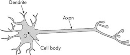
The cell body contains the nucleus and all the usual organelles found in the cytoplasm. Dendrites are short extensions of the cell body that receive stimuli. The axon is a long, slender extension that transmits an impulse from the cell body to another neuron or to an organ. A nerve impulse begins at the top of the dendrites, passes through the dendrites to the cell body, and moves down the axon.
Types of Neurons
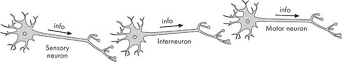
Neurons can be classified into three groups: sensory neurons, motor (effector) neurons, and interneurons. Sensory neurons receive impulses from the environment and bring them to the body. For example, sensory neurons in your hand are stimulated by touch. A motor neuron transmits the impulse to muscles or glands to produce a response. The muscle will respond by contracting or the gland will respond by secreting a substance (e.g., a hormone). Interneurons are the links between sensory neurons and motor neurons. They’re found in the brain or spinal cord:
How Neurons Communicate
Before we talk about the events related to the transmission of a nerve impulse, let’s review how neurons interact. There are billions of neurons running throughout the body, firing all the time. More often than not, one or more neurons are somewhat “connected.” This means that one neuron has its dendrites next to another neuron’s axon. In this way, the dendrites of one cell can pick up the impulse sent from the axon of another cell. The second neuron can then send the impulse to its cell body and down its axon, passing it on to yet another cell.
Resting Potential
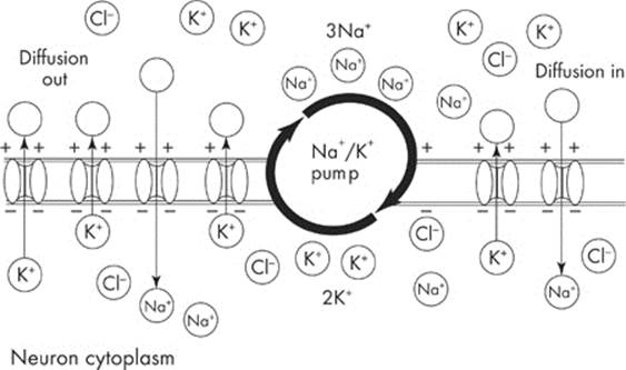
Neurons are not always transmitting signals. The transmission of an impulse depends on the ionic gradients that exist across the axonal membrane. In humans, the concentration of sodium ions is higher in the extracellular fluid than the concentration of potassium ions. The reverse is true inside the axonal membrane. The concentration of potassium ions is higher in the cytosol than the concentration of sodium ion. Because there are many potassium channels that are open but only a relatively small number of sodium channels open the resting potential results from the diffusion of sodium and potassium ions through open ions channels.
The resting potential arises from two activities:
- The Na+K+ ATPase—This pump pushes two potassium ions (K+) into the cell for every three sodium ions (Na+) it pumps out of the cell which leads to a net loss of positive charges within the cell.
- Leaky protein channels—Some potassium channels in the plasma membrane are “leaky” allowing a slow diffusion of K+ out of the cell.
Both the Na+K+ ATPase pump and the leaky channels cause a potential difference between the inside of the neuron and the surrounding interstitial fluid. The membrane potential is always negative inside the cell, and the neuronal membrane is said to be polarized. In humans, the negative charge is –70mV.
Action Potential
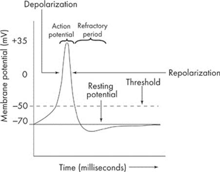
Here’s what a neuron does in response to a stimulus. If a stimulus has enough intensity to excite a neuron, the cell reaches its threshold—the minimum amount of stimulus a neuron needs to respond. This creates what we call an action potential, that is, a change in the membrane potential that produces a nerve impulse. The action potential is an all-or-none response—it doesn’t fire “part way.”
Depolarization
At the point where the axon connects to the cell body, tiny gated sodium ion channels open up and allow sodium ions to rush into the cell. So many sodium ions rush in that the cell now becomes more positive inside than outside. This change is known as depolarization: The interior of the cell has “switched” its polarity from a negative to a positive charge. For now, just remember that an action potential makes the cell depolarize.
The net change is substantial. The charge has shifted from the –70 mV we saw earlier to about +35 mV:
Let’s recap what’s happened so far. In an action potential:
- The cell’s tiny “gates” open up.
- Sodium ions rush in.
- The polarity of the cell changes: The axon is now positive on the inside and negative on the outside.
Repolarization
Once sodium ions have flooded the neuron, the sodium channels close. At this point, the potassium channels open. The potassium ions, which are on the inside of the axon, now rush out. As the potassium ions move out of the cell, the electrical charges reverse again. The inside of the cell becomes more negative than the outside of the cell.
We can now say that this section of the neuron has been repolarized. In other words, the charge has returned to its original polarization.
The Refractory Period
Here’s one thing you should remember: Although the charge has returned to its original state, at the end of the action potential the ions are now on the wrong side of the axonal membrane. Sodium ions are on the inside and potassium ions are on the outside of the axonal membrane. Originally, sodium ions were on the outside and potassium ions were on the inside. The neuron reestablishes the order of the ions, and this process is carried out by the sodium-potassium pump.
This pump reestablishes the original ion distribution by kicking three sodium ions out of the cell for every two potassium ions it brings into the cell. The period after an action potential is known as the refractory period.
During this period, the sodium channels are now reset and are able to open, but the cell membrane potential is further from the threshold. A greater stimulus is required to reach the threshold, so it is more difficult to initiate another action potential.
To summarize:
- A “resting” neuron is polarized; that is, it is more negative on the inside than on the outside.
- When an action potential comes along, the neuron transmits an impulse down its axon.
- First, voltage-gated sodium channels open, allowing sodium ions to rush in. This is known as depolarization: The neuron becomes more positive on the inside and more negative on the outside.
- Sodium channels close and potassium channels open, which restores its negative charge. This is known as repolarization.
- The neuron enters a refractory period.
- The neuron reestablishes the ion distribution thanks to the sodium-potassium pump.
When one small area is depolarized, it causes a “domino effect.” The action potential spreads to the rest of the axon. The impulse is transmitted down the axonal membrane until it reaches the end of the axon called the axon bulb. Now, the neuron wants to pass the impulse to the next neuron. How does it manage this?
When an impulse reaches the end of an axon, the axon releases a chemical called a neurotransmitter into the space between the two neurons. This space is called a synapse. The neurotransmitter diffuses across the synaptic cleft and binds to receptors on the dendrites of the next neuron:
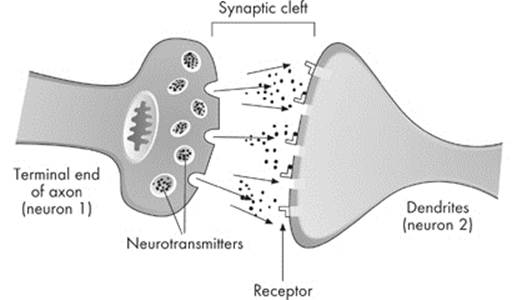
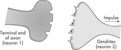
This usually triggers an action potential in the second neuron if the synaptic membrane is excited. Now, the impulse moves along the second neuron from dendrites to axon:
There are many neurotransmitters, but the most important one for the AP Biology Exam is called acetylcholine. Acetylcholine is a neurotransmitter that
- is released from the end of an axon, when Ca2+ moves into the terminal end of the axon
- is picked up almost instantly by the dendrites of the next neuron
- can stimulate muscles to contract or inhibit postsynaptic potential
- is released between neurons in the parasympathetic system, which we will discuss shortly
The extra acetylcholine in the synaptic cleft is broken down by the enzyme acetylcholinesterase.
Other important neurotransmitters include norepinephrine and GABA. Norepinephrine is a peptide neurotransmitter that is released between neurons within the central nervous system. GABA is secreted in the central nervous system and acts as an inhibitor.
Speed of an Impulse
Sometimes a neuron has supporting cells that wrap around its axon. These cells are called Schwann cells. Schwann cells produce a substance called the myelin sheath, which insulates the axon:
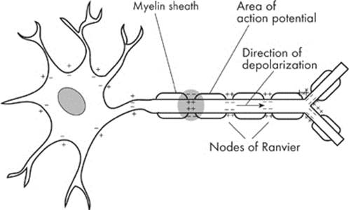
As you can see from the illustration above, the whole axon isn’t covered with myelin sheaths. The spaces between myelin sheaths—the exposed regions of the axon—are called the nodes of Ranvier. Myelin sheaths speed up the propagation of an impulse. Instead of the standard “domino effect” that occurs during an action potential, the impulse can now jump from node to node. This form of conduction is called saltatory conduction.
Thanks to the myelin sheath, the neuron can transmit an impulse down the axon far more rapidly than it could without its help.
PARTS OF THE NERVOUS SYSTEM
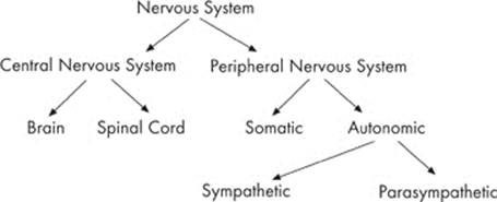
The nervous system can be divided into two parts: the central nervous system and the peripheral nervous system.
Central Nervous System
All of the neurons within the brain and spinal cord make up the central nervous system. All of the other neurons lying outside the brain and the spinal cord—in our skin, our organs, and our blood vessels—are collectively part of the peripheral nervous system. Although both of these systems are really part of one system, we still use the terms central and peripheral.
So keep them in mind:
- The central nervous system includes the neurons in the brain and spinal cord.
- The peripheral nervous system includes all the rest.
Peripheral Nervous System
The peripheral nervous system is further broken down into the somatic nervous system and the autonomic nervous system.
- The somatic nervous system is the part that controls voluntary activities. For example, the movement of your eyes across the page as you read this line is under the control of your somatic nervous system.
- The autonomic nervous system is the part that controls involuntary activities. Your heartbeat and your digestive system, for example, are under the control of the autonomic nervous system.
The interesting thing about these two systems is that they sometimes overlap. For instance, you can control your breathing if you choose to. Yet most of the time you do not think about it: Your somatic system hands control of your respiration over to the autonomic system.
The autonomic system is broken down even further to the sympathetic nervous system and the parasympathetic nervous system. These two systems actually work antagonistically.
The sympathetic system controls the “fight-or-flight” response, which occurs when an organism confronted with a threatening situation prepares to fight or flee. To get ready for a quick, effective action, whether that be brawling or bolting, the sympathetic nervous system raises your heart and respiration rates, causes your blood vessels to constrict, increases the levels of glucose in your blood, and produces “goose bumps” on the back of your neck. It even reroutes your blood sugar to your skeletal muscles in case you need to make a break for it. After the threat has passed, the parasympathetic nervous system brings the body back to homeostasis—that is, back to normal. It lowers your heart and respiratory rates and decreases glucose levels in the blood.
The flow chart below will give you a nice overview of the different parts of the nervous system:
PARTS OF THE BRAIN
The brain can also be divided into parts. Here’s a summary of the major divisions within the brain.
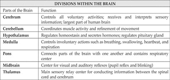
The cerebrum consists of outer gray matter (the cerebral cortex) and inner white matter. One structure that is often mentioned on the AP biology test is the corpus callosum. The corpus callosum is a thick band of nerve fibers of the white matter that enable the right and left side of the cerebral hemispheres to communicate.
NERVOUS SYSTEM QUIZ
Directions: Each of the questions or incomplete statements below is followed by five suggested answers or completions. Select the answer that is best in each case. Answers can be found here.
1. Which description correctly identifies myelin sheath in its role of nerve impulse transmission?
(A) It inactivates Na+ gates of Na channels during an action potential.
(B) It releases neurotransmitters into the synapse.
(C) It completely insulates the axon of neurons.
(D) It slows down the conduction of nerve impulses.
(E) Its presence leads to a concentration of voltage-gated Na+ and K+ channels at the nodes of Ranvier.
Directions: Each group of questions consists of five lettered headings followed by a list of numbered phrases or sentences. For each numbered phrase or sentence, select the one heading that is the most closely related to it and fill in the corresponding oval on the answer sheet. Each heading may be used once, more than once, or not at all in each group.
Questions 2–6 refer to the following parts of the brain
(A) Cerebral cortex
(B) Spinal cord
(C) Medulla oblongata
(D) Cerebellum
(E) Hypothalamus
2. Controls many vital functions such as heartbeat, respiration, and blood pressure
3. Integrates simple motor responses
4. Reflex center for muscular coordination
5. Composed of gray matter
6. Most complex part of the mammalian brain