CONCEPTS IN BIOLOGY
PART VI. PHYSIOLOGICAL PROCESSES
26. The Body's Control Mechanisms and Immunity
26.5. Sensory Input
The activities of the nervous and endocrine systems are often responses to input received from the sense organs. Sense organs of various types are located throughout the body. Many of them are located on the surface, where environmental changes can be detected easily. Hearing, sight, and touch are good examples. Other sense organs are located within the body and indicate to the organism how its various parts are changing. For example, pain and pressure are often used to monitor internal conditions. The sense organs detect changes, but the brain is responsible for perception—the recognition that a stimulus has been received. Sensory input is detected through many kinds of mechanisms, including chemical recognition, the detection of energy changes, and the monitoring of physical forces.
Chemical Detection
All cells have receptors on their surfaces that can bind selectively to molecules they encounter. This binding process can cause changes in the cells in several ways. In some cells, it causes depolarization. When this happens, the binding of molecules to the cell can stimulate neurons and cause messages to be sent to the central nervous system, informing it of a change in the surroundings. Many internal sense organs respond to specific molecules. For example, the brain and aorta contain cells that respond to concentrations of hydrogen ions, carbon dioxide, and oxygen in the blood. In other cases, a molecule binding to the cell surface may cause certain genes to be expressed, and the cell responds by changing the molecules it produces. This is typical of the way the endocrine system receives and delivers messages. Taste and smell are two ways of detecting chemicals in our surroundings.
Taste
Most cells have specific binding sites for specific molecules. Some, such as the taste buds on the tongue, soft palate, and throat appear to respond to classes of molecules. Traditionally, four kinds of tastes have been identified: sweet, sour, salt, and bitter. However, recently, a fifth kind of taste, umami (meaty), has been identified. Umami receptors respond to the amino acid glutamate, which is present in many kinds of foods and often is added to food as a flavor enhancer (monosodium glutamate—MSG).
The taste buds that give us the sour sensation respond to the presence of hydrogen ions (H+). (Acidic foods taste sour.) The hydrogen ions stimulate the cells in two ways: They enter the cell directly or they alter the normal movement of sodium and potassium ions across the cell membrane. In either case, the cell depolarizes and stimulates a neuron. In similar fashion, sodium ions (Na+) stimulate the taste buds that give us the sensation of a salty taste by directly entering the cell, which causes the cell to depolarize.
However, the sensations of sweetness, bitterness, and umami occur when molecules bind to specific surface receptors on the cell. Sweetness can be stimulated by many kinds of organic molecules, including sugars and artificial sweeteners, as well as by inorganic lead compounds. When a molecule binds to a sweetness receptor, a series of molecular changes occurs within the cell that leads to the depolarization of the cell. The sweet taste of lead salts in old paints partly explains why children sometimes eat paint chips. Because the lead interferes with normal brain development, this behavior can have disastrous results.
The cells that respond to bitter sensations have a variety of receptor molecules on their surface that bind to many kinds of compounds. When a substance binds to one of the receptors, the cell depolarizes. In the case of umami, it is the glutamate molecule that binds to receptors on the receptor cells.
Each of these tastes has a significance from an evolutionary point of view. Carbohydrates are a major food source, and many carbohydrates taste sweet; therefore, this sense is useful in identifying foods that have high food value. Similarly, proteins and salts are necessary in the diet. Therefore, being able to identify these items in potential foods is extremely valuable. This is true for salt, which must often be obtained from mineral sources. On the other hand, many bitter and sour materials are harmful. Many plants produce bitter-tasting toxic materials, and acids are often the result of the bacterial decomposition (spoiling) of foods. Being able to identify bitter and sour allows organisms to avoid potentially harmful foods.
Much of what we often refer to as taste involves such inputs as appearance, temperature, texture, and smell. If food does look appealing, it will probably influence how it tastes to a person. Cold coffee has a different taste than hot coffee, even though they are chemically the same. Lumpy, cooked cereal and smooth cereal have different tastes. If we are unable to smell food, it doesn’t taste as it should, which is why we sometimes lose our appetite when we have a stuffy nose. There still is much to learn about how taste cells detect chemicals and the role of associated senses in modifying taste.
Smell
The sense of smell is much more versatile than taste; it can detect thousands of different molecules at very low concentrations. The cells that make up the olfactory epithelium, the lining of the nasal cavity, which responds to smells, bind molecules to receptors on their surfaces. Recent research shows that each receptor cell binds to only one kind of odor molecule. The difference in cells is determined by the expression of genes. Each cell contains thousands of genes for detecting odors but only one of those is activated in each receptor cell and is expressed as a particular receptor molecule on the surface of the cell. The receptor cells are extremely sensitive. In some cases, a single molecule of a substance is sufficient to cause a receptor cell to send a message to the brain, where the sensation of odor is perceived. These sensory cells also fatigue rapidly. For instance, when we first walk into a room, we readily detect specific odors; however, after a few minutes, we are unable to detect them. Most perfumes and aftershaves are undetectable after 15 minutes of continuous exposure to them.
Vision
The eyes primarily respond to changes in the flow of light energy. The curved surfaces of the cornea and the lens focus light on a light-sensitive layer of the back of the eye, known as the retina (figure 26.13). Muscles attached to the lens allow it to change shape so that we can focus on both near and far objects.
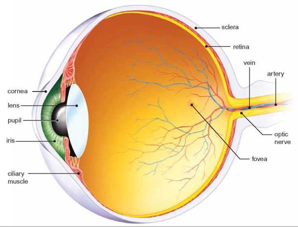
FIGURE 26.13. Structure of the Eye
Light enters the eye through the an opening in the iris known as the pupil. The cornea and lens focus light on the retina where the light is detected.
There are two kinds of receptors in the retina of the eye (figure 26.14). The cells called rods respond to a broad range of wavelengths of light and are responsible for black-and-white vision. Because rods are very sensitive to light, they are useful in dim light. Rods are located over most of the retinal surface, except for the area of most acute vision, known as the fovea centralis. The other kind of receptor cells, called cones, are not as sensitive to light, but they can detect different wavelengths of light. They are found throughout the retina but are concentrated in the fovea centralis. This combination of receptors gives us the ability to detect color when light levels are high, but we rely on black-and-white vision at night. There are three varieties of cones: One type responds best to red light, one type responds best to green light, and the third type responds best to blue light. The stimulation of various combinations of these three kinds of cones allows us to detect different shades of color (figure 26.15).
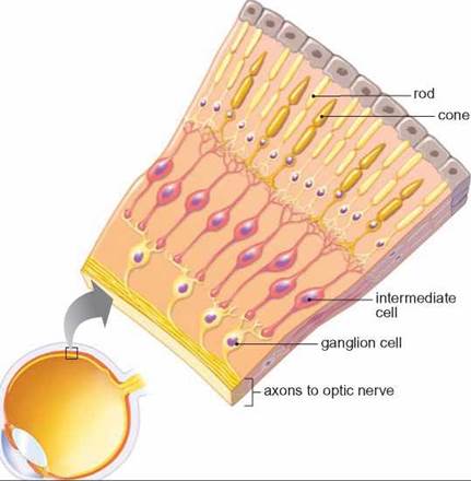
FIGURE 26.14. Structure of the Retina
The retina contains two kinds of receptor cells—rods and cones. When light strikes the rods and cones, they depolarize and stimulate nerve cells that send impulses to the brain.
Rods and the three kinds of cones each contain a pigment that decomposes when struck by light of the proper wavelength and sufficient strength. Rhodopsin is the pigment found in rods. This change in the structure of rhodopsin causes the rod to depolarize. Cone cells have a similar mechanism of action, and each of the three kinds of cones has a different pigment. Because rods and cones synapse with neurons, they stimulate a neuron when depolarized and cause a message to be sent to the brain. Thus, the pattern of color and light intensity recorded on the retina is detected by rods and cones and converted into a series of nerve impulses, which the brain receives and interprets.
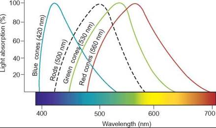
FIGURE 26.15. Light Reception by Cones
There are three different kinds of cones. Each responds differently to red, green, and blue wavelengths of light. Stimulation of combinations of these three kinds of cones gives us the ability to detect many different shades of color.
Hearing and Balance
Sound is produced by the vibration of molecules. The ears respond to changes in sound waves. Consequently, the ears detect changes in the quantity of energy and the quality of sound waves. Sound has several characteristics. Loudness, or volume, is a measure of the intensity of sound energy that arrives at the ear. Very loud sounds literally vibrate the body and can cause hearing loss if they are too intense. Pitch is a quality of sound that is determined by the frequency of the sound vibrations. High-pitched sounds have short wavelengths; low-pitched sounds have long wavelengths.
The sound that arrives at the ear is first funneled by the external ear to the tympanum, also known as the eardrum. The cone shape of the external ear focuses sound on the tympanum and causes it to vibrate at the same frequency as the sound waves reaching it. Attached to the tympanum are three tiny bones: the malleus (hammer), incus (anvil), and stapes (stirrup). The malleus is attached to the tympanum, the incus is attached to the malleus and stapes, and the stapes is attached to a small, membrane-covered opening called the oval window, in a snail-shaped, fluid-filled structure known as the cochlea. The vibration of the tympanum causes the tiny bones (malleus, incus, and stapes) to vibrate; in turn, they cause a corresponding vibration in the membrane of the oval window.
The cochlea detects sound. When the oval window vibrates, the fluid in the cochlea begins to move, causing the basilar membrane to vibrate. Cells on this membrane depolarize when they are stimulated by its vibrations. Because they synapse with neurons, messages can be sent to the brain (figure 26.16).
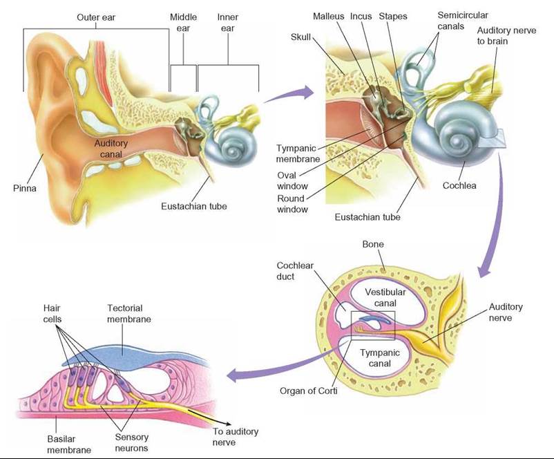
FIGURE 26.16. The Anatomy of the Ear
The ear consists of an external cone, which directs sound waves to the tympanum. Vibrations of the tympanum move the ear bones and vibrate the oval window of the cochlea, where the sound is detected. The semicircular canals monitor changes in the position of the head, helping us maintain balance.
Most sounds consist of a mixture of pitches. High-pitched, short-wavelength sounds cause the basilar membrane to vibrate at the base of the cochlea near the oval window. Low-pitched, long-wavelength sounds vibrate the basilar membrane far from the oval window. Loud sounds cause the basilar membrane to vibrate more vigorously than do faint sounds. Thus, the brain can perceive the loudness of various sounds as well as their pitch.
Associated with the cochlea are two fluid-filled chambers and a set of fluid-filled tubes, called the semicircular canals. The semicircular canals are not involved in hearing but are involved in maintaining balance and posture. In the walls of these canals and chambers are cells similar to those found on the basilar membrane. These cells are stimulated by movements of the head and by the position of the head with respect to the force of gravity. The head’s constantly changing position gives sensory input that is important in maintaining balance.
Touch
What we normally call the sense of touch consists of a variety of kinds of input, which are responded to by three kinds of receptors: Some receptors respond to pressure, others to temperature, and others, which we call pain receptors, to cell damage (figure 26.17). When these receptors are appropriately stimulated, they send a message to the brain. Because receptors are stimulated in particular parts of the body, the brain can localize the sensation. However, not all parts of the body are equally supplied with these receptors. The finger tips, lips, and external genitals have the highest density of these nerve endings, whereas the back, legs, and arms have far fewer receptors.
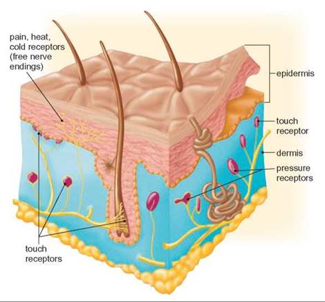
FIGURE 26.17. The Sense of Touch
The sense of touch is really a mixture of sensory cells located in the skin, muscles, joints, and certain internal organs. They send impulses to the brain, which interprets the input and generates responses to the stimuli.
Some internal receptors, such as pain and pressure receptors, are important in allowing us to monitor our internal activities. Many pains generated by the internal organs are often perceived as if they were somewhere else. For example, the pain associated with a heart attack is often perceived to be in the left arm or jaw. Pressure receptors in joints and muscles provide information about the degree of stress being placed on a portion of the body. This is also important information to send back to the brain so that adjustments can be made in movements to maintain posture. If you have ever had your foot “go to sleep” because the nerve stopped functioning, you have experienced what it is like to lose this constant input of nerve messages from the pressure sensors that assist in guiding the movements you make. Your movements become uncoordinated until the nerve function returns to normal.
26.5. CONCEPT REVIEW
11. What is detected by the nasal epithelium, the cochlea of the ear, and the retina of the eye?
12. Name the five kinds of taste that humans are able to detect. What other factors are involved in taste?
13. List three kinds of receptors associated with touch.