CONCEPTS IN BIOLOGY
PART III. MOLECULAR BIOLOGY, CELL DIVISION, AND GENETICS
9. Cell Division—Proliferation and Reproduction
9.3. Mitosis—Cell Replication
When eukaryotic cells divide, two events occur. (1) The replicated genetic information of a cell is equally distributed in mitosis. (2) After mitosis, the cytoplasm of the cell also divides into two new cells. This division of the cell’s cytoplasm is called cytokinesis—cell splitting.
The individual stages of mitosis transition seamlessly from one to the next. Because there are no clear-cut beginning or ending points for each stage, scientists use key events to identify the different stages of mitosis. The four phases are prophase, metaphase, anaphase, and telophase.
Prophase
Key events:
• Chromosomes condense.
• Spindle and spindle fibers form.
• Nuclear membrane disassembles.
• Nucleolus disappears.
As the G2 phase of interphase ends, mitosis begins. Prophase is the first stage of mitosis. One of the first visible changes that identifies when the cell enters prophase is that the thin, tangled chromatin present during interphase gradually coils and thickens, becoming visible as separate chromosomes consisting of 2 chromatids (figure 9.5). As the nucleus disassembles during prophase, the nucleolus is no longer visible.

FIGURE 9.5. Early Prophase
Chromosomes begin to appear as thin, tangled threads and the nucleolus and nuclear membrane are present. The two sets of microtubules, known as the centrioles, begin to separate and move to opposite poles of the cell. A series of fibers, known as the spindle, will shortly begin to form.
As the cell moves toward the end of prophase, a number of other events also occur in the cell (figure 9.6). One of these events is the formation of the spindle and its spindle fibers. The spindle is a structure, made of microtubules, that spans the cell from one side to the other. The spindle fibers consist of microtubules and are the individual strands of the spindle. As prophase proceeds and the nuclear membrane gradually disassembles, the spindle fibers attach to the chromosomes. Spindle fibers must attach to the chromosomes so that the spindle fibers can move chromosomes during later stages of mitosis.
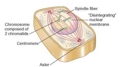
FIGURE 9.6. Late Prophase
In late prophase, the chromosomes appear as 2 chromatids connected at a centromere. The nucleolus and the nuclear membrane have disassembled. The centrioles have moved farther apart, the spindle is produced, and the chromosomes are attached to the spindle fibers.
One difference between plant and animal cell division can be observed in prophase. In animal cells, the spindle forms between centrioles. In plants, the spindle forms without centrioles. Centrioles are cellular organelles comprised of microtubules. Centrioles replicate during the G2 stage of interphase and begin to move to opposite sides of the cell during prophase. As the centrioles migrate, the spindle is formed between them and eventually stretches across the cell, so that spindle fibers encounter chromosomes when the nuclear membrane disassembles. Plant cells do not form their spindle between centrioles, but the spindle still forms during prophase.
Another significant difference between plant and animal cells is the formation of asters during mitosis. Asters are microtubules that extend outward from the centrioles to the plasma membrane of an animal cell. Whereas animal cells form asters, plant cells do not. Some scientists hypothesize that asters help brace the centriole against the animal plasma membrane by making the membrane stiffer. This might help in later stages of mitosis, when the spindle fibers and centrioles may need firm support to help with chromosome movement. It is believed that plant cells do not need to form asters because this firm support is provided by their cell walls.
Metaphase
Key event:
• Chromosomes align at the equatorial plane of the cell.
During metaphase, the second stage of mitosis, the chromosomes align at the equatorial plane. There is no nucleus present during metaphase because the nuclear membrane has disassembled, and the spindle, which started to form during prophase, is completed. The chromosomes are at their most tightly coiled, are attached to spindle fibers and move along the spindle fibers until all their centromeres align along the equatorial plane of the cell (figure 9.7). At this stage in mitosis, each chromosome still consists of 2 chromatids attached at the centromere.
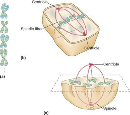
FIGURE 9.7. Metaphase
(a) During metaphase, the chromosomes are moved by the spindle fibers and align at the equatorial plane. The equatorial plane is the region in the middle of the cell. Notice that each chromosome still consists of 2 chromatids. (b) When viewed from the edge of the plane, the chromosomes appear to be lined up. (c) When viewed from another angle, the chromosomes appear to be spread apart, as if on a tabletop.
To understand the arrangement of the chromosomes during metaphase, keep in mind that the cell is a three-dimensional object. A view of a cell in metaphase from the side is an equatorial view. From this perspective, the chromosomes appear as if they were in a line. If we viewed the cell from a pole, looking down on the equatorial plane, the chromosomes would appear scattered about within the cell, even though they were all in a single plane.
Anaphase
Key event:
• Sister chromatids move toward opposite ends of the cell.
Anaphase is the third stage of mitosis. The nuclear membrane is still absent and the spindle extends from pole to pole. The sister chromatids of each chromosome separate as they move along the spindle fibers toward opposite poles (figure 9.8). When this separation of chromatids occurs, the chromatids become known as separate daughter chromosomes.
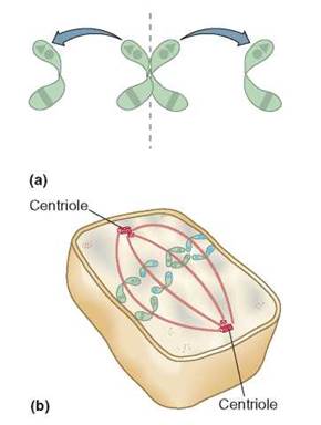
FIGURE 9.8. Anaphase
(a) The pairs of chromatids separate after the centromeres replicate. (b) The chromatids, now called daughter chromosomes, are separating and moving toward the poles.
The sister chromatids separate because two important events occur. The first is that enzymes in the cell digest the portions of the centromere that holds the 2 chromatids together. The second event is that the chromatids begin to move. The kinetochore is a multi-protein complex attached to each chromatid at the centromere (figure 9.9). The kinetochore causes the shortening of the spindle fibers that are attached to it. By shortening the spindle fibers, the kinetochore pulls its chromatid toward the pole.
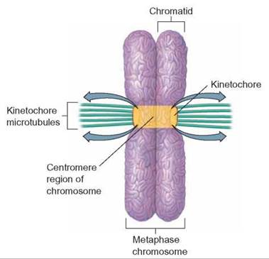
FIGURE 9.9. Kinetochore
The kinetochore on the chromosome is where the spindle fibers bind to the chromosome. During anaphase, the two chromatids separate from each other as (each) kinetochore shortens the spindle fiber (to which it is attached), pulling the chromosome toward the centrioles.
The two sets of daughter chromosomes migrating to opposite poles during anaphase have equivalent genetic information. This is true because the two chromatids of each chromosome, now called daughter chromosomes, were produced by DNA replication during the S stage of Interphase. Thus there are two equivalent sets of genetic information. Each set moves toward opposite poles.
Telophase
Key events:
• Spindle fibers dissasemble.
• Nuclear membrane re-forms.
• Chromosomes uncoil.
• Nucleolus re-forms.
During telophase, the cell finishes mitosis. The spindle fibers disassemble. The nuclear membrane forms around the two new sets of chromosomes, and the chromosomes begin to uncoil back into chromatin, so that the genetic information found on their DNA can be read by transcriptional enzymes. The nucleolus re-forms as the cell begins to make new ribosomes for protein synthesis. The cell is preparing to reenter interphase. With the separation of genetic material into two new nuclei, mitosis is complete (figure 9.10).

FIGURE 9.10. Telophase
During telophase, the spindle disassembles and the nucleolus and nuclear membrane reforms.
Cytokinesis
At the end of telophase a cell has two nuclei. The process of mitosis has prepared the two nuclei to be passed on to the daughter cells. Next, the process of cytokinesis creates the daughter cells. Cytokinesis is the process during which the cell contents are split between the two new daughter cells.
Different cell types use different strategies for achieving cytokinesis (figure 9.11). In animal cells, cytokinesis results from the formation of a cleavage furrow. The cleavage furrow is an indentation of the plasma membrane that pinches in toward the center of the cell, thus splitting the cytoplasm in two. In an animal cell, cytokinesis begins at the plasma membrane and proceeds to the center. In plant cells, a cell plate begins to form at the center of the cell and grows out to the plasma membrane. The cell plate is made of normal plasma membrane components. It is formed by both daughter cells, so that, when complete, the two cells have separate membranes. The cell wall is then formed between the newly formed cells.
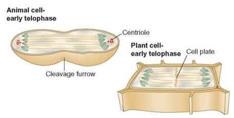
FIGURE 9.11. Cytokinesis: Animal and Plant
In animal cells, there is a pinching in of the cytoplasm, which eventually forms two daughter cells. Daughter cells in plants are formed when a cell plate separates the cell into two cells.
The completion of mitosis and cytokinesis marks the end of one round of cell division. Each of the newly formed daughter cells then starts the cell’s cycle over by entering interphase at G1. These cells can grow, replicate their DNA, and enter another round of mitosis and cytokinesis to continue the cell cycle or can stay metabolically active without dividing by staying in G0.
Summary
Mitosis is much more than splitting the cytoplasm of a cell into two parts (table 9.1). Much of the process is devoted to ensuring that the genetic material is split appropriately between the daughter cells. The sister chromatids formed during DNA replication, contain identical genetic information. The sister chromatids are separated to each of the resulting daughter cells. By dividing the genetic information as sister chromatids, the daughter cells inherit the same genetic information that was present in the parent cell. Because the daughter cells have the same genetic information as the parent, they can replace lost cells and have access to all the same genetic information as the parent cell. With the same genetic information, the daughter cells can have the same function.
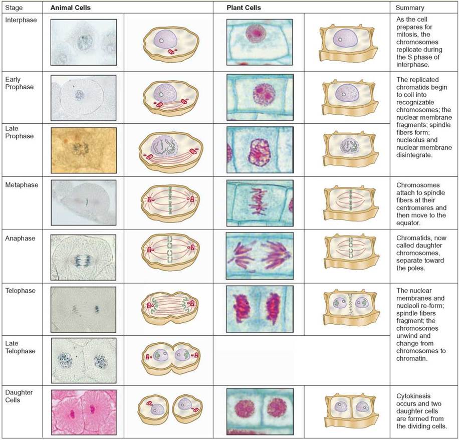
TABLE 9.1. Summary of the Cell Cycle
The stages of the cell cycle are shown in photographs and drawings for both animal and plant cells. The photographed animal cells are from whitefish blastulas. The photographed plant cells are from onion root tips.
9.3. CONCEPT REVIEW
5. Name the four stages of mitosis and describe what occurs in each stage.
6. During which stage of a cell’s cycle does DNA replication occur?
7. At what phase of mitosis does a chromosome become visible?
8. List five differences between an interphase cell and a cell in mitosis.
9. Define the term cytokinesis.
10. What are the differences between plant and animal mitosis?
11. What is the difference between cytokinesis in plants and animals?