CONCEPTS IN BIOLOGY
PART III. MOLECULAR BIOLOGY, CELL DIVISION, AND GENETICS
9. Cell Division—Proliferation and Reproduction
9.8. Meiosis—Gamete Production
Consider a cell that has only 4 chromosomes (figure 9.22). The two from the father are shown in blue and the two from the mother are in green. Notice in figure 9.22 that there are two pairs of homologous chromosomes. Each pair consists of a green chromosome and a blue chromosome. One pair is long. The other pair is short.
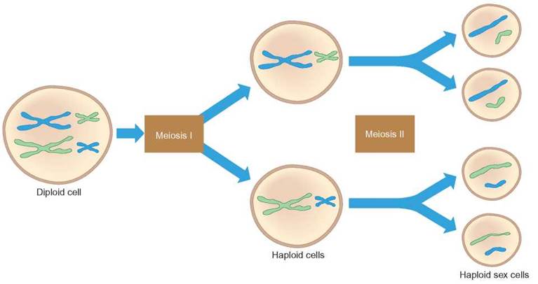
FIGURE 9.22. Meiosis
The cell division process of meiosis occurs in organisms that reproduce sexually. Meiosis occurs in two stages. The first stage, meiosis I, results in the formation of two cells. After each of these cells divides during meiosis II, four gametes are produced. The father’s chromosomes are shown in blue and the two from the mother are in green.
Meiosis involves two cell divisions and produces four cells. Meiosis I consists of the processes that occur during the first division, and meiosis II consists of the processes that occur during the second division. Before meiosis occurs, the cell is in interphase of the cell cycle. As with mitosis, the interphase that precedes meiosis includes DNA replication. Before DNA replication, chromosomes have only one chromatid. After DNA replication, chromosomes consist of two chromatids.
Meiosis I
Meiosis I is a reduction division, in which the chromosome number in the two cells produced is reduced from diploid to haploid. The sequence of events in meiosis I is divided into four phases: prophase I, metaphase I, anaphase I, and telophase I.
Prophase I
Key events:
• Chromosomes condense.
• Spindle and spindle fibers form.
• Nuclear membrane disassembles.
• Synapsis and crossing-over occur.
A number of important events occur during Prophase I. Several of these events also occur during prophase of mitosis: the nuclear membrane disassembles; the spindle fibers form; and the chromosomes condense. However, in meiosis, once the chromosomes are fully condensed, synapsis causes homologous chromosomes to move toward one another, so that the chromosomes lie next to each other. While the chromosomes are synapsed, crossing-over occurs. Crossing-over is the exchange of equivalent sections of DNA on homologous chromosomes. Crossing-over is shown in figure 9.23 as bits of blue on the green chromosome and bits of green on the blue chromosome. The crossing-over process is carefully regulated to make sure that the DNA sections that are exchanged contain equivalent information. This means that usually no information is lost or gained by either chromosome; genetic information is simply exchanged. Because the two members of each homologous pair of chromosomes came from different parents (one from the mother and one from father), there are minor differences in the DNA present on the two chromosomes. Crossing-over happens many times along the length of the homologous chromosomes.
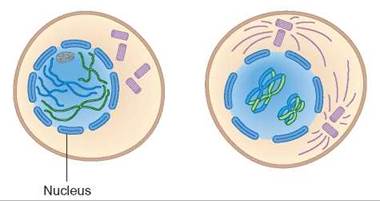
FIGURE 9.23. Prophase I
During prophase I, several visible changes occur as the cell prepares for division. The nuclear membrane is being broken down and the spindle begins to form. As the nuclear membrane disintegrates, the chromosomes can be moved throughout the cell. As the cell advances through prophase, the chromosomes also become more condensed and are paired as homologous pairs.
Crossing-over is very important, because it allows a more thorough mixing of genes from one generation to the next. Without crossing-over, each of the chromosomes an organism inherits in the mother’s egg would be passed on exactly as it was to the organism’s offspring.
Metaphase I
Key event:
• Chromosomes align on equatorial plane as synapsed pairs.
In metaphase I, the centromere of each chromosome attaches to the spindle. The synapsed pair of homologous chromosomes moves into position on the cell’s equatorial plane as a single unit. The orientation of the members of each pair of chromosomes is random with regard to the cell’s poles. Figure 9.24 shows only one possible arrangement. An equally likely arrangement of chromosomes during this stage would be to flip the positions of two identically sized chromosomes. In the figure, this flipped arrangement would place all of the green chromosomes on one side of the cell. The number of possible arrangements increases with the number of chromosomes present in the cell. The arrangement is determined by chance.
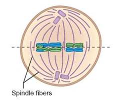
FIGURE 9.24. Metaphase I
Notice that the homologous chromosome pairs are arranged on the equatorial plane in the synapsed condition. The dotted line represents the equatorial plane. This cell shows one way the chromosomes could be lined up; however, a second arrangement is possible.
Compare metaphase I of meiosis with metaphase of mitosis (figure 9.24 and figure 9.7). Note the different ways the chromosomes are arranged. In mitosis, each chromosome is arranged at the equator independently of the others and the chromatids will separate. In meiosis I the chromosomes are arranged at the equator in homologous pairs and the homologous chromosomes (each consisting of two chromatids) separate.
Anaphase I
Key events:
• Homologous chromosomes separate from each other.
• Chromosomes move toward cell’s poles.
• Reduction occurs (diploid-2w to haploid-w).
Anaphase I is the stage during which homologous chromosomes separate (figure 9.25). During this stage, the chromosome number is reduced from diploid to haploid. The two members of each pair of homologous chromosomes move away from each other toward opposite poles. The direction each takes is determined by how each pair was originally oriented on the spindle.
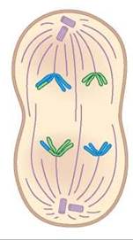
FIGURE 9.25. Anaphase I
During this phase, one member of each homologous pair is segregated from the other member of the pair. Notice that the chromatids of the chromosomes do not separate.
This arrangement of chromosomes in anaphase I, causes the key difference between mitosis and meiosis. In anaphase of mitosis, chromatids separate from each other. In anaphase I of meiosis, homologous chromosomes separate from each other. Each chromosome is independently attached to a spindle fiber at its centromere. Unlike the anaphase stage of mitosis, in anaphase I of meiosis the centromeres that hold the chromatids together do not divide. The chromosomes are still in their replicated form, consisting of 2 chromatids in anaphase I.
Because the homologous chromosomes and the genes they carry are being separated from one another, this process is called segregation. The way in which a single pair of homologous chromosomes segregates does not influence how other pairs of homologous chromosomes segregate. That is, each pair segregates independently of other pairs. This is known as independent assortment of chromosomes. Both segregation and independent assortment are key components in understanding how to solve genetics problems.
Telophase I
Key events:
• Spindle fibers disassemble.
• Chromosomes uncoil.
• Nuclear membrane re-forms.
• Nucleoli reappear.
Telophase I consists of changes that return the cell to an interphase-like condition (figure 9.26). The chromosomes uncoil and become long, thin threads; the nuclear membrane reforms around them; and nucleoli reappear. Following this activity, cytokinesis divides the cytoplasm into two separate cells.
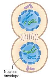
FIGURE 9.26. Telophase I
Cytokinesis occurs during telophase I. During cytokinesis, two cells are formed. Each cell is haploid, containing one set of chromosomes.
Because of meiosis I, the total number of chromosomes is divided equally, so that each daughter cell has one member of each homologous chromosome pair. This means that each cell receives one-half the genetic information of the parent cell, but it has 1 chromosome of each kind and thus has one full set of chromosomes. Each chromosome is still composed of 2 chromatids joined at the centromere. The chromosome number for the cells is reduced from diploid (2n) to haploid (n). In the cell we have been using as our example, the number of chromosomes is reduced from 4 to 2. The four pairs of chromosomes have been distributed to the two daughter cells.
Depending on the type of cell, there may be a time following telophase I when the cell engages in normal metabolic activity corresponding to an interphase stage. Figure 9.27 summarizes the events in meiosis I.
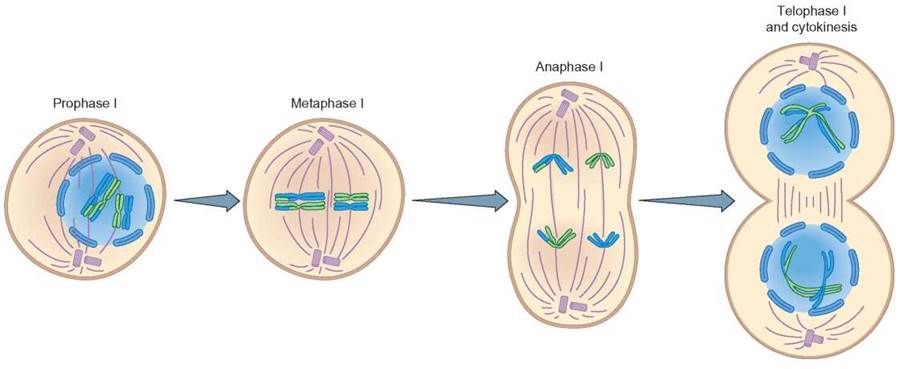
FIGURE 9.27. Meiosis I
The stages in meiosis I result in reduction division. This reduces the number of chromosomes in the parental cell from the diploid number to the haploid number in each of the two daughter cells.
Meiosis II
Meiosis II includes four phases: prophase II, metaphase II, anaphase II, and telophase II. The two daughter cells formed during meiosis I both continue through meiosis II, so that four cells result from the two divisions. During the time between telophase I and the beginning of meiosis II, no DNA replication occurs. The genetic information in cells starting meiosis II is the same as that in cells ending meiosis I. The events in the division sequence of meiosis II are the same as those that occur in mitosis.
Prophase II
Key events:
• Chromosomes condense.
• Spindle and spindle fibers form.
• Nuclear membrane disassembles.
• Nucleoli disassemble.
Prophase II is similar to prophase in mitosis; the nuclear membrane is disassembled and the spindle apparatus begins to form. However, it differs from prophase I in that the cells are haploid, not diploid (figure 9.28).
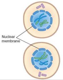
FIGURE 9.28. Prophase II
The two daughter cells are preparing for the second division of meiosis.
Metaphase II
Key event:
• Chromosomes align at the equator in unpaired manner.
Metaphase II is typical of any metaphase stage, because the chromosomes attach by their centromeres to the spindle at the equatorial plane of the cell. Because pairs of homologous chromosomes are no longer together in the same cell, each chromosome moves as a separate unit (figure 9.29).
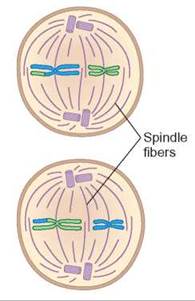
FIGURE 9.29. Metaphase II
During metaphase II, each chromosome lines up on the equatorial plane. Each chromosome is composed of 2 chromatids (a replicated chromosome) joined at a centromere.
Anaphase II
Key event:
• Chromatids separate and begin to move to cell’s poles.
Anaphase II of meiosis differs from anaphase I of meiosis in that, during anaphase II, the centromere of each chromosome divides, and the chromatids, now called daughter chromosomes, move to opposite poles. This is similar to mitosis (figure 9.30). There are no paired homologous chromosomes in this stage; therefore, segregation and independent assortment cannot occur as in meiosis I.
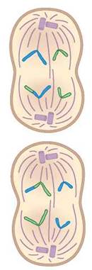
FIGURE 9.30. Anaphase II
Anaphase II is very similar to anaphase of mitosis. The centromere of each chromosome divides and 1 chromatid separates from the other. As soon as this happens, they are no longer referred to as chromatids; each strand of nucleoprotein is now called a daughter chromosome.
Telophase II
Key events:
• Nuclear membrane re-forms.
• Chromosomes uncoil.
• Nucleoli reappear.
• Spindle fibers disassemble.
During telophase II, the cell returns to a nondividing condition. New nuclear membranes form, nucleoli reappear, chromosomes uncoil, the spindles disappear and cytokinesis occurs (figure 9.31).
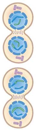
FIGURE 9.31. Telophase II
During the telophase II stage, the nuclear membranes form, chromosomes uncoil. Cytokinesis occurs.
Telophase II is followed by the maturation of the four cells into gametes—either sperm or eggs. In many organisms, including humans, egg cells are produced in such a manner that three of the four cells resulting from meiosis in a female disintegrate. However, because the one that survives is randomly chosen, the likelihood of obtaining any particular combination of genes is not affected. The events of meiosis II are shown in figure 9.32.
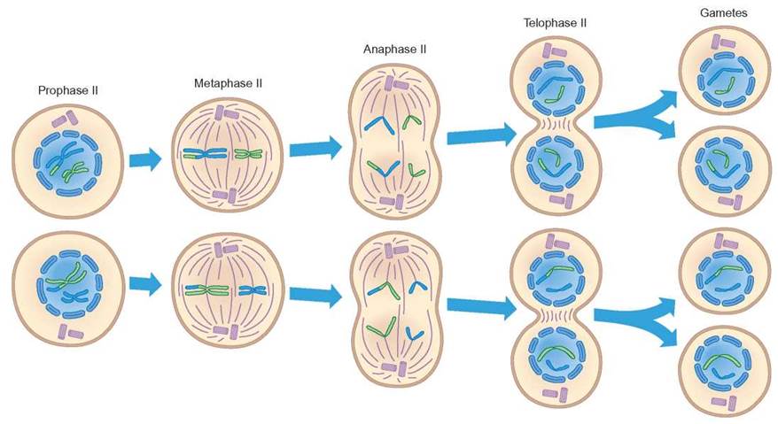
FIGURE 9.32. Meiosis II
During meiosis II, the centromere of each chromosome splits and each chromosome divides into separate chromatids. Four haploid cells are produced, each with 1 chromatid of each kind. These four
haploid cells are gametes.
The stages of meiosis I and meiosis II are summarized in table 9.3. The differences between mitosis and meiosis have been identified throughout this chapter. A comparison of these two processes appears in table 9.4.
TABLE 9.3. Stages of Meiosis
Interphase |
|
Diploid |
As the diploid (2n) cell moves from G0 into meiosis, the chromosomes replicate during the S phase of interphase. |
Prophase I |
|
Diploid |
The replicated chromatin begins to coil into recognizable chromosomes and the homologous chromosomes synapse; chromatids may cross-over; the nuclear membrane and nucleoli fragment; centrioles move to form the cell's poles; spindle fibers are formed. |
Metaphase I |
|
Diploid |
Synapsed homologous chromosomes attach to the spindle fibers at their centromeres. Pairs of homologous chromosomes align at the equator. |
Anaphase I |
|
Transition |
The two members of homologous pairs of chromosomes separate from each other as they move toward the poles of the cell. |
Telophase I |
|
Haploid |
The two newly forming daughter cells are now haploid (n) because each contains only one of each pair of homologous chromosomes; the nuclear membranes and nucleoli re-form; spindle fibers fragment; the chromosomes unwind and change from chromosomes (composed of 2 chromatids) to chromatin. |
Prophase II |
|
Haploid |
Each of the two haploid (n) daughter cells from meiosis I undergoes chromatin coiling to form chromosomes, each of which is composed of 2 chromatids; the nuclear membrane fragments; centrioles move to form the cell's poles; spindle fibers form. |
Metaphase II |
|
Haploid |
Chromosomes attach to the spindle fibers at the centromeres and move to the equator of the cell. |
Anaphase II |
|
Haploid |
Centromeres separate, allowing the 2 chromatids of a chromosome to separate toward the poles. |
Telophase II |
|
Haploid |
Four haploid (n) cells are formed from the division of the two meiosis I cells; the nuclear membranes and nucleoli re-form; spindle fibers fragment; the chromosomes unwind and change from chromosomes to chromatin; these cells become the sex cells (egg or sperm). |
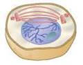
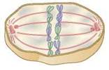
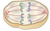
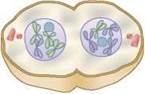
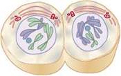
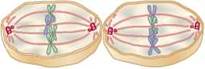
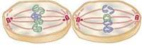
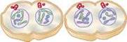
TABLE 9.4. Comparison of Mitosis and Meiosis
Mitosis |
Meiosis |
1. One division completes the process. |
1. Two divisions are required to complete the process. |
2. Chromosomes do not synapse. |
2. Homologous chromosomes synapse in prophase I. |
3. Homologous chromosomes do not cross-over. |
3. Homologous chromosomes cross-over in prophase I. |
4. Centromeres divide in anaphase. |
4. Centromeres divide in anaphase II but not in anaphase I. |
5. Daughter cells have the same number of chromosomes as the parent cell (2n → 2n or n → n). |
5. Daughter cells have half the number of chromosomes as the parent cell (2n → n). |
6. Daughter cells have the same genetic information as the parent cell. |
6. Daughter cells are genetically different from the parent cell. |
7. Mitosis generates body cells. |
7. Meiosis generates sex cells. |
8. Mitosis results in growth, the replacement of worn-out cells, and the repair of damage. |
8. Meiosis is necessary for sexual reproduction. |
9.8. CONCEPT REVIEW
22. Diagram the metaphase I stage of a cell with the diploid number of 8.
23. What is unique about prophase I?
24. In which phase of meiosis do daughter chromosomes form?
25. Why is it impossible for synapsis to occur during meiosis II?
26. Can a haploid cell undergo meiosis? Why or why not?
27. List three differences between mitosis and meiosis.