CONCEPTS IN BIOLOGY
PART III. MOLECULAR BIOLOGY, CELL DIVISION, AND GENETICS
9. Cell Division—Proliferation and Reproduction
9.10. Nondisjunction and Chromosomal Abnormalities
In the normal process of meiosis, the number of chromosomes in diploid cells is reduced to haploid. This involves segregating homologous chromosomes into separate cells during the first meiotic division. Occasionally, a pair of homologous chromosomes does not segregate properly and both chromosomes of a pair end up in the same gamete. Nondisjunction occurs when homologous chromosomes do not separate during meiosis. In figure 9.37, two cells are missing a chromosome and the genes that were carried on it. This condition usually results in the death of the cells. The other cells have an extra copy of a chromosome. This extra genetic information may also lead to the death of the cell. Some of these abnormal cells, however, do live and develop into sperm or eggs.
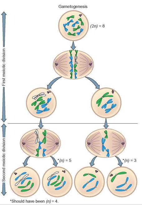
FIGURE 9.37. Nondisjunction During Gametogenesis
When a pair of homologous chromosomes fails to separate properly during meiosis I, gametogenesis results in gametes that have an abnormal number of chromosomes. Notice that two of the cells have an additional chromosome, whereas the other two are deficient by the same chromosome.
If one of these abnormal sperm or eggs unites with a normal gamete, the offspring will have an abnormal number of chromosomes. In monosomy, instead of the normal two chromosomes, a cell has just one of the pair of homologous chromosomes. In trisomy, a chromosome is present in three copies. All the cells that develop by mitosis from such zygotes will also have an abnormal number of chromosomes.
It is possible to examine cells and count chromosomes. Among the easiest cells to view are white blood cells. They are dropped onto a microscope slide, so that the cells are broken open and the chromosomes are separated. Photographs are taken of chromosomes from cells in the metaphase stage of mitosis. The chromosomes in the pictures can then be cut and arranged for comparison with known samples (figure 9.38). This picture of an individual’s chromosomal makeup is referred to as a karyotype.
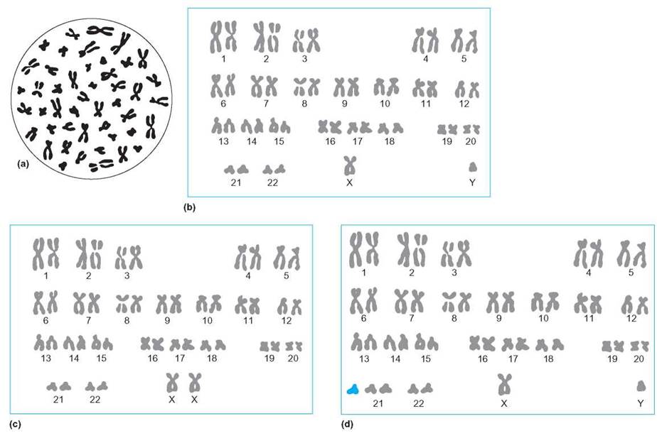
FIGURE 9.38. Human Male and Female Chromosomes
The randomly arranged chromosomes shown in the circle simulate metaphase cells spattered onto a microscope slide (a). Those in parts (b) and (c) have been arranged into homologous pairs. Part (b) shows a male karyotype, with an X and a Y chromosome, and (c) shows a female karyotype, with two X chromosomes. (d) Notice that each pair of chromosomes is numbered and that the person from whom these chromosomes were taken has an extra chromosome number 21. The person with this trisomic condition might display a variety of physical characteristics, including slightly thickened eyelids, flattened facial features, a large tongue, and short stature and fingers. Most individuals also display some mental retardation. This condition is known as Down syndrome.
One example of the effects of nondisjunction is the condition known as Down syndrome. If a gamete with 2 number 21 chromosomes has been fertilized by a gamete containing the typical one copy of chromosome number 21, the resulting zygote has 47 chromosomes—one more than the expected count of 46 chromosomes (figure 9.38d). The child who developed from this fertilization has 47 chromosomes in every cell of his or her body as a result of mitosis and thus can have the symptoms characteristic of Down syndrome. These include thickened eyelids, a large tongue, flattened facial features, short stature and fingers, some mental impairment, and faulty speech (figure 9.39).

FIGURE 9.39. Down Syndrome
Every cell in the body of a person with Down Syndrome has 1 extra chromosome. With special care, planning, and training, people with this syndrome can lead happy, productive lives.
In the past, it was thought that the mother’s age at childbirth played an important role in the occurrence of trisomies, such as Down syndrome. In women, gametogenesis begins early in life, but cells destined to become eggs are put on hold during meiosis I. Beginning at puberty and ending at menopause, one of these cells completes meiosis I monthly. This means that cells released for fertilization later in life are older than those released earlier in life. Therefore, it was believed that the chances for abnormalities, such as nondisjunction, increase as the mother ages. However, the evidence no longer supports this age-egg link. Currently, the increase in the frequency of trisomies with age has been correlated with a decrease in the activity of a woman’s immune system. As she ages, her immune system is less likely to recognize the difference between an abnormal and a normal embryo. This means that miscarriage is less common and she is more likely to carry an abnormal fetus to full term.
Figure 9.40 illustrates the frequency of the occurrence of Down syndrome births at various ages in women. Notice that the frequency increases very rapidly after age 37. Physicians normally encourage older women who are pregnant to have the cells of their fetus checked to see if they have the normal chromosome number. Nondisjunction can occur in either the production of eggs or sperm, so either parent can be the cause of an abnormal chromosome number.
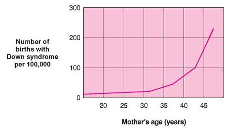
FIGURE 9.40. Down Syndrome as a Function of a Mother's Age
Notice that, as the age of the woman increases, the frequency of births of children with Down Syndrome increases only slightly until the age of approximately 37. From that point on, the rate increases drastically. This increase is thought to occur because older women experience fewer miscarriages of abnormal embryos.
9.10. CONCEPT REVIEW
30. Define the term nondisjunction.
31. What is the difference between monosomy and trisomy?
Summary
Cell division is necessary for growth, repair, and reproduction. Mitosis and meiosis are two important forms of cell division. Cells go through a cell cycle, a nondividing period when normal cell activities take place followed by DNA replication, and cell division (mitosis and cytokinesis). Interphase is the period of growth and preparation for division. Mitosis is divided into four stages: prophase, metaphase, anaphase, and telophase. During mitosis, two daughter nuclei are formed from one parent nucleus. These nuclei have identical sets of chromosomes and genes that are exact copies of those of the parent. The regulation of mitosis is important if organisms are to remain healthy. Regular divisions are necessary to replace lost cells and to allow for growth. However, uncontrolled cell division may result in cancer and disruption of the total organism’s well-being.
Meiosis is a specialized process of cell division, resulting in the production of four cells, each of which has the haploid number of chromosomes. The total process involves two sequential divisions, during which one diploid cell reduces to four haploid cells. Mutations and various processes of meiosis, such as crossing-over, segregation, and independent assortment, ensure that all sex cells are unique. The various mechanisms that generate genetic diversity in sexually reproducing organisms assure that when two gametes unite, the individual offspring is genetically unique.
Basic Review
1. What is the key difference between mitosis and meiosis?
a. Mitosis involves two rounds of cell division, whereas meiosis involves one round of cell division.
b. DNA is not split between cells in meiosis, but this does occur during mitosis.
c. Mitosis produces cells genetically identical to the parent, whereas meiosis produces cells with half the genetic information as the parent.
d. None of the above is correct.
2. Which of the following is true of interphase?
a. The chromosomes line up on the equatorial plane.
b. DNA replication occurs in this phase.
c. The DNA in the cell halves.
d. All of the above are true.
3. Chromosomes are most likely to appear to be lining up near the middle of the cell during which phase of mitosis?
a. interphase
b. prophase
c. metaphase
d. telophase
4. Which of the following types of information do cells use to determine if they will divide?
a. genetic health
b. their current location
c. the need for more cells
d. All of the above are correct.
5. p53 mutations lead to cancer because
a. DNA damage is not repaired.
b. mutated cells are allowed to grow.
c. multiple mutations in the cell’s regulatory proteins occur.
d. All of the above are correct.
6. Haploid cells
a. carry two copies of the genetic information.
b. carry one copy of the genetic information.
c. carry partial copies of the genetic information.
d. are mutant.
7. Reduction division occurs
a. in meiosis II.
b. in meiosis I.
c. in mitosis.
d. after fertilization.
8. Genetic diversity in the gametes of an individual is generated through:
a. mitosis.
b. independent assortment.
c. crossing-over.
d. both b and c.
9. Trisomy means
a. that three copies of a chromosome are present.
b. Down syndrome.
c. that only three cells are present.
d. none of the above.
10. A nondisjunction event occurs when
a. homologous chromosomes did not separate correctly.
b. non-homologous chromosomes did not separate correctly.
c. daughter cells did not undergo cytokinesis correctly.
d. None of the above is correct.
11. Chemical changes of chromatin (DNA and histones) that do not alter the nucleotide sequence are called _____ changes.
12. Mutagens can be carcinogens. (T/F)
13. _____ is the cellular process of deciding which genes a cell will express when mature.
14. The gonads in females are known as _____.
15. These features characterize which kind of cell division?
(a) Homologous chromosomes do not cross-over.
(b) Centromeres divide in anaphase.
Answers
1. c 2. b 3. c 4. d 5. d 6. b 7. b 8. d 9. a 10. a 11. epigenetic 12. T 13. Determination 14. ovaries 15. mitosis.
Thinking Critically
Cancer, p53, Antibodies and Nanoparticles
A molecular oncologist and her colleagues at Georgetown University have developed a nanoparticle that is coated with a tumor-targeting antibody. The nanoparticle is able to locate primary and hidden metastatic tumor cells and deliver a fully functioning copy of the p53 tumor-suppressor gene. The presence of the p53 gene improves the efficacy of conventional cancer therapies such as chemo- and radiation therapy and reduces their side effects. Review the material on cell membranes, antibodies, cancer, and the role of p53 and explain the details of this treatment to a friend. (You might explore the Internet for further information.)