Biology For Dummies
Part IV Systems Galore! Animal Structure and Function
Chapter 15
Going with the Flow: Respiratory and Circulatory Systems
In This Chapter
Discovering the four ways animals exchange gases
Understanding how circulatory systems feed cells and take out their trash
Examining the heart and circulatory systems of not-so-complex animals
Tracing the path of blood through your heart and blood vessels
Surveying the details of your body’s most important fluid — blood
Every living thing needs to be able to exchange materials (think food, oxygen, and waste products) with the environment and circulate these materials around their bodies. Complex animals use respiratory systems to exchange gases such as oxygen and carbon dioxide with their environment and circulatory systems (often called cardiovascular systems when they’re in animals with backbones) to move nutrients and gases around their bodies.
In this chapter, we present some of the different processes used by animals to exchange and move important materials. In particular, we focus on the details of the human respiratory and circulatory systems.
Passing Gas: How Animals “Breathe”
All animals, from worms and fish to insects and humans, must exchange gases between themselves and their environment on a continual basis throughout every moment of their lives. Simple animals exchange gases throughout their entire body surfaces, whereas more complex animals have respiratory systems that process the air from the environment. This latter group relies on breathing to simultaneously move oxygen gas from the air into the body and remove carbon dioxide from the body and return it to the air. Ultimately, whichever way an animal does it, the actual exchange of oxygen and carbon dioxide gases between the animal and its environment occurs across a moist surface.
 Respiration is the entire process of taking air in, exchanging needed gases for unnecessary gases, using the needed gases, and releasing the waste form of gases.
Animals use four types of gas-exchange systems, which we cover in detail in the next sections:
Integumentary exchange occurs through the outer surface of an animal. Worms and amphibians employ this system.
Gills are external structures that exchange gases in watery environments, which is why you find gills on many forms of marine life.
Tracheal exchange systems rely on a network of tubes that end in holes to move oxygen and carbon dioxide throughout the bodies of certain types of insects.
Lungs are internal structures that use diffusion to transport gases into and out of the body. Land animals, including humans, and marine mammals, such as dolphins and whales, have lungs.
Integumentary exchange
To understand integumentary exchange, you first need to know what the heck an integument is. The integument is the outer covering of an animal. In worms, frogs, and salamanders — all of which respire through integumentary exchange — the integument is more of an outer membrane; in you, the integument is your skin.
Small animals that constantly stay moist may “breathe” right through their skin. Oxygen from the air diffuses through such an animal’s moist surface and into the fluids in its body; at the same time, carbon dioxide diffuses out. (Refer to Chapter 4 for a full explanation of diffusion.) Because oxygen and carbon dioxide are swapped across the integument, this process is called integumentary exchange.
Earthworms are a perfect example of integumentary exchange in action. They have small blood vessels called capillaries right under their “skin.” As an earthworm moves through the soil, it loosens the soil, creating air pockets. The worm takes in oxygen from the air pockets and releases carbon dioxide right through its outer surface.
You know how worms get flooded out of the ground when it rains and end up all over your driveway and sidewalk? Well, they head right back into the soil as soon as they can (and not just because they’re potential bird food). If they lingered on your driveway, their outer surfaces would dry out, preventing them from taking in oxygen and getting rid of carbon dioxide. When this happens, they die. (This is also the reason why pouring salt on a slug stops it in its slimy tracks. The salt dehydrates its outer surface, which prevents it from exchanging gases.)
Gills
Animals that live in water, including lobsters and starfish, have gills, which are extensions of their outer membranes. The membranes in gills are very thin (usually just one cell thick), which allows for easy gas exchange. Capillaries connect to the cells in the gills so that gases can be taken in from the water and passed into the bloodstream of the aquatic animal. Also, gaseous waste can diffuse from the capillaries into the cells of the gills and pass out into the watery environment.
The type of gill you’re probably most familiar with is that of a fish. In fish, the gills are membranous filaments covered by a flap called an operculum, which is the flap you can see opening and closing on the head of a fish. A fish opens and closes the flap by opening and closing its mouth. After water enters the mouth, it’s forced over the gills and then out the back of the operculum. As the water passes over the gills in one direction, the blood inside the gills moves in the opposite direction. Oxygen from the water diffuses into the capillaries in the gills, and carbon dioxide diffuses out of the capillaries in the gills. After the oxygen is in the capillaries, it can be transported throughout the fish’s body so all the cells can get some of the needed gas.
Because the water outside the gill and blood inside the gill are moving in opposite directions, the exchange of gases in a fish gill is referred to as countercurrent exchange. Countercurrent exchange improves the efficiency of gas exchange.
Tracheal exchange systems
Tracheal exchange systems are made possible by a network of tubes called a trachea. The holes at the ends of the tubes, which open to the outside surface, are called spiracles. You can find this system in insects.
The trachea found in insects is different from the trachea found in your body. In insects, the trachea is the network of tubes that runs through the entire body and opens to the air; in humans, the trachea is a tube that carries air down into the lungs.
In a tracheal exchange system, oxygen diffuses directly into the trachea, and carbon dioxide exits through the spiracles. The cells of the body exchange air directly with the tracheal system, and the oxygen and carbon dioxide don’t need to be carried through a circulatory system because the tracheal system runs through all parts of the insect’s body.
 Some insects, such as bees and grasshoppers, combine a breathing process with a tracheal exchange system. They contract muscles to pump air in and out of their tracheal systems. A grasshopper even has air sacs on some of the air tubes in its tracheal system. The bags “pump” like fireplace bellows after pressure from muscles is applied.
Lungs
Lungs are the opposite of gills (which we describe earlier in this chapter). Gills extend out off of an organism, and lungs are internal growths of the surface of the body. Animals’ lungs are housed inside their bodies in order to keep them moist. They basically work by providing lots of moist surface area for the diffusion of oxygen and carbon dioxide. Lungs may be different shapes and sizes in various land animals, but they function essentially the same as they do in humans. We use humans as the model for the mechanics of the lungs in order to give you a better understanding of how your body works.
Humans have a pair of lungs that lie in the chest cavity (as shown in Figure 15-1); one lung is on the left side of the trachea (the tube that connects the nose and mouth to the lungs), and the other is on the right side of it. Inside the lungs, the trachea branches off into bronchi (small passageways that move air into the lungs), which then branch and rebranch off into smaller bronchioles (smaller versions of bronchi). The bronchioles end in little clusters of sacs called alveoli (tiny, moist sacs where gas exchange occurs in the lungs) that look a little bit like raspberries. Each alveolus (that’s the singular word for alveoli) is wrapped with capillaries so that gas exchange can occur between the lungs and the blood. The muscle found underneath the lungs is called the diaphragm, and it’s responsible for contracting and creating negative pressure to draw air into the lungs. A pair of ribs surrounds the chest cavity to protect the lungs (and heart) and to assist in the motions of breathing.
Because lungs are the most complex gas-exchange system employed by animals, we break down the details for you in the following sections.
Figure 15-1:Anatomic structures of the human respiratory system.
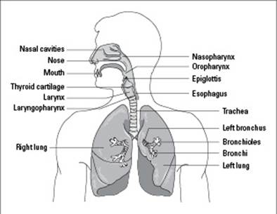
From LifeART®, Super Anatomy 1, © 2002, Lippincott Williams & Wilkins
Taking a peek at what happens when you breathe
When you breathe in, called inhalation or inspiration, the diaphragm muscle contracts (meaning it becomes smaller and moves down), allowing your rib cage to move upward and outward. Because the lungs have more room when your chest is expanded, they open up, similar to how a balloon blows up when it’s filled with air. The opening up of the lungs means there’s more room in the lungs, so air rushes in to fill the space. When your diaphragm relaxes, your rib cage moves back downward and inward, increasing air pressure inside your lungs and forcing air out. The process of breathing out is called exhalation or expiration.
Following is a breakdown of how oxygen passes through all the branches of your respiratory system when your lungs fill up:
1. Oxygen enters through your nostrils and flows through the top part of your throat.
Inside your nasal cavity, hair, cilia, and mucus trap dust and dirt particles, purifying the air that enters your lungs. Occasionally, you must cough and either spit or swallow to move the trapped particles out of your throat. (Don’t worry about swallowing dirt; it enters your stomach where it’s digested and excreted.)
2. Oxygen then moves into the middle part of your throat and through the space around your vocal cords.
When you eat, food passes through your throat (or pharynx) on its way to your stomach. When you breathe, air passes through your pharynx on its way to the lungs. So, both your mouth and your nose connect to the pharynx. The place where your mouth connects is called the oropharynx; the place where your nose connects is called the nasopharynx.
3. Next, oxygen enters your trachea, flows through the bronchi and bronchioles, and then flows into the alveoli.
Capillaries surround the alveoli, ready to accept oxygen and give up carbon dioxide (see Figure 15-2). Oxygen and carbon dioxide diffuse across the membranes of the alveoli, and the capillaries move the freshly oxygenated blood into your circulatory system.
Gas exchange in the lungs occurs only in the alveoli.
Figure 15-2:Oxygenation of blood at the respiratory membrane.
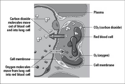
From LifeART®, Super Anatomy 2, © 2002, Lippincott Williams & Wilkins
Understanding the concept of diffusion
As explained in Chapter 4, diffusion is when a substance moves from an area of higher concentration to an area of lower concentration. Gas exchange in the lungs is an example of diffusion.
Oxygen is more concentrated within each of the 300 million alveoli in your lungs than it is in the capillaries surrounding the alveoli. Your capillaries in turn have a higher concentration of carbon dioxide than oxygen. The oxygen in the alveoli diffuses across the alveoli’s membranes and the capillaries’ membranes and into the capillaries. Meanwhile, the carbon dioxide diffuses across the capillaries’ membranes and the alveoli’s membranes into the lungs for expulsion from your body. After oxygen is in the capillaries, your red blood cells snatch it up and transport it throughout your body. While this happens, your diaphragm relaxes, and the carbon dioxide waste leaves your body as you exhale.
Circulation: Nutrients In, Garbage Out
Every animal alive possesses a circulatory system. This system makes sure that all the animal’s cells obtain the nutrients they need to function and that all the cells dispose of waste so it doesn’t build up within the animal’s body and cause illness (or, yikes, death). Some of the additional responsibilities of a circulatory system include
Delivering oxygen to cells and picking up carbon dioxide
Distributing hormones to cells
Maintaining body temperature by transporting heat
Transporting cells to fight infection (more on this in Chapter 17)
Two kinds of circulatory systems exist: open and closed. We fill you in on both types in the next sections.
Open circulatory systems
In an open circulatory system, the animal’s heart pumps a bloodlike fluid called hemolymph into an open cavity (called a hemocoel) through openings in the heart called ostia. When the hemolymph flows into the hemocoel, it directly bathes the tissues of the organism with nutrients — no blood vessels are involved. Muscle contractions push the hemolymph back toward the heart so it can be circulated throughout the animal again and again. Insects and some mollusks (specifically snails and clams) possess an open circulatory system.
In insects, hemolymph carries nutrients but not oxygen to the cells. Oxygen is circulated via the tracheal exchange system (which we describe earlier in this chapter).
Closed circulatory systems
A closed circulatory system is the type of circulatory system you’re most familiar with. It has a network of vessels (think of them as highways that connect one organ to another) that perform the transportation and keep your blood from seeping out. In animals, each blood vessel in the network is responsible for transporting nutrients and oxygen to the cells and removing wastes and carbon dioxide from them.
The three types of blood vessels are
Arteries
Veins
Capillaries
Animals with backbones, called vertebrates, possess a closed circulatory system. Some animals without backbones, called invertebrates, also have one; these include some worms, octopuses, and squids.
Closed circulatory systems, which are said to be closed because they have vessels that contain the fluid, are more efficient than open circulatory systems because they need to meet the dual demand of delivering both oxygen and nutrients to cells.
Getting to the Heart of Simpler Animals
Hearts come in different sizes and shapes, but a heart’s function remains the same in all organisms that have one: It pumps fluid throughout the circulatory system. That fluid is either hemolymph or blood, depending on the type of circulatory system an animal has, and it transports either nutrients or a combination of nutrients and oxygen to the animal’s cells.
In animals with an open circulatory system, this process occurs the same way. However, in animals with a closed circulatory system, the process happens differently depending on how the system is set up. We give you a peek at how the process occurs in two of the simpler animals — worms and fish — in the sections that follow.
A worm’s heart and circulatory system
Although you may think earthworms are insects that possess an open circulatory system, they’re not (technically they’re annelids). These little guys and gals have a closed circulatory system that’s a bit simpler in design than that of a human.
Earthworms have just one dorsal (top side) blood vessel and one ventral (bottom side) blood vessel, plus a network of capillaries. The heart of an earthworm is a series of muscular rings near the thicker tip of the worm. It pumps blood away from the heart through the ventral blood vessel. From there, the blood oozes into all the capillaries to reach all the cells of the worm before traveling back to the heart through the dorsal blood vessel.
A fish’s heart and circulatory system
A fish’s heart has two separate chambers, one that receives blood from the body and another that pumps the blood out over the gills. Its closed circulatory system makes a single loop through the body of the fish. Overall, the process of how blood passes through a fish is rather simple.
1. When a fish’s heart pumps, blood leaves the heart through the ventral aorta that runs along the underside of the fish.
2. The ventral aorta carries the blood to the gills, and the blood then passes through the capillaries along the gills to pick up oxygen.
This part of the circulatory loop is called gill circulation.
3. The oxygenated blood immediately flows from the gills into the dorsal aorta, which runs along the upper side of the fish.
4. The dorsal aorta carries the oxygenated blood to the rest of the fish’s capillaries.
This part of the loop is referred to as systemic circulation.
5. After the blood has reached all the cells within the fish, it returns to the heart.
The circulatory system in a fish is simple and effective, but because the blood passes through the heart only once during its travels, a fish’s blood pressure is quite low. (Blood pressure is the force that sends blood through an animal’s circulatory system.)
Exploring the Human Heart and Circulatory System
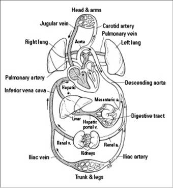
The heart and circulatory system of a human, as well as some other mammals, are complex. Because these animals are larger, they need to have a higher blood pressure to push the blood throughout their entire bodies. This need results in a two-circuit circulatory system, a system that has two distinct pathways (as you can see in Figure 15-3):
One pathway is for pulmonary circulation, which first delivers deoxygenated blood to the lungs so it can become oxygenated and then delivers oxygenated blood back to the heart.
The other circuit is for systemic circulation, which carries oxygenated blood from the heart to the rest of the body.
Figure 15-3:Pulmonary circulation and systemic circulation work together.
The human heart (depicted in Figure 15-4) has four chambers: two ventricles, muscular chambers that squeeze blood out of the heart and into the blood vessels, and two atria, muscular chambers that drain and then squeeze blood into the ventricles. The heart is divided into left and right halves, so there’s a left atrium and a left ventricle, as well as a right atrium and a right ventricle. The two atria reside at the top of the heart; the two ventricles are at the bottom.
Note: If Figure 15-4 seems confusing to you because it looks as if the right atrium is on the left side of the heart, pretend you’re looking at someone’s heart through her chest. Her right atrium is on the right side of her body, which is how we’ve depicted it in the figure.
Figure 15-4: The structures of the human heart and the flow of blood through them.
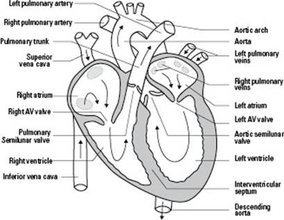
Your heart is divided into halves because of your two-circuit circulatory system. The right side of your heart pumps blood to your lungs, while the left side of your heart pumps blood to the rest of your body. Blood goes into both pathways with each and every pump.
Valves separate one chamber of the heart from another. Each valve consists of a pair of strong flaps of muscle tissue, called cusps or leaflets. When your heart is working properly, the valves open and close fully so blood can flow through it in only one direction.
Four valves separate the four chambers of your heart from one another and from the major blood vessels that are connected to it (refer to Figure 15-4 for the visual):
The right atrioventricular (AV) valve is located between the right atrium and right ventricle. This valve is also referred to as the tricuspid valve because it has three flaps in its structure.
The pulmonary semilunar valve separates the right ventricle from the pulmomary artery. Semilunar means “half-moon” and refers to the shape of the valve.
The left atrioventricular (AV) valve is between the left atrium and left ventricle. This valve is also called the bicuspid valve because it has only two flaps in its structure.
The aortic semilunar valve separates the left ventricle from the aorta. Like the pulmonary semilunar valve, this valve has a half-moon shape.
The sections that follow take you on a journey through the human circulatory system. You find out just how blood moves through your heart and the rest of your body, and you discover what gets your heart beating in the first place. Bon voyage!
The many faces of heart disease
Heart disease is the top cause of death in the United States. Deaths from heart disease are usually attributed to heart attacks, but heart attacks can be caused by many factors, including the following:
Atherosclerosis: Blockages in the arteries occur when fats, especially cholesterol, accumulate in the lining of the arteries. Cholesterol serves a necessary function within the body, but when too much cholesterol is present — which can be caused by diet or by genetic factors — it starts to stick to the vessels instead of passing through them. These fatty deposits are called plaques. As the plaques increase in size, they fill more and more of the artery, eventually affecting blood flow.
Hypertension: More commonly referred to as “high blood pressure,” hypertension puts added stress on the arteries of the heart, increasing the risk of damage and fatty deposits along artery walls.
Ischemic heart disease: Ischemia is a lack of oxygen, and this type of heart disease can occur when the arteries are partially blocked. People with this condition have difficulty breathing during exercise or times of stress because the blocked arteries slow the flow of blood, preventing enough oxygen from being delivered to the heart muscle’s tissues. This lack of oxygen can cause a pain in the chest that radiates to the left arm; this pain is called angina pectoris.
Thromboembolism: If an artery is blocked by a plaque, blood cells can stick to the plaque, eventually forming a blood clot that can travel through the bloodstream and block blood vessels. A blood clot stuck in a blood vessel is called a thrombus. If a thrombus breaks free and moves around the bloodstream, it’s called an embolism.
Entering the cardiac cycle
Your heart is an impressive little organ. Even though it’s only as big as a clenched adult fist, it pumps 5 liters of blood (the equivalent to 21//2 big bottles of soda) throughout your body 70 times a minute. Did we mention it’s a hard worker too? Your heart never stops working from the time it starts beating (when you were nothing but a wee little embryo in your mother’s womb) until the moment you die. It doesn’t even get an entire second to rest. It beats continuously every 0.8 seconds of your life; this is known as the cardiac cycle.
During the cardiac cycle, your heart forces blood into your blood vessels and takes a quick nap. Here’s exactly what happens:
The left and right atria contract.
The left and right ventricles contract.
The atria and ventricles rest (for just 0.4 seconds).
When the atria and ventricles are resting, the muscle fibers within them aren’t contracting. Therefore, the relaxed atria allow the blood within them to drain into the ventricles beneath them. With most of the blood from the atria now in the ventricles, the atria contract to squeeze any remaining blood down into the ventricles. Then the ventricles immediately contract to force blood into the blood vessels.
The period of relaxation in the heart muscle is referred to as diastole, and the period of contraction in the heart muscle is called systole. If these terms sound familiar, it’s probably because you’ve heard them used in terms of blood pressure.
In a blood pressure reading, such as the normal value of 120/80 mmHg, 120 is the systolic blood pressure, the pressure at which blood is forced from the ventricles into the arteries when the ventricles contract, and 80 is the diastolic blood pressure, the pressure in the blood vessels when the muscle fibers are relaxed. The abbreviation mmHg stands for millimeters of mercury (Hg is the chemical symbol for mercury).
If your blood pressure is 140/90 mmHg, which is the borderline value between normal and high blood pressure, that means your heart is working harder to pump blood through your body and not relaxing as well between pumps. This reading indicates that something is causing your heart to have to work at a much higher level all the time to keep blood flowing through your body, which stresses it out. The culprit could be a hormonal imbalance, a dietary problem (too much sodium or caffeine), a mechanical problem in the heart, a side effect of medication, or blockages in your blood vessels.
Navigating the path of blood through the body
The cardiac cycle, which describes the rhythmic contraction and relaxation of the heart muscle (see the preceding section), coincides with the path of blood through your body. As each atrium and ventricle contracts, blood is pumped into certain major blood vessels that connect to your heart and then continues flowing throughout your circulatory system. In other words, here’s where your two-circuit circulatory system really comes into play.
The following sections describe the process of pulmonary and systemic circulation, as well as the process of capillary exchange (which gets nutrients into and wastes out of your cells).
Oxygenating the blood: Pulmonary circulation
Pulmonary circulation, the first pathway of your two-circuit circulatory system, brings blood to your lungs for oxygenation. Following is a rundown of how blood moves during pulmonary circulation (trace the path in Figure 15-4 as you read):
1. Deoxygenated blood from your body enters the right atrium of your heart through the superior vena cava and the inferior vena cava.
Superior means “higher,” and inferior means “lower,” so the superior vena cava is at the top of the right atrium, and the inferior vena cava is at the bottom of the right atrium.
2. From the right atrium, the deoxygenated blood drains into the right ventricle through the right AV valve.
When the ventricles contract, the right AV valve closes off the opening between the ventricle and the atrium so blood doesn’t flow back up into the atrium.
3. The right ventricle then contracts, forcing the deoxygenated blood through the pulmonary semilunar valve and into the pulmonary artery.
The pulmonary semilunar valve keeps blood from flowing back into the right ventricle after it’s in the pulmonary artery.
4. The pulmonary artery carries the blood that’s very low in oxygen to the lungs, where it becomes oxygenated.
Spreading oxygenated blood around: Systemic circulation
Systemic circulation brings oxygenated blood to the cells of your body. Here’s how blood moves through this pathway (you can follow along by tracing the path in Figure 15-4):
1. Freshly oxygenated blood returns from the lungs to the heart via the pulmonary veins.
Note that your pulmonary veins are the only veins in your body that contain oxygenated blood; all of your other veins contain deoxygenated blood.
2. The pulmonary veins push the oxygenated blood into the left atrium, which then relaxes, allowing the blood to drain into the left ventricle through the left AV valve.
3. As the left ventricle contracts, the oxygenated blood is pumped into the main artery of the body — the aorta.
To get to the aorta, blood passes through the aortic semilunar valve, which serves to keep blood in the aorta from flowing back into the left ventricle.
4. The aorta branches into other arteries, which then branch into smaller arterioles, carrying oxygenated blood all around your body.
Throughout your body, arterioles meet up with capillaries where oxygen is exchanged for carbon dioxide.
The blood vessels in order of decreasing oxygen content are as follows:
Arteries
Arterioles
Capillaries
Venules
Veins
Exchanging the good and the bad: Capillary exchange
For being the teeniest of blood vessels, capillaries, which bridge the smallest of the arteries and the smallest of the veins, have a rather important role: to facilitate the exchange of materials between capillaries and cells via diffusion — in other words, to make capillary exchange possible.
Your capillaries are only as thick as one cell, so the contents within them can easily exit by diffusing through the capillaries’ membranes (see Chapter 4 for more on diffusion). And, because the capillaries’ membranes touch the membranes of other cells all over the body, the capillaries’ contents can easily continue moving through adjacent cells’ membranes.
Through capillary exchange, oxygen leaves red blood cells in the bloodstream and enters all the other cells of the body. Capillary exchange also allows nutrients to diffuse out of the bloodstream and into other cells. At the same time, the other cells expel waste products, including carbon dioxide, that then enter the capillaries.
After the capillaries “pick up” the garbage from other cells, they carry the wastes and carbon dioxide through the deoxygenated blood to the smallest of the veins, which are called venules. The venules branch into bigger vessels called veins, which then carry the deoxygenated blood toward the main vein — the vena cava. The two branches of the vena cava enter the right atrium, which is where pulmonary circulation begins.
The pressure created when the ventricles contract is what forces blood through the arteries. However, this pressure declines as the blood gets farther away from the heart and into the capillaries. Blood pressure doesn’t force blood through the veins as it does through the arteries. What makes blood travel through the veins are contractions of your skeletal muscles. As your limbs and trunk move, deoxygenated blood is pushed farther along the venules and veins, eventually returning to the heart. Without movement, blood pools in the veins, creating poor circulation.
Seeing what makes your ticker tick
Electrical impulses from your heart muscle cause your heart to beat. These impulses begin in special areas of tissue within the heart, called nodes, that are infused with nerves. The nodes send out signals that stimulate contraction of the heart muscle cells, causing your heart to beat. For particulars on the process, check out the following:
1. Each beat of your heart is started by an electrical signal from the sinoatrial (SA) node in your right atrium.
The SA node is also called your natural pacemaker because it sets the pace of your heartbeats. (Yes, your heart already contains a pacemaker. People who have pacemakers “installed” through surgery have it done because their natural pacemaker has stopped working correctly.)
2. The signal from the SA node spreads across the left and right atria, causing them to contract and push the blood into the ventricles.
3. As the electrical impulse passes through the atria, it signals the atrioventricular (AV) node to take action.
The AV node is located in the lower part of the right atrium. The signal is temporarily slowed in this node so that your ventricles have time to fill with blood.
4. The signal in the AV node stimulates an area of tissue called the bundle of His.
The bundle of His lies between the right and left ventricles and connects with specialized fibers called Purkinje fibers.
5. When the impulse reaches the Purkinje fibers, it causes the ventricles to contract, completing the heartbeat.
The sound your heart makes — described as lub-dub, lub-dub — is attributed to the closing of the heart’s valves. The first heart sound, the lub, is caused by AV valves closing to prevent backflow from the ventricles into the atria. The second heart sound, the dub, occurs when the semilunar valves close to keep blood in the aorta from flowing back into the left ventricle.
A Bloody-Important Fluid
Blood is the fluid that sustains life in animals with a closed circulatory system — including you. Some blood cells carry oxygen, which is necessary for metabolic reactions; some blood cells fight off invading substances that could destroy your cells; and other blood cells help form clots, which keep your body from losing too much of this precious fluid and assist with wound healing. The following sections introduce you to the elements that make up your blood and the special process that keeps you from losing too much blood when you cut yourself.
The solids found in your essential fluid
Believe it or not, your blood, although it’s a fluid, contains solid parts called formed elements. These solid parts are your red blood cells, white blood cells, and platelets. You can find out more about all three in the sections that follow.
Note: We use the word solid simply to differentiate platelets and blood cells from the liquid portion of blood. Platelets and blood cells definitely aren’t hard and solid. If they were, they wouldn’t be able to squeeze through your capillaries.
Red blood cells
Your red blood cells, which are also called erythrocytes, have the important responsibility of carrying oxygen throughout your body. Hemoglobin, the iron-containing molecule that harnesses oxygen, exists in the red blood cells. It not only binds oxygen and transports it to capillaries but it also helps transport carbon dioxide from the capillaries back to the lungs to be exhaled. By transporting oxygen and hemoglobin, your red blood cells are an extremely important part of homeostasis — how your body tries to constantly achieve and maintain balance.
If a person has too few red blood cells, as determined by a lab test that measures red blood cell count, or if her red blood cells don’t have enough hemoglobin, she has anemia, a disease characterized by too few red blood cells or a hemoglobin deficiency that often leads to feelings of fatigue. Anemia can be caused by dietary deficiencies, metabolic disorders, hereditary conditions, or damaged bone marrow.
Red blood cells are created in the red bone marrow. They live about 120 days shuttling oxygen and carbon dioxide, and then certain white blood cells destroy them in the liver and spleen. As the red blood cells are destroyed, the iron they contain is recycled back to the red bone marrow to be used in new cells. The rest of the material in the old red blood cells is degraded and transported to the digestive system, where much of it ends up in fecal matter.
White blood cells
Your white blood cells, which are also called leukocytes, are involved in functions controlled by your immune system, which is responsible for fighting infections (head to Chapter 17 for details on the immune system). If a person has a low white blood cell count, her immune system isn’t functioning properly. If her white blood cell count is too high, that indicates she has some type of infection.
Following are the five important types of white blood cells you should know:
Basophils release histamines, those annoying little chemical molecules that cause you to swell up with hives, itch like crazy, sneeze, wheeze, and get teary-eyed when you’re around something you’re allergic to. All of these reactions are side effects of inflammation, a very important defensive process that helps you clear damaging agents out of your body.
Eosinophils help defend the body against invading organisms, particularly parasitic worms.
Lymphocytes are key players in your adaptive immune response, the response your body makes to defend you against invading microbes. Two of their important functions are to destroy virally infected cells and to make defensive proteins called antibodies.
Monocytes are precursors to macrophages. Macrophages digest bacteria and viruses (macro- means “big,” and phago means “to eat,” so a macrophage is literally a big eater).
Neutrophils are the most abundant white blood cells in the body. These cells eat bacteria; in doing so, they keep your system from being overrun by every germ with which it comes in contact.
Platelets
Platelets, which are also called thrombocytes, are pieces of cells that work to form blood clots (we detail the clotting process later in this chapter). Platelets form when pieces are torn off of cells called megakaryocytes. Because they’re just cell fragments, platelets are smaller than red or white blood cells. They survive in the blood for about ten days.
The number of platelets in the blood is often determined as part of a complete blood count. Low numbers of platelets can indicate certain cancers and chronic bleeding disorders. Increased numbers of platelets may be a sign of chronic infection or certain blood diseases.
The plasma “stream” in your bloodstream
The liquid portion of your blood is plasma. Your blood cells and platelets flow in your plasma much like leaves float in a stream. In fact, when you think about it, plasma literally puts the “stream” in bloodstream.
Plasma contains many important proteins, without which you’d die. Two major proteins found in plasma are
Gamma globulin: Also called immunoglobulin, gamma globulin is a broad term for a class of defensive proteins that make up the different types of antibodies. The production of antibodies, which help to fight infections, is controlled by your immune system (as explained in Chapter 17).
Fibrinogen: This protein is involved in blood clotting.
How blood clots form
When you cut your finger chopping an onion or picking up a piece of broken glass, your body embarks on a mission to form a blood clot (a semisolid plug made of blood cells trapped in a protein mesh) to prevent you from bleeding to death. First, the injured blood vessel constricts, reducing blood flow to the injured blood vessel, which helps limit blood loss. (Tourniquets help squeeze off blood flow in much the same way when major blood vessels are damaged.) With the injured blood vessel constricted, the platelets present in the blood that’s passing through that vessel start to stick to the collagen fibers that are part of the blood vessel wall. Eventually, a platelet plug forms, and it fills small tears in the blood vessel.
After the platelet plug is formed, enzymes called clotting factors (your body has 12 of them) initiate a chain of reactions to create a clot. The process is rather complex, but you don’t need to know all the nitty-gritty details; just focus on these highlights:
After a platelet plug forms, the coagulation phase begins, which involves a cascade of enzyme activations that lead to the conversion of inactive prothrombin to active thrombin. (Calcium is required for this reaction to occur.)
Thrombin itself acts as an enzyme and causes fibrinogen — one of the two major plasma proteins — to form long fibrin threads.
Fibrin threads entwine the platelet plug, forming a meshlike framework.
The fibrin framework traps red blood cells that flow toward it, forming a clot. (Note: Because red blood cells are tangled in the meshwork, clots appear to be red. As the red blood cells trapped on the outside dry out, the color turns a brownish red, and a scab forms.)