Biology For Dummies
Part IV Systems Galore! Animal Structure and Function
Chapter 18
The Nervous and Endocrine Systems, Messengers Extraordinaire
In This Chapter
Diving into the ins and outs of nervous systems
Figuring out how the brain and senses work
Seeing how a nerve impulse travels through your nervous system
Discovering how the endocrine system uses hormones to regulate your inner workings
With all the metabolic processes and reactions going on in living things, organisms need to be able to exert some control in order to avoid chaos. Enter the nervous and endocrine systems. The nervous system, which consists of a brain and nerves, is responsible for picking up information from the organism’s sense organs, interpreting that information, and coordinating a response. The endocrine system releases chemical hormones that travel throughout the body and regulate metabolic cycles. In this chapter, we fill you in on the structure and function of both systems and explore how the human body responds to their signals.
The Many Intricacies of Nervous Systems
Animals are the only living things on Earth with complex nervous systems that first receive and interpret sensory signals from the environment and then send out messages to direct the animal’s response. The complexity of an animal’s nervous system depends on its lifestyle and body plan.
Animals whose bodies don’t have a defined head or tail have nerve nets, which are weblike arrangements of nerve cells that extend throughout the body. A starfish is a good example of an animal with a nerve net.
Animals with a defined head (such as worms, insects, reptiles, mammals, and birds) possess a two-part nervous system:
• The central nervous system (CNS) consists of the animal’s brain and central neurons. It’s housed in the head.
• The peripheral nervous system (PNS) consists of all the nerves that travel to the rest of the animal’s body.
 The tendency in animals to have neurons concentrated in the head end of the body is called cephalization. The trend toward having a central nervous system is referred to as centralization.
In the following sections, we give you more detail about the CNS and PNS so you can tell the two apart aside from where they’re located. We also get you acquainted with the worker bees of the nervous system — neurons — and explain how your neurons can bypass your brain on special occasions.
Distinguishing between the CNS and PNS
In all animals with a backbone, including you, the CNS (pictured in Figure 18-1) consists of a brain and a spinal cord. The brain contains centers that process information from the sense organs, centers that control emotions and intelligence, and centers that control homeostasis of the body (see Chapter 13 for more on homeostasis). The spinal cord controls the flow of information to and from the brain; it sits within a liquid called cerebrospinal fluid that guards the CNS against shocks caused by movement and helps supply nutrients and remove wastes.
Both the brain and the spinal cord are highly protected. One layer of protection is the blood-brain barrier, which is created by the capillaries surrounding the brain. These capillaries are highly selective about what they allow to enter the brain or cerebrospinal fluid. The second layer of protection is the meninges, two layers of connective tissue that surround the brain and spinal cord.
 From there the nervous system branches off into the PNS (also shown in Figure 18-1), which is divided into two systems:
Somatic nervous system: This part of the PNS carries signals to and from the skeletal muscles. It controls many of an animal’s voluntary responses to signals in its environment. In your case, the movements you make when walking, throwing a baseball, or driving a car are all controlled by your somatic nervous system.
Autonomic nervous system: This part of the PNS controls the (mostly involuntary) internal processes in the body, such as heartbeat and digestion. It has two divisions that work opposite each other to maintain homeostasis:
• The sympathetic nervous system automatically stimulates the body when action is required. This is the part of the nervous system responsible for the fight-or-flight response, which stimulates a surge of adrenaline to give the body quick energy so it can escape danger. The sympathetic nervous system also quickens the heart rate to move blood through the blood vessels faster and releases sugar from the liver’s glycogen stores into the blood so fuel is readily available to the cells.
• The parasympathetic nervous system stimulates more routine functions, such as the secretion of digestive enzymes or saliva. In contrast to the sympathetic nervous system, the parasympathetic nervous system slows down the heart rate after the fight-or-flight response is no longer needed.
Figure 18-1: The human nervous system.
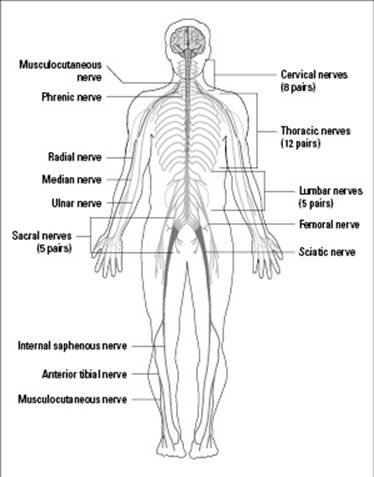
From LifeART®, Super Anatomy 1, © 2002, Lippincott Williams & Wilkins
Branching out to study neuron structure
The nervous system contains two types of cells: neurons and neuroglial cells. Neurons are the cells that receive and transmit signals; neuroglial cells are the support systems for the neurons (in other words, they protect and nourish the neurons).
Neurons have a rather elongated structure, as you can see in Figure 18-2.
Each neuron contains a nerve cell body with a nucleus and organelles such as mitochondria, endoplasmic reticulum, and a Golgi apparatus (see Chapter 4 for more on cells and organelles).
Tiny projections called dendrites branch off the nerve cell body at the receiving end of the neuron. The dendrites act like tiny antennae that pick up signals from other cells.
The opposite side of the neuron extends into a long, thin, branching fiber called an axon. The axon is insulated by a myelin sheath made up of segments called Schwann cells.
Figure 18-2: The basic structure of a motor neuron (left) and a sensory neuron (right), including the path of an impulse.
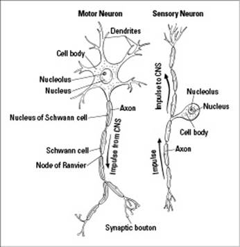
Nerve impulses enter a neuron through the dendrites. They then travel down the branches of the dendrites to the nerve cell body before being carried along the axon. When the impulses reach the branches at the end of the axon, they’re transmitted to the next neuron. Impulses continue to be carried in this fashion until they reach their final destination.
Processing signals with the three types of neurons
The three major functions of a nervous system are to collect, interpret, and respond to signals. Different types of neurons carry out each of these functions.
Sensory neurons collect sensory information and bring it to the CNS. Also called afferent neurons, sensory neurons are designed primarily to receive initial stimuli from sense organs — the eyes, ears, tongue, skin, and nose. However, they’re also responsible for receiving internally generated impulses regarding adjustments that are necessary for the maintenance of homeostasis. For example, if you touch the tip of a knife, the sensory neurons in your finger will transmit impulses to other sensory neurons until the impulse reaches an interneuron.
Interneurons within the CNS integrate the sensory information and send out responding signals. Also called connector neurons or association neurons, interneurons “read” impulses received from sensory neurons. When an interneuron receives an impulse from a sensory neuron, the interneuron determines what (if any) response to generate. If a response is required, the interneuron passes the impulse on to motor neurons. To continue with the example from the preceding bullet, the interneurons in the cerebral cortex of your brain will process the incoming sensory information and send out responding signals.
Motor neurons carry the responding signals from the CNS to the cells that are to carry out the response. Also called efferent neurons, motor neurons stimulate effector cells that generate reactions. To conclude our example, responding signals from your brain travel through motor neutrons until they reach your muscles, signaling your muscles to contract and pull your finger away from the sharp knife.
Acting without thinking
Sometimes the nervous system can work without the brain, as in a reflex arc. A reflex arc gives sensory nerves direct access to motor nerves so information can be transmitted immediately.
When you touch a hot stovetop, your sensory nerves detect the excessive heat and instantly fire off a message to your motor nerves that says, “Pull your hand away!” The motor nerves call the proper muscles into action to move your hand away before you can even “think” about it. Your brain comes into play after the fact when you sense the pain of the burn or think to yourself how silly it was to touch a hot stovetop in the first place.
What a Sensation! The Brain and the Five Senses
The brain is the master organ of the body in animals with a central nervous system because it takes in all the information received by the animals’ sense organs and produces the appropriate responses. The brain consists of three main parts: the forebrain (uppermost part), the midbrain (middle brain), and the hindbrain (lowermost part). These three parts are further organized into four major regions that we present in the following list:
Cerebrum: Also called the telencephalon, the cerebrum is the largest part of the brain and is responsible for consciousness. It’s located at the uppermost part of the brain and divided into left and right halves, which are called cerebral hemispheres. Each cerebral hemisphere has four lobes named for the bones of the skull that cover them: frontal, parietal, temporal, and occipital. Specific areas of the lobes are responsible for certain functions, such as concentration, speech recognition, memory, and so on.
Diencephalon: Found at the center of the brain, the diencephalon is a structure that consists of the thalamus and hypothalamus. The thalamus processes information going to and from the spinal cord, and the hypothalamus controls homeostasis by regulating hunger, thirst, sleep, body temperature, water balance, and blood pressure. At the base of the hypothalamus is the pituitary gland, which helps maintain homeostasis in the body by secreting many important hormones.
Cerebellum: The cerebellum, which is found at the base of the brain, coordinates muscle functions such as maintaining normal muscle tone and posture.
Brain stem: Located below the cerebellum, the brain stem is made up of three structures: the midbrain, the pons, and the medulla oblongata. The brain stem controls critical functions such as breathing and your heartbeat.
 Bet you didn’t know that the spinal cord is actually a continuation of the brain stem. You can thank us for this bit of knowledge when you ace that bonus question on your next biology test.
The human sense organs — eyes, ears, tongue, skin, and nose — help to protect the body. They’re filled with receptors that relay information through sensory neurons to the appropriate places within the nervous system. Each sense organ contains different receptors.
General receptors are found throughout the body. They’re present in skin, visceral organs (visceral meaning in the abdominal cavity), muscles, and joints.
Special receptors include chemoreceptors (chemical receptors) found in the mouth and nose, photoreceptors (light receptors) found in the eyes, and mechanoreceptors (movement receptors) found in the ears.
Table 18-1 compares the various types of receptors found in an animal’s nervous system.
|
Table 18-1 Types of Receptors & Their Functions |
||
|
Receptor |
Location |
Function |
|
Chemoreceptor |
Taste buds, cilia in nasal cavity |
Detect chemicals in food and air |
|
Mechanoreceptor |
Cilia in ear |
Detect movement of ear drum and ossicles (ear bones) |
|
Osmoreceptor |
Hypothalamus |
Detect concentration of solutes in the bloodstream |
|
Photoreceptor |
Retina of eye |
Detect light |
|
Proprioceptor |
Muscles |
Detect positioning and movement of limbs |
|
Stretch receptor |
Lungs, tendons, ligaments |
Detect expansion or elongation of muscle tissue |
We delve into the five senses in greater detail in the sections that follow, with a focus on how these senses operate in humans.
Oooh, that smell: Olfaction
If you walk in the door of your home and smell an apple pie baking and peppers and onions sautéing, how do you know that the apple pie is apple pie and that the pepper and onions are in fact peppers and onions and not eggplant and zucchini? The key lies in your olfactory cells (you can remember the name of these cells by thinking that they “smell like an old factory”); they’re responsible for your sense of smell.
Olfactory cells line the top of your nasal cavity. They have cilia on one end that project into the nasal cavity (see Chapter 4 for more on cilia). On the other end, they have olfactory nerve fibers that pass through the ethmoid bone and into the olfactory bulb, which is directly attached to the cerebral cortex of your brain.
As you breathe, anything that’s in the air you take in enters your nasal cavity. You don’t “smell” air or dust or pollen, but you do smell chemicals. The olfactory cells are chemoreceptors, which means they have protein receptors that can detect subtle differences in chemicals.
As you breathe upon walking into your kitchen, the chemicals from the apple pie, peppers, and onions waft into your nasal cavity. There, the chemicals bind to the cilia, generating a nerve impulse that’s carried through the olfactory cells, into the olfactory nerve fiber, up to the olfactory bulb, and directly to your brain. Your brain then determines what you’re smelling. If the scent is something you’ve smelled before and are familiar with, your brain recalls the information that has been stored in your memory. If you’re sniffing something that you haven’t experienced before, you need to use another sense, such as taste or sight, to make an imprint on your brain’s memory.
Mmm, mmm, good: Taste
The senses of smell and taste work closely together. If you can’t smell something, you can’t taste it either. That’s because taste buds and olfactory cells (described in the preceding section) are chemoreceptors designed to detect chemicals. Your tongue is covered with taste buds, clusters of cells that specialize in recognizing tastes. The taste receptor cells within your taste buds allow you to detect five different types of taste: sweet, sour, bitter, salty, and umami (savory).
A lot of people think that taste buds are the little bumps on the tongue, but they’re wrong. Those little bumps are actually papillae; the taste buds exist in the grooves between each papilla.
Foods contain chemicals, and when you put something into your mouth, the taste receptor cells in your tongue can detect what chemicals you’re ingesting. Each taste bud has a pore at one end that allows the chemicals in the food to enter the taste bud and reach the taste receptor cells. The taste receptor cells are connected at the other end to sensory nerve fibers that carry the taste signals to the brain.
The sense of taste allows you to enjoy food, which you must ingest to live, but it also serves a higher function. When your taste buds detect chemicals, they send the signal to your brain that sets in motion the production and release of the proper digestive enzymes necessary for breaking down the food you’re ingesting. This function allows your digestive system to work optimally to retrieve as many nutrients as possible from food.
Now hear this: Sound
Your ears are the sense organ for your sense of hearing, which kicks into high gear when sound waves are shuttled through your ear canal and into your middle ear — the place where your eardrum is located. When a sound wave hits your eardrum, the eardrum moves tiny bones (specifically the malleus, incus, and stapes). The movement of these bones is picked up by the mechanoreceptors in your inner ear, which exist on cilia-containing hair cells between the end of the semicircular canals and the vestibule. When the cilia move, they create an impulse that’s sent through the cochlea to the eighth cranial nerve, which carries the impulse to the brain so it can interpret the information as a specific sound.
Your ears do something else for you that’s not in any way related to your sense of hearing: They help you maintain your equilibrium. In other words, they keep you from falling over because they contain fluid within the semicircular canals of the inner ear. When this fluid moves, that movement is ultimately detected by the cilia. The cilia transmit impulses to your brain about angular and rotational movement, as well as movement through vertical and horizontal planes, all of which helps your body keep its balance.
When the fluid in your ear doesn’t stop moving, you can develop motion sickness.
Seeing is believing: Sight
Vision is perhaps the most complex of all the senses because it depends upon the correct structure and function of all the parts of your eye.
The colored part of the eye is the iris, which is actually a pigmented muscle that controls the size of your pupil. The pupil is the “black hole” at the very center of your iris; it dilates to allow more light into the eye and contracts to allow less light into the eye.
The cornea covers and protects the iris and pupil.
The lens of the eye is located just behind the pupil. It’s connected to the iris by a small muscle called the ciliary muscle, which changes the shape of the lens to help you see things that are both far and near. Specifically, the lens flattens so you can see farther away, and it becomes rounded so you can see things that are near to you. The process of changing the shape of the lens is called accommodation. (People lose the ability of accommodation as they grow older, prompting the need for glasses.)
The vitreous body is located behind the lens and is filled with a gelatinous material called vitreous humor. This substance gives shape to the eyeball and also transmits light to the very back of the eyeball, where the retina lies.
The retina contains two types of photoreceptors that detect light:
• Rods detect motion and can function in low light.
• Cones detect fine detail and color and work best in bright light. The three types of cones each detect a different color — red, blue, or green. (Color blindness occurs when one type of cone is lacking. For example, if you’re missing your red cones, you can’t see red.)
Light strikes the rods and cones, generating nerve impulses that travel to two types of neurons: first to the bipolar cells and then to the ganglionic cells. The axons of the ganglionic cells form the optic nerve, which carries the impulses directly to the brain.
Approximately 150 million rods are in a retina, but only 1 million ganglionic cells and nerve fibers are there, which means many more rods can be stimulated than there are cells and nerve fibers to carry the impulses. Your eye must therefore combine “messages” before the impulses can be sent to the brain.
A touchy-feely subject: Touch
Your eyes, ears, nose, and tongue all contain special receptors. Your skin, on the other hand, contains only general receptors, but it possesses a combination of general receptors that allow you to distinguish between different types of touches, from fine touches and pressure to vibration and painful sensations:
Pain receptors make it possible for you to feel pain, like what you experience when you slam your finger in a car door.
Mechanoreceptors allow you to feel pressure or fine touches, like a hug, a handshake, or a gentle stroke.
Thermoreceptors help you feel temperature, like heat on a hot summer day.
These three types of general receptors are interspersed throughout your skin’s surface, but they’re not distributed evenly. For one thing, you have more of some types of receptors than others (as in you have many more receptors for pain than you do for temperature). For another thing, some parts of your skin have more receptors than other parts. Case in point: You have many more receptors in the skin covering the tips of your fingers than you do in the skin on your back.
When your general receptors are activated, they generate an impulse that’s carried to your spinal cord and then up to your brain.
Following the Path of Nerve Impulses
Nerve impulses pass through a neuron in about seven milliseconds — that’s faster than a lightning strike! The sections that follow explain how a nerve impulse moves through a neuron and how it then makes its way from one neuron to the next.
Traveling from one end to the other
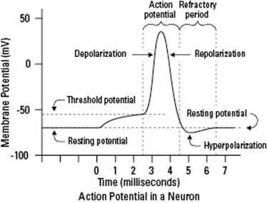
Before a nerve impulse can enter the brain, it must first pass from one side of a neuron to the other. When a neuron is inactive, just waiting for a nerve impulse to come along, the electrical difference across the nerve cell membrane is called its resting potential. In this state, the neuron is polarized with sodium on the outside of its membrane and potassium on the inside. (A neuron is polarized when the electrical charge on the outside of its membrane is positive and the charge on the inside is negative.)
The outside of a polarized neuron contains excess sodium ions (Na+); the inside of it contains excess potassium ions (K+). How can the charge inside the cell be negative if positive ions are present, you ask? Well, the cell pushes out greater numbers of Na+ than the number of K+ it pulls in. So, the cell becomes more positive outside and less positive — or negative — inside.
Following is a description of the process that causes a resting neuron to send a nerve impulse and then return to a resting state; Figure 18-3 gives you the visual:
1. Sodium ions move inside the neuron’s membrane, causing an action potential.
An action potential is the wave of electrical activity that represents the nerve impulse. When a stimulus reaches a resting neuron, the gated ion channels on the resting neuron’s membrane open suddenly, allowing the Na+ that was on the outside of the membrane to rush into the cell. As this happens, the neuron becomes depolarized (more positive ions go charging to the inside of the membrane), making the inside of the cell positive as well.
Each neuron has a threshold level. The threshold is the point at which there’s no holding back. After the stimulus goes above the threshold level, more gated ion channels open, allowing more Na+ inside the cell. This causes complete depolarization of the neuron, creating an action potential. In this state, the neuron continues to open Na+ channels all along the membrane, creating an all-or-none phenomenon. All-or-none means that if a stimulus doesn’t exceed the threshold level and cause all the gates to open, no action potential results; after the threshold is crossed, there’s no turning back — complete depolarization occurs, and the stimulus is transmitted.
When an impulse travels down an axon covered by a myelin sheath, the impulse must move between the uninsulated gaps that exist between each Schwann cell. These gaps are called the nodes of Ranvier. During the action potential, the impulse undergoes saltatory conduction (think salt, as in the sodium ions that allow this to happen) and jumps from one node of Ranvier to the next node of Ranvier, increasing the speed at which the impulse can travel.
2. Potassium ions move outside the membrane, and sodium ions stay inside the membrane, repolarizing the cell.
After the inside of the neuron is flooded with Na+, the gated ion channels on the inside of the membrane open to allow the K+ to move to the outside of the membrane. With K+ moving to the outside, the cell’s balance is restored by repolarizing the membrane. (The result is a polarization that’s opposite of the initial polarization that had Na+ on the outside and K+ on the inside.) Just after the K+ gates open, the Na+ gates close; otherwise, the membrane wouldn’t be able to repolarize.
3. The neuron becomes hyperpolarized when there are more potassium ions on the outside than there are sodium ions on the inside.
When the K+ gates finally close, the neuron has slightly more K+ on the outside than it has Na+ on the inside. This causes the cell’s potential to drop slightly lower than the resting potential, and the membrane is said to be hyperpolarized. This period doesn’t last long, though. After the impulse has traveled through the neuron, the action potential is over, and the cell membrane returns to normal.
4. The neuron enters a refractory period, which returns potassium to the inside of the cell and sodium to the outside of the cell.
The refractory period is when the Na+ and K+ are returned to their original sides (that means Na+ on the outside and K+ on the inside). A protein called the sodium-potassium pump returns the ions to their rightful sides of the neuron’s cell membrane. The neuron is then back to its normal polarized state, which is where it stays until another impulse comes along.
Figure 18-3: The transmission of a nerve impulse.
Jumping the gap between neurons
Nerve impulses can’t move directly from one neuron into another one because neurons don’t actually touch each other. Gaps exist between the axon of one neuron and the dendrites of the next neuron. Nerve impulses must therefore move through that gap to continue on their path through the nervous system.
The gap between two neurons is called a synaptic cleft or synapse.
In many invertebrate animals and in the brains of vertebrate animals, impulses are carried across synapses by electrical conduction. However, outside of the brain, such as when a neuron sends a signal to a muscle cell, nerve impulses are conducted across synapses by a series of chemical changes, which occur as follows:
1. Calcium gates open.
At the end of the axon from which the impulse is coming (called the presynaptic cell because the axon precedes the synapse), the membrane depolarizes, allowing the gated ion channels to open so they can let in calcium ions (Ca2+).
2. Synaptic vesicles release a neurotransmitter.
When the Ca2+ rushes into the end of the presynaptic axon, synaptic vesicles connect to the presynaptic membrane. The synaptic vesicles then release a chemical called a neurotransmitter into the synapse.
3. The neurotransmitter binds with receptors on the postsynaptic neuron.
The chemical that serves as the neurotransmitter diffuses across the synapse and binds to proteins on the membrane of the neuron that’s about to receive the impulse. The proteins serve as the receptors, and different proteins serve as receptors for different neurotransmitters — that is, neurotransmitters have specific receptors.
4. Excitation or inhibition of the postsynaptic membrane occurs.
Whether excitation or inhibition occurs depends on what chemical served as the neurotransmitter and the result that it had. For example, if the neurotransmitter causes the Na+ channels to open, the membrane of the postsynaptic neuron becomes depolarized, and the impulse is carried through that neuron. If the K+ channels open, the membrane of the postsynaptic neuron becomes hyperpolarized, and the impulse is stopped dead.
If you’re wondering what happens to the neurotransmitter after it binds to the receptor, here’s the story: After the neurotransmitter produces its effect, whether that effect is excitation or inhibition, it’s released by the receptor and goes back into the synapse. In the synapse, enzymes degrade the chemical neurotransmitter into smaller molecules. Then the presynaptic cell “recycles” the degraded neurotransmitter, sending the chemicals back into the presynaptic membrane so that during the next impulse, when the synaptic vesicles bind to the presynaptic membrane, the complete neurotransmitter can again be released.
Table 18-2 lists some common neurotransmitters and their functions.
|
Table 18-2 Characteristics of Common Neurotransmitters |
||
|
Neurotransmitter |
Source |
Function |
|
Acetylcholine |
Secreted at gaps between the neurons and muscle cells |
Stimulates or inhibits contraction of muscles, depending on receptor |
|
Dopamine |
Created from amino acids |
Affects movement, emotion, and feelings of pleasure, and plays an important role in drug addiction |
|
Epinephrine |
Created from amino acids |
Responsible for fight-or-flight response |
|
Norepinephrine |
Released by postganglionic axons |
Increases blood pressure |
|
Serotonin |
Produced through enzymatic reaction involving tryptophan |
Regulates sleep, calms anxiety, and affects sexual behavior |
The Endocrine System: All Hormones Are Not Raging
The endocrine system (shown in Figure 18-4) is the system that handles hormone production and secretion within an organism. It keeps a check on cellular processes and components of the bloodstream and can make adjustments as necessary.
Figure 18-4: The endocrine system.
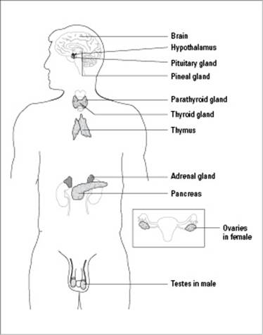
From LifeART®, Super Anatomy 1, © 2002, Lippincott Williams & Wilkins
The endocrine system contains organs called endocrine glands that produce hormones — chemical messengers that coordinate the activities of specific cells in certain areas of the body. Hormones are carried in the bloodstream to a target tissue elsewhere in the body, where they must be absorbed into the tissue before they can have an effect.
 The word endocrine stems from a Greek word meaning within. Endocrine glands secrete their products into the bloodstream, which remains within the body. On the other hand, exocrine glands secrete products to the outside of the body. Examples of exocrine gland secretions are sweat and saliva.
The next sections fill you in on how hormones operate and get you acquainted with some of the important jobs they perform.
Seeing how hormones work
Hormones are long-distance messengers, carrying their message through the bloodstream to target cells, the cells that respond to the hormone, throughout the body. In order for a cell to respond to a particular hormone, it must have receptors (molecules that bind to signaling molecules like hormones and cause changes in the behavior of cells) for that hormone. (Cells that don’t have receptors for a particular hormone don’t respond to that hormone.) When the hormone binds to the receptor on target cells, the receptor is activated and causes a change in the behavior of the cell.
Hormones in vertebrates can be divided into two groups:
Peptide hormones, such as insulin, are short chains of amino acids; you can think of them as very small proteins. Peptide hormones are hydrophilic (water loving), so they don’t pass easily through cell membranes.
The receptors for peptide hormones are embedded in the plasma membranes of target cells. Peptide hormones bind to their receptors at the cell surface, activating the receptor and causing a change in the part of the receptor that extends into the inside of the cell. The internal part of the receptor interacts with molecules inside the cell to cause a change in behavior. Because the message from the hormone is passed through the plasma membrane, this signaling process is called signal transduction.
Steroid hormones, such as testosterone and estrogen, are lipids, so they’re hydrophobic (water fearing) and can pass easily through the hydrophobic layer of the plasma membrane and enter cells. Thus, the receptors for steroid hormones are located inside the cell.
Once inside the cell, steroid hormones pass through the cytoplasm of the cell and diffuse into the nucleus. Inside the nucleus, steroid hormones bind to receptor proteins, forming an activated complex. The activated hormone-receptor complex directly causes a change in the behavior of the cell, often by acting as transcription factors that turn on the transcription of certain genes. After these genes are transcribed and translated, the newly made proteins perform the function that represents the cell’s response to the hormone (see Chapter 8 for more on the transcription and translation of proteins).
Surveying the general functions of hormones
Hormones play several important roles, whether they come from a plant, an invertebrate animal, or a vertebrate animal. Following are just a few examples of the many functions that hormones serve within an organism:
Assuring that growth occurs properly: In humans, growth hormones must be secreted at normal levels by the pituitary gland throughout childhood and adolescence. The extremes of having too many or too few growth hormones are obvious — giants or midgets, respectively. In invertebrate animals, such as insects, growth hormones are responsible for molting, which is the shedding of the outer layer (the exoskeleton). Hormones also regulate growth in plants, and you can find out more about that in Chapter 21.
Ensuring that development and maturation occur properly and on time: In insects, metamorphosis — the process of changing body forms during developmental stages — is controlled by a substance called juvenile hormone. (Metamorphosis is the process that changes a larva or caterpillar into a pupa and then into a moth or a butterfly.) In vertebrates, hormones trigger the transition from juvenile to adult forms and cause the onset of sexual maturity. Plant hormones regulate developmental events such as seed germination and flowering.
Making sure that reproduction occurs at the best possible time: For humans, who have steady supplies of food year-round and sheltered environments in which to live, reproduction can occur whenever the urge hits (see Chapter 19 for the specifics on human reproduction). But for other animals and plants, reproduction needs to occur during certain seasons of the year when climate and food supplies are optimal. Consequently, hormones stimulate these organisms’ reproductive urges only when the climate and food conditions are just right.
So many different hormones are found in animals, that we can’t possibly tell you about them all. However, we do want to give you a peek into the functions of specific hormones, which is why Table 18-3 highlights some of the many hormones found in mammals like you, along with the glands that produce them and their major functions.
|
Table 18-3 Several Important Mammalian Hormones |
||
|
Hormone |
Gland |
Function |
|
Adrenaline (epinephrine) |
Adrenal gland |
Increases metabolism and glucose in the blood; constricts some blood vessels |
|
Aldosterone (mineralocorticoids) |
Adrenal gland |
Regulates the balance of salt and water in the body by causing kidneys to reabsorb sodium and release potassium |
|
Antidiuretic hormone |
Pituitary gland |
Signals kidneys to retain water |
|
Estrogen |
Ovaries |
Stimulates growth of the uterine lining; triggers and maintains secondary sex characteristics in females |
|
Follicle-stimulating hormone (FSH) |
Pituitary gland |
Stimulates production of eggs and sperm |
|
Glucagon |
Pancreas |
Raises glucose (sugar) in the blood |
|
Growth hormone |
Pituitary gland |
Stimulates growth of bones; promotes metabolic functions |
|
Insulin |
Pancreas |
Lowers glucose (sugar) in the blood |
|
Luteinizing hormone (LH) |
Pituitary gland |
Stimulates ovaries and testes |
|
Melatonin |
Pineal gland |
Regulates sleep and wake cycles |
|
Oxytocin |
Pituitary gland |
Triggers contraction of the uterus and mammary glands (to release milk) |
|
Progesterone |
Ovaries |
Supports growth of the uterine lining |
|
Prolactin |
Pituitary gland |
Stimulates milk production |
|
Testosterone |
Testes |
Stimulates sperm formation; triggers and maintains secondary sex characteristics in males |
|
Thyroid-stimulating hormone |
Pituitary gland |
Stimulates the thyroid gland |
|
Thyroxine (T4) |
Thyroid |
Stimulates and maintains metabolism |