Biology of Humans
3. The Cell
In Chapter 2a we learned how some foodborne illnesses are caused by singlecelled microorganisms we call bacteria. Bacteria are examples of prokaryotic cells, and they differ in several key ways from the cells in our bodies, which are eukaryotic. In this chapter we first compare these two basic types of cells, and then we discover how eukaryotic cells work by examining the structures that all cells in the human body have in common. We begin with the plasma membrane, the outer boundary of a cell, and work inward. After examining the various organelles—small structures within cells that have specialized functions—we explore the ways that cells obtain the energy they need to do their work of running the body.
Eukaryotic Cells Compared with Prokaryotic Cells
The cell theory is a fundamental organizing principle of biology that guides the way biologists think about living things. A theory, you may recall from Chapter 1, is a well-researched and well- supported explanation for some aspect of the physical universe. The cell theory states that (1) a cell is the smallest unit of life; (2) cells make up all living things, from unicellular to multicellular organisms; and (3) new cells can arise only from preexisting cells.
· Microscopes allow us to see cells and the diverse organelles inside them. Each organelle performs a specific function for the cell.
As we mentioned, there are two basic types of cells—eukaryotic cells and prokaryotic cells. Prokaryotic cells are structurally simpler and typically smaller than eukaryotic cells. They are limited to bacteria and another group of microscopic organisms called Archaea. You are probably already aware of bacteria, some of which inhabit your body. Many bacterial inhabitants are harmless, but others can cause illness (see Chapters 2a and 13a). Archaea may be less familiar to you. They include species that inhabit extreme environments such as the high-saline Great Salt Lake or the hot sulfur springs of Yellowstone National Park. Most prokaryotic cells are surrounded by a rigid cell wall, as shown in Figure 3.1.
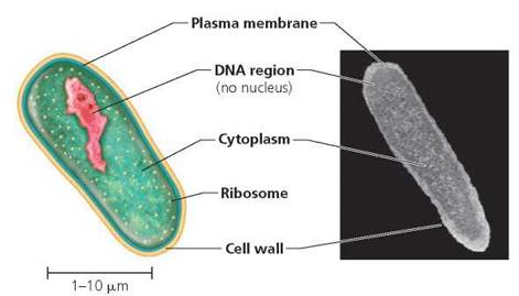
FIGURE 3.1. Prokaryotic cells, such as a bacterium, lack internal membrane-bound organelles.
The cells of plants, animals, and all other organisms except bacteria and Archaea are eukaryotic. All the cells that make up your body, therefore, are eukaryotic. The difference between eukaryotic and prokaryotic cells relates to the presence or absence of membrane-bound organelles. An organelle or "little organ" is a component within a cell that carries out specific functions. Some organelles have membranes, others do not. Nonmembranous organelles such as ribosomes and cytoskeletal elements are found in both prokaryotic and eukaryotic cells. Unique to eukaryotic cells, however, are membrane-bound organelles such as mitochondria and endoplasmic reticulum (Figure 3.2). Another of the membrane-bound organelles found in all typical eukaryotic cells is a well-defined nucleus containing DNA. Note that in prokaryotes, a membrane does not surround the DNA (refer, again, to Figure 3.1). Among eukaryotes, plant cells have cell walls, but animal cells do not. Table 3.1 reviews the major differences between eukaryotic and prokaryotic cells.
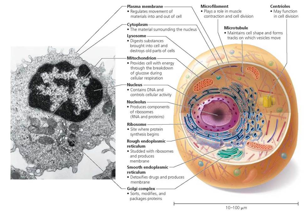
FIGURE 3.2. Eukaryotic cells, such as the generalized animal cell shown here, have internal membrane-bound organelles.
The nucleus is a membrane-bound organelle. Look closely at the other organelles in the cell and read their functions. Based on the structure and functions of the organelles shown, list those that you think are membrane-bound organelles and those that are nonmembranous organelles.
The membrane-bound organelles include the nucleus, rough endoplasmic reticulum, smooth endoplasmic reticulum, Golgi complex, mitochondrion, and lysosome. The nonmembranous organelles include ribosomes, microfilaments, centrioles, and microtubules.
TABLE 3.1. Review of Features of Prokaryotic and Eukaryotic Cells
Feature |
Prokaryotic Cells |
Eukaryotic Cells |
Organisms |
Bacteria, Archaea |
Plants, animals, fungi, protists |
Size |
1-10 pm across |
10-100 pm across |
Membrane-bound organelles |
Absent |
Present |
DNA form |
Circular |
Coiled, linear strands |
DNA location |
Cytoplasm |
Nucleus |
Internal membranes |
Rare |
Many |
Cytoskeleton |
Present |
Present |
Cell Size and Microscopy
Most eukaryotic and prokaryotic cells are so small that they are typically measured in micrometers (pm), which are equal to 1/1000 meters (m). (An obvious exception is the chicken egg.) The small size of cells is dictated by a physical relationship known as the surface-to-volume ratio. This relationship says that as a cell gets larger, its surface area increases much more slowly than its volume (Figure 3.3). Nutrients enter a cell, and wastes leave a cell, at its surface. Therefore, a large cell would have difficulty moving all the nutrients it needs and all the wastes it produces across its inadequate surface and would die. A small cell, on the other hand, has sufficient surface for the uptake and removal of substances and would survive.
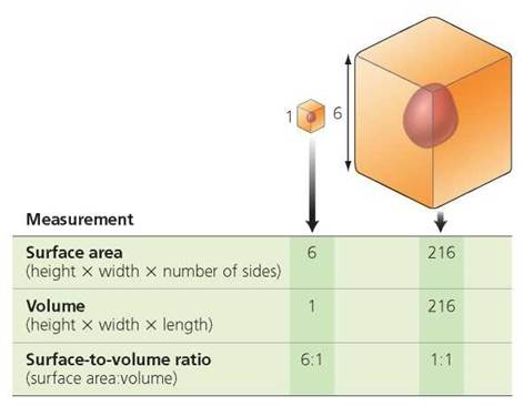
FIGURE 3.3. Cells must remain small In size because the ratio of surface area to volume decreases rapidly as cell size Increases.
Due to the small size of most cells, you need a microscope to see them. Throughout this book you will see micrographs, which are photographs obtained using a microscope (Figure 3.4). Microscopic specimens can be imaged using beams of either light or electrons. Light microscopes, which are used in many classrooms, have the advantage that they are relatively inexpensive and simple to operate. Electron microscopes, though more complex and expensive, have the capacity to reveal finer details because the wavelength of an electron beam is smaller than the wavelengths of visible light. Whether using light or electrons, the beam can be either transmitted through a thinly sliced specimen or bounced off of the specimen's surface.
Figure 3.4a is a transmitted light micrograph through three striated muscle cells that have been stained with biological dyes to increase the contrast between different cellular components. The three nuclei visible in this picture are colored dark purple because the dye used has an affinity for acidic components in the cell such as DNA. Figure 3.4b is a transmitted electron micrograph that shows the structure of striated muscle cells in more detail than is possible using light to image the tissue. In this case, the contrast between different cellular components is produced by staining the tissue with heavy metals such as lead and uranium. Different components of the cells absorb different amounts of these heavy metals. Components that readily absorb the metals differentially block the electron beam from passing through the sample. Figure 3.4c is a scanning electron micrograph produced by bouncing an electron beam off the surface of several striated muscle cells. The beam is scanned across the surface of the sample, and electrons that bounce off the surface are collected by a detector. For every point that is scanned, the number of electrons reaching the detector is used to calculate the relative brightness of that spot on the sample. This information is used to construct the image. Images produced with electron beams are not in color. The pictures shown in Figures 3.4b and 3.4c have been colored to highlight certain features, an improvement made possible by computer-assisted processing of images. Other micrographs in the text have also been colored.
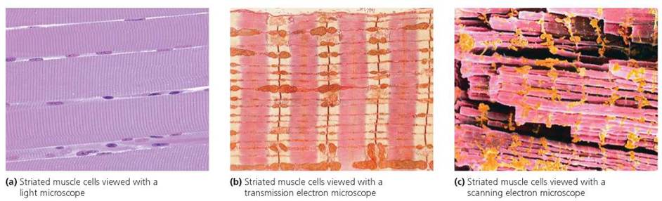
FIGURE 3.4. Micrographs are photographs taken through a microscope. Here, striated muscle cells have been photographed using three different types of microscope. Electron microscopes use beams of electrons to produce images with finer details than those viewed with light microscopes.
Cell Structure and Function
The structure of a cell exquisitely reflects its functions. For example, few human cells are more specialized than sperm or eggs, the cells that carry genetic information and other materials needed to make a new individual of the next generation. A sperm is specialized for swimming to the egg and fertilizing it. As such, a sperm is streamlined and equipped with a whiplike tail. In the head of the sperm is an enzyme-containing sac that spills open to release enzymes that digest a path through the layers of cells surrounding the egg. In contrast, the egg is immobile and much larger than a typical cell because it is literally packed with nutrients and other materials needed to initiate development. A mature red blood cell is another example of a cell whose structure reflects its function. As the red blood cell matures, it extrudes its nucleus and most organelles, leaving more space for hemoglobin, the protein that transports oxygen. A mature red blood cell is thus an exception to the rule that eukaryotic cells have a well-defined nucleus and other membrane-bound organelles. Consider, also, a cardiac muscle cell. This cell is specialized for contraction, and for propagating the signal for contraction from one muscle cell to the next. Thus it is filled with contractile proteins and joined to adjacent cells by specialized junctions that strengthen cardiac tissue and promote rapid conduction of impulses throughout the heart. In each of these cases, careful study of the cell's structure provides excellent clues to its function, and vice versa (Figure 3.5).
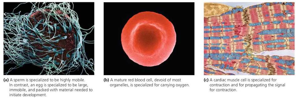
FIGURE 3.5. A cell's structure reflects its specific function. These cell types from the human body illustrate the close tie between structure and function.
Plasma Membrane
We begin our examination of the cell at its outer surface—the plasma membrane. This remarkably thin outer covering controls the movement of substances both into and out of the cell. Because the concentrations of substances in a cell's interior are critically balanced, molecules and ions are not permitted to move randomly in and out.
Both prokaryotic and eukaryotic cells have a plasma membrane, but only eukaryotic cells also contain internal membranes that divide the cell into many compartments. Each compartment contains its own assortment of enzymes and is specialized for particular functions. In general, the principles described for the plasma membrane also apply to the membranes inside the cell.
Plasma Membrane Structure
The plasma membrane is made of lipids, proteins, and carbohydrates. Recall from Chapter 2 that phospholipids are the major components of the plasma membrane. These molecules, with their hydrophilic (water-loving) heads and hydrophobic (water-fearing) tails, form a double layer—called the phospholipid bilayer—at the surface of the cell (Figure 3.6). The hydrophilic heads facing outside the cell interact with the extracellular fluid (also known as interstitial fluid), which is the watery solution outside cells. The hydrophilic heads facing inside the cell interact with the cytoplasm, which is the jellylike solution inside the cell. The cytoplasm includes all contents of the cell between the plasma membrane and the nucleus. Within the phospholipid bilayer, the hydrophobic tails point toward each other and hold the plasma membrane together.
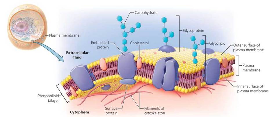
FIGURE 3.6. The structure of the plasma membrane of a cell according to the fluid mosaic model
Interspersed in the phospholipid bilayer are proteins, as seen in Figure 3.6. Some proteins are embedded in the membrane, and some of these span the bilayer completely. Other proteins are simply attached to the inner or outer surface of the membrane. Molecules of cholesterol are also scattered throughout the bilayer.
As you can see in Figure 3.6, carbohydrates attach only to the outer surface of the plasma membrane. Most of these carbohydrates are attached to proteins, forming glycoproteins. We will see that glycoproteins often function in cell recognition. Other carbohydrates are attached to lipids, forming glycolipids.
The structure of the plasma membrane is often described as a fluid mosaic. The proteins are interspersed among the lipid molecules like tiles of different colors within a mosaic. Many of the proteins are able to move sideways through the bilayer to some degree, giving the membrane its fluid quality.
Plasma Membrane Functions
The plasma membrane performs several vital functions for the cell. First, by imposing a boundary between the cell's internal and external environment, the plasma membrane maintains the cell's structural integrity. Second, the structure of the plasma membrane regulates the movement of substances into and out of the cell, permitting entry to some substances but not others. For this reason, the membrane is often described as being selectively permeable. You will read more about the transport of materials across the plasma membrane in the next section of this chapter.
The plasma membrane also functions in cell-cell recognition. Cells distinguish one type of cell from another by recognizing molecules—often glycoproteins—on their surface. Membrane glycoproteins differ from one species of organism to another and among individuals of the same species. Even different cell types within an individual have different membrane glycoproteins. This variation allows the body to recognize foreign invaders such as bacteria. Your own body, for example, would recognize such invaders because the bacteria lack the surface molecules found on your cells. The bacteria, in turn, "read" the different surface molecules of your cells to settle preferentially on some kinds of cells but not others.
Another important function of the plasma membrane is communication between cells. Such communication relies on receptors, specialized proteins in the plasma membrane (or inside the cell) that bind particular substances that affect cell activities. For example, hormones secreted by one group of cells may bind to receptors in the plasma membranes of other cells. The receptors then relay a signal to proteins inside the cell, which transmit the message to other nearby molecules. Through a series of chemical reactions, the hormone's "message" ultimately initiates a response by the recipient cell, perhaps causing it to release a certain chemical.
Finally, the plasma membrane plays an important role in binding pairs or groups of cells together. Cell adhesion molecules (CAMs) extend from the plasma membranes of most cells and help attach the cells to one another, especially during the formation of tissues and organs in an embryo. The functions of the plasma membrane are as follows:
• Maintain structural integrity of the cell
• Regulate movement of substances into and out of the cell
• Provide recognition between cells
• Provide communication between cells
• Stick cells together to form tissues and organs
Stop and think
Of the five functions of the plasma membrane listed previously, which might explain the difficulty of transplanting tissues and organs successfully from one body to another? Why would rejection of such transplants occur? Under what circumstances might one body accept a tissue graft or organ from another?
Movement Across the Plasma Membrane
Recall that an important function of the plasma membrane is to control which substances move into and out of the cell. Substances cross the plasma membrane in several ways. These methods are described as either active—requiring the cell to expend energy—or passive—requiring no energy expenditure by the cell.
Simple diffusion. Some materials cross the plasma membrane passively through simple diffusion, the random movement of a substance from a region of higher concentration to a region of lower concentration. Concentration is the number of molecules of a substance in a particular volume, while a concentration gradient is a difference in the relative number of molecules or ions of a given substance in two adjacent areas. The end result of simple diffusion is an equal distribution of the substance in the two areas; in other words, diffusion tends to eliminate the concentration gradient. Consider what happens when someone is cooking bacon in the kitchen. At first, the smell of bacon is localized in the kitchen. Soon, however, the smell permeates adjoining rooms, too, as odor molecules move from where they are more concentrated (the kitchen) to where they are less concentrated (other parts of the house). Eventually the odor molecules are equally distributed, but they still move randomly in all directions. Likewise, when a substance diffuses across a membrane from a region of higher concentration to a region of lower concentration, the movement of its molecules does not stop once the concentration has equalized. Instead, the molecules continue to move randomly back and forth across the membrane. The rate of movement in each direction, however, is now the same. Substances such as carbon dioxide and oxygen diffuse through the plasma membrane of our cells (Figure 3.7).
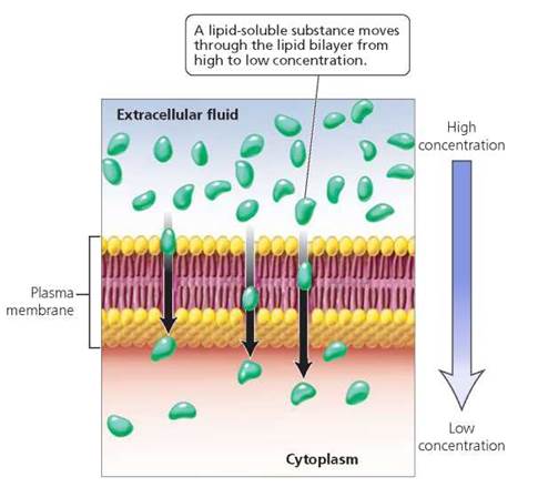
FIGURE 3.7. Simple diffusion is the random movement of a substance from a region of higher concentration to a region of lower concentration.
Facilitated diffusion. Water-soluble substances are repelled by lipids, so they cannot move through the phospholipid bilayer by simple diffusion. If they are to cross a cell membrane, their transport must be assisted, or "facilitated," by certain proteins within the membrane. Some of these proteins, called carrier proteins, bind to a particular water-soluble substance. Such binding prompts a change in the protein's shape and has the effect of carrying the substance to the other side of the membrane. Other proteins form channels through which certain water-soluble substances can move. Facilitated diffusion is the movement of a substance from a region of higher concentration to a region of lower concentration with the aid of a membrane protein. Molecules of glucose, for example, enter fat cells by facilitated diffusion. In this example, a molecule of glucose in the extracellular fluid binds to a carrier protein in the plasma membrane, which helps to move the glucose molecule from outside to inside the fat cell (Figure 3.8). Facilitated diffusion does not require energy and is thus a form of passive transport.
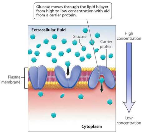
FIGURE 3.8. Facilitated diffusion is the movement through the plasma membrane of a substance from a region of higher concentration to a region of lower concentration with the aid of a membrane protein that acts as a channel or a carrier protein.
Osmosis. Osmosis is a type of diffusion in which water moves across a plasma membrane or any other selectively permeable membrane from a region of higher water concentration to a region of lower water concentration. The movement of water occurs in response to a concentration gradient of a dissolved substance (solute). Consider what happens when a substance such as table sugar (in this case, our solute) is dissolved in water (our solvent) in a membranous bag through which water, but not sugar, can move. Keep in mind that when solute concentration is low, water concentration is high; and when solute concentration is high, water concentration is low. If the membranous bag is placed into a hypertonic solution, meaning a solution whose solute concentration is higher than that inside the bag, more water moves out of the bag than in, causing the bag to shrivel (Figure 3.9a). If, on the other hand, the bag is placed into an isotonic solution, one with the same solute (sugar) concentration as inside the bag, there is no net movement of water in either direction, and the bag maintains its original shape (Figure 3.9b). When the bag is placed into a hypotonic solution, in which the concentration of solute is lower than that inside the bag, more water moves into the bag than out, causing the bag to swell and possibly burst (Figure 3.9c). Osmosis does not require energy and is thus a form of passive transport.
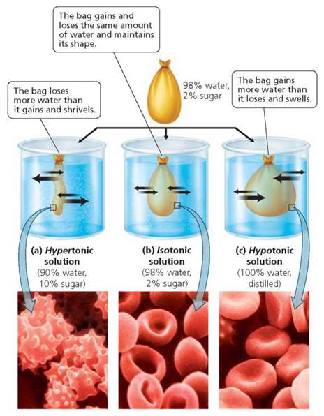
FIGURE 3.9. Osmosis is the diffusion of water across a selectively permeable membrane. The drawings show what happens when a membranous bag through which water but not sugar can move is placed in solutions that are (a) hypertonic, (b) isotonic, or (c) hypotonic to the solution inside the bag. The width of the black arrows corresponds to the amount of water moving into and out of the bag. The photographs show what happens to red blood cells when placed in the three kinds of solutions. Red blood cells are normally shaped like flattened disks, as in part b.
Red blood cells behave the same way the bag in our example does, as shown at the bottom of Figure 3.9. Red blood cells move through a fluid, called plasma. As the figure illustrates, the shape of red blood cells responds to different levels of solute concentration in the plasma.
Active transport. Active transport is a mechanism that moves substances across plasma membranes with the aid of a carrier protein and energy supplied by the cell (through the breakdown of ATP; see Chapter 2). So far in our discussion of movement across plasma membranes, we have described substances moving from regions of higher concentration to regions of lower concentration. However, in most cases of active transport, substances are moved from regions of lower concentration to higher concentration, as shown in Figure 3.10. This type of movement is described as going "against the concentration gradient" and occurs when cells need to concentrate certain substances. For example, the cells in our bodies contain higher concentrations of potassium ions (K+) and lower concentrations of sodium ions (Na+) than their surroundings. Through active transport, proteins in the plasma membrane help maintain these conditions, pumping potassium ions into the cell and sodium ions out of the cell. In this example, both potassium and sodium are moving from regions of lower concentration to regions of higher concentration.
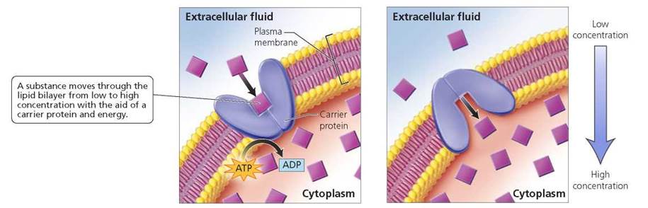
FIGURE 3.10 Active transport is the movement of molecules across the plasma membrane, often from an area of lower concentration to one of higher concentration with help from a carrier protein and energy, usually in the form of ATP.
Endocytosis. Most small molecules cross the plasma membrane by simple diffusion, facilitated diffusion, or active transport. Large molecules, single-celled organisms such as bacteria, and droplets of fluid containing dissolved substances enter cells through endocytosis (Figure 3.11). In endocytosis, a region of the plasma membrane engulfs the substance to be ingested and then pinches off from the rest of the membrane, in this way enclosing the substance in a saclike structure called a vesicle. The vesicle then travels through the cytoplasm. Two types of endocytosis are phagocytosis ("cell eating") and pinocytosis ("cell drinking"). In phagocytosis, cells engulf large particles or bacteria (Figure 3.11a). In pinocytosis, they engulf droplets of fluid (Figure 3.11b), thus bringing all of the substances dissolved in the droplet into the cell.
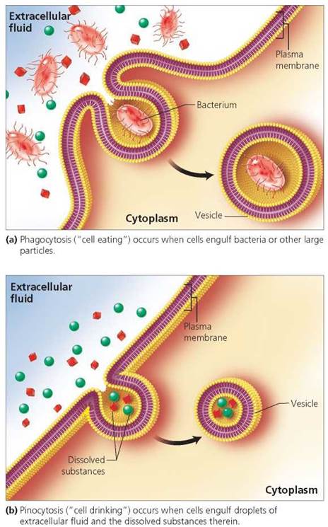
FIGURE 3.11. Endocytosis—phagocytosis or pinocytosis—occurs when a localized region of the plasma membrane surrounds a bacterium, large molecule, or fluid containing dissolved substances and then pinches inward to form a vesicle that moves into the cell.
Exocytosis. The process by which large molecules leave cells is exocytosis. In a cell that produces hormones, for example, the hormones are enclosed in membrane-bound vesicles that travel through the cell's cytoplasm toward the plasma membrane. When the vesicle reaches the plasma membrane, the vesicle membrane fuses with the plasma membrane and then the vesicle opens up to release the hormone outside the cell. Nerve cells also release chemicals by exocytosis. Exocytosis is shown in Figure 3.12.
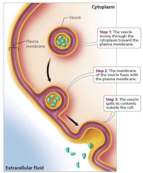
FIGURE 3.12. Cells package large molecules In membrane-bound vesicles, which then spill their contents by exocytosis.
Table 3.2 reviews the ways in which substances move across the plasma membrane.
TABLE 3.2. Review of Mechanisms of Transport across the Plasma Membrane
Mechanism |
Description |
Simple diffusion |
Random movement from region of higher concentration to region of lower concentration |
Facilitated diffusion |
Movement from region of higher concentration to region of lower concentration with the aid of a carrier or channel protein |
Osmosis |
Movement of water from region of higher water concentration (lower solute concentration) to region of lower water concentration (higher solute concentration) |
Active transport |
Movement often from region of lower concentration to region of higher concentration with the aid of a carrier protein and energy usually from ATP |
Endocytosis |
Process by which materials are engulfed by plasma membrane and drawn into cell in a vesicle |
Exocytosis |
Process by which a membrane-bound vesicle from inside the cell fuses with the plasma membrane and spills contents outside of cell |
Stop and think
People with kidney disorders may need dialysis to remove waste products from their blood. The blood is passed through a long, coiled tube submerged in a tank filled with dialyzing fluid. The tubing is porous, allowing small molecules to diffuse out of the blood into the dialyzing fluid. Urea is a waste product that must be removed from the blood. Glucose molecules, on the other hand, should remain in the blood. To achieve these goals, what should the concentrations of urea and glucose be like in the dialyzing fluid?
Organelles
Inside the eukaryotic cell, the primary role of membranes is to create separate compartments where specific chemical processes critical to the life of the cell are carried out. The membrane- bound organelles distributed in the cells' cytoplasm have different functions—just like the different offices within a large company, some of which are responsible for production, some for purchasing, and others for shipping. The compartmentalization allows segregated combinations of molecules to carry out specific tasks (see Figure 3.2). Some organelles give directions for manufacturing cell products. Others make or modify the products or transport them. Still other organelles process energy or break down substances for use or disposal. Nonmembranous organelles also perform specific functions for the cell.
Nucleus
The cell nucleus contains almost all of the cell's genetic information (Figure 3.13). The DNA within the nucleus controls cellular structure and function because it contains a code for the production of proteins. All our cells contain the same genetic information. The characteristics of a particular cell—what makes it a muscle cell or a liver cell—are determined largely by the specific directions it receives from its nucleus.
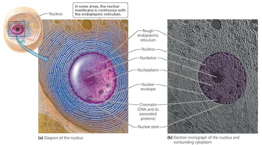
FIGURE 3.13. The nucleus contains almost all the genetic information of a cell.
A double membrane called the nuclear envelope surrounds the nucleus and separates it from the cytoplasm, as shown in Figure 3.13. Communication between the nucleus and cytoplasm occurs through openings in the envelope called nuclear pores. The traffic of selected materials across the nuclear envelope allows the contents of the cytoplasm to influence the nucleus and vice versa.
Genetic information within the nucleus is organized into chromosomes, threadlike structures made of DNA and associated proteins. The number of chromosomes varies from one species to another. For example, humans have 46 chromosomes (23 pairs), house mice have 40 chromosomes, and domestic dogs have 78. Individual chromosomes are visible with a light microscope during cell division, when they shorten and condense (Figure 3.14a). At all other times, however, the chromosomes are extended and not readily visible. In this dispersed state, the genetic material is called chromatin (Figure 3.14b). The chromatin and other contents of the nucleus constitute the nucleoplasm. We will discuss chromosomes and cell division in Chapter 19.
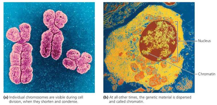
FIGURE 3.14. Chromosomes are composed of DNA and associated proteins.
The nucleolus, a specialized region within the nucleus (see Figure 3.13), forms and disassembles during the course of the cell cycle (see Chapter 19). It is not surrounded by a membrane but is simply a region where DNA has gathered to produce a type of RNA called ribosomal RNA (rRNA). Ribosomal RNA is a component of ribosomes, which are sites where protein synthesis begins. Ribosomes may be suspended in the cytoplasm (free ribosomes) or attached to the endoplasmic reticulum (bound ribosomes).
Endoplasmic Reticulum
The endoplasmic reticulum (ER) is part of an extensive network of channels connected to the nuclear envelope and certain organelles (Figure 3.15). In some regions, the ER is studded with ribosomes and because of this is called rough endoplasmic reticulum (RER). The amino acid chains made by the attached ribosomes are threaded through the RER's membrane to its internal spaces. There the chains are processed and modified, enclosed in vesicles formed from the RER membrane, and transferred to the Golgi complex (discussed shortly) for additional processing and packaging. Proteins made by ribosomes bound to ER will be incorporated into membranes or eventually secreted by the cell. Proteins produced by free ribosomes will remain in the cell.
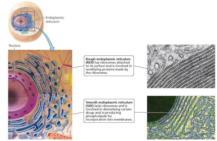
FIGURE 3.15. The endoplasmic reticulum (ER) is continuous with the nuclear membrane and consists of two regions: rough ER and smooth ER.
Smooth endoplasmic reticulum (SER) lacks ribosomes. The SER (particularly in liver cells) detoxifies alcohol and other drugs. Typically, enzymes of SER modify the drugs to make them more water soluble and easier to eliminate from the body. Another function of SER is the production of phospholipids. These phospholipids, along with proteins from the RER, are used to make the RER membrane. Because the RER membrane is continually used to form vesicles for shipping, it must be replenished constantly.
Golgi Complex
The Golgi complex consists of a series of interconnected, flattened membranous sacs. This organelle is the cell's protein processing and packaging center (Figure 3.16). Protein-filled vesicles from the RER arrive at the "receiving side" of the Golgi complex, fuse with its membrane, and empty their contents inside. The Golgi complex then chemically modifies many of the proteins as they move, by way of vesicles, from one membranous disk in the stack to the next. When the processing is finished, the Golgi complex sorts the proteins, much as a postal worker sorts letters, and sends them to their various destinations. Some of the proteins emerging from the "shipping side" are packaged in vesicles and sent to the plasma membrane for export from the cell or to become membrane proteins. Other proteins are packaged in lysosomes. Figure 3.17 on page 56 summarizes the movement of protein-filled vesicles from the rough endoplasmic reticulum to the Golgi complex for processing and eventual release.
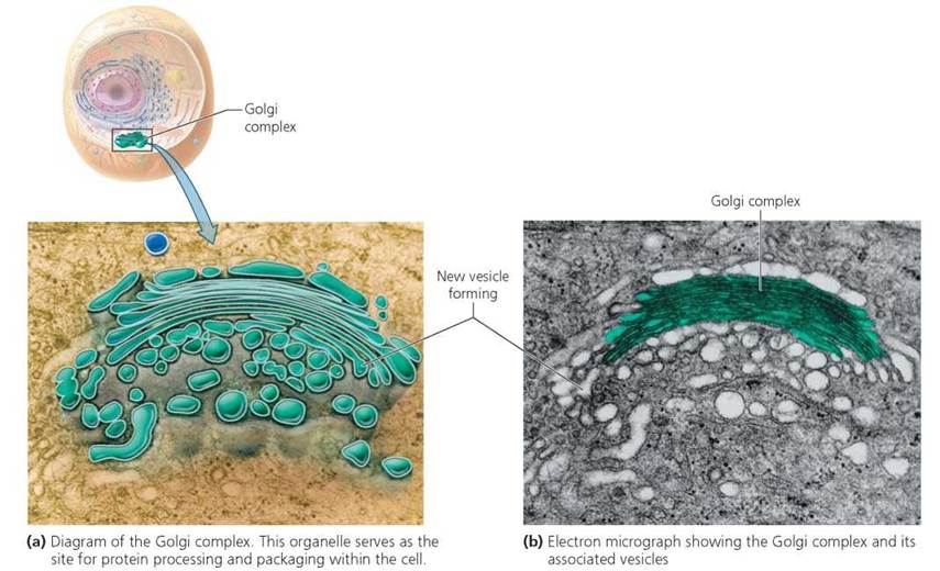
FIGURE 3.16. The Golgi complex
Lysosomes
How does the cell break down worn-out parts or digest large objects that it takes in by phagocytosis? If it simply released digestive enzymes into its cytoplasm, for example, it would soon destroy itself. Instead, intracellular digestion occurs mainly within lysosomes. Lysosomes are roughly spherical organelles consisting of a single membrane packed with about 40 different digestive enzymes. The enzymes and membranes of lysosomes are made by the RER and then sent to the Golgi complex for additional processing. Eventually, enzyme-filled lysosomes bud and then pinch off from the Golgi complex (see Figure 3.17) and begin their diverse roles in digestion within the cell. These roles include destroying foreign invaders such as bacteria, and breaking down worn-out organelles.
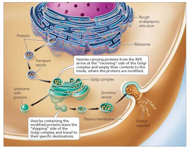
FIGURE 3.17. The route by which protein-filled vesicles from the rough endoplasmic reticulum travel to the Golgi complex for processing and eventual release.
Consider, for example, what happens when a cell engulfs a bacterium. You can follow this process in Figure 3.18 (see pathway on right). During the process of phagocytosis (Step 1), a vesicle encircles the bacterium. A lysosome released from the Golgi complex then fuses with the vesicle (Step 2), and the lysosome's digestive enzymes break the bacterium down into smaller molecules. These molecules diffuse out of the vesicle into the cytoplasm, where they can be used by the cell (Step 3). Indigestible residues may be expelled from the cell by exocytosis (Step 4), or they may be stored indefinitely in vesicles inside the cell (Step 5).
Lysosomes also break down obsolete parts of the cell itself. Worn-out organelles and macromolecules are broken down into smaller components that can be reused (see Figure 3.18, pathway on left). For example, an organelle called a mitochondrion (discussed later) lasts only about 10 days in a typical liver cell before being destroyed by lysosomes. After worn-out mitochondria are destroyed, their component monomers, such as amino acids, are returned to the cytoplasm for reuse. Such "housecleaning" keeps the cell functioning properly and promotes the recycling of essential materials.
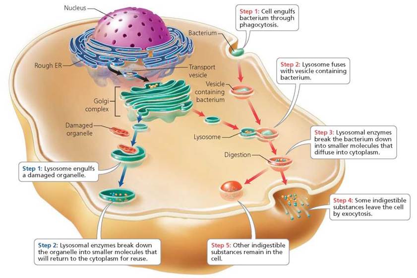
FIGURE 3.18. Lysosome formation and function in intracellular digestion. Lysosomes, released from the Golgi complex, digest a bacterium engulfed by the cell (see pathway on right). Lysosomes also digest obsolete parts of the cell itself (see pathway on left).
The absence of a single kind of lysosomal enzyme can have devastating consequences. Molecules that would normally be broken down by the missing enzyme start to collect in the lysosomes and cause them to swell. Ultimately, the accumulating molecules interfere with cell function. These lysosomal storage diseases are inherited and progress with age.
Tay-Sachs disease is a lysosomal storage disease caused by the absence of the lysosomal enzyme hexosaminidase (Hex A), which breaks down lipids in nerve cells. When Hex A is missing, the lysosomes swell with undigested lipids. Infants with Tay-Sachs disease appear normal at birth but begin to deteriorate by about 6 months of age as abnormal amounts of lipid accumulate in the nervous system. By the age of 4 or 5, Tay-Sachs causes paralysis and death. At present there is no cure for this disease. However, there is a blood test to detect individuals who carry the gene for Tay-Sachs. Called carriers, these individuals do not have the disease but could pass the gene to their offspring.
What would you do?
Imagine that you and your spouse want to start a family, but both of you are carriers of Tay-Sachs disease and could pass the gene to your children. The possible outcomes for any child you might conceive are as follows: the child may not have the gene for Tay-Sachs and may be healthy, may have the disease and die in early childhood, or may be a carrier as you are. Your parents urge adoption. Your spouse prefers not to adopt but to use prenatal screening to check if your fetus has the disease. What would you do?
Certain environmental factors cause disease by interfering with lysosomes. In the Environmental Issue essay, The Deadly Interaction between Asbestos and Lysosomes, we describe the impact of asbestos on health.
Mitochondria
Most cellular activities require energy. Energy is needed to transport certain substances across the plasma membrane and to fuel many of the chemical reactions that occur in the cytoplasm. Specialized cells such as muscle cells and nerve cells require energy to perform their particular activities. The energy needed by cells is provided by mitochondria (singular, mitochondrion), the organelles within which most of cellular respiration occurs. Cellular respiration, discussed later in the chapter, is a four-phase process in which oxygen and an organic fuel such as glucose are consumed and energy in the form of ATP is released. The first phase takes place in the cytoplasm. The remaining three phases occur in the mitochondria.
The number of mitochondria varies considerably from cell to cell and is roughly correlated with a cell's demand for energy. Most cells contain several hundred to thousands of mitochondria. Like the nucleus, but unlike other organelles, mitochondria are bounded by a double membrane (Figure 3.19). The inner and outer membranes create two separate compartments that serve as sites for some of the reactions in cellular respiration.
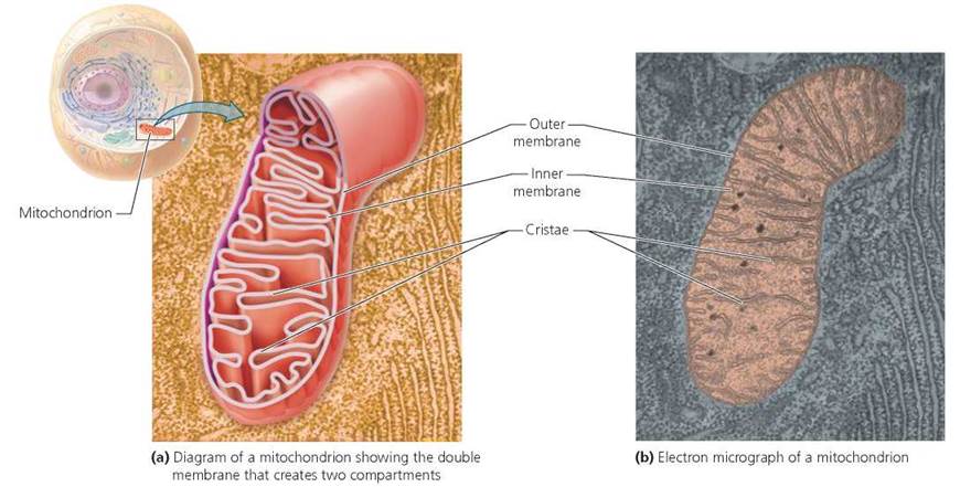
FIGURE 3.19. Mitochondria are sites of energy conversion in the cell.
The infoldings of the inner membrane of a mitochondrion are called cristae, and these are the sites of the last phase of cellular respiration. Finally, mitochondria contain ribosomes and a small percentage of a cell's total DNA (the rest being found in the nucleus, as noted earlier). Mitochondria contain ribosomes and DNA because they are likely descendants of once free-living bacteria that invaded or were engulfed by ancient cells (see Chapter 22). Table 3.3 reviews the functions of organelles.
TABLE 3.3. Review of Major Organelles and Their Functions
Organelle |
Function |
Nucleus |
Contains almost all the genetic information and influences cellular structure and function |
Rough endoplasmic reticulum (RER) |
Studded with ribosomes (sites where the synthesis of proteins begins); produces membrane |
Smooth endoplasmic reticulum (SER) |
Detoxifies drugs; produces membrane |
Golgi complex |
Sorts, modifies, and packages products of RER |
Lysosomes |
Digest substances imported from outside the cell; destroy old or defective cell parts |
Mitochondria |
Provide cell with energy through the breakdown of glucose during cellular respiration |
Stop and think
We have discussed the nucleus, endoplasmic reticulum (including rough and smooth), ribosomes, Golgi complex, lysosomes, and mitochondria. Assign each of these organelles to one of the following main functions within a cell: manufacturing, breakdown, or energy processing.
Cytoskeleton
Traversing the cytoplasm of the cell is a complex network of fibers called the cytoskeleton. The fibers are divided into three types: microtubules are the thickest; microfilaments are the thinnest; and intermediate filaments are the diverse group in between. Microtubules and microfilaments disassemble and reassemble, whereas intermediate filaments tend to be more permanent.
Microtubules are straight, hollow rods made of the protein tubulin. Some microtubules near the plasma membrane maintain cell shape. Microtubules also form tracks along which organelles or vesicles travel. For example, secretory vesicles (membrane-bound vesicles containing material to be released from the cell) that have budded from the Golgi complex make their way to the plasma membrane by moving along a microtubule track. Finally, microtubules play a role in the separation of chromosomes during cell division. A microtubule-organizing center located near the nucleus contains a pair of centrioles, each composed of nine sets of three microtubules arranged in a ring (Figure 3.20). Centrioles may function in cell division and in the formation of cilia and flagella.
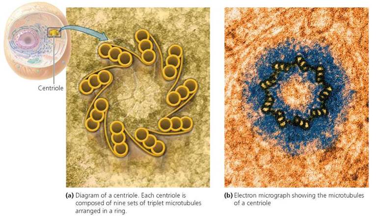
FIGURE 3.20. Centrioles may play a role In cell division.
Microtubules serve as the working parts of two types of cell extensions called cilia (singular, cilium) and flagella (singular, flagellum). Cilia are numerous, short extensions on a cell that move with the back-and-forth motion of oars. They are found, for example, on the surfaces of cells lining the respiratory tract (Figure 3.21a), where they sweep debris trapped in mucus away from the lungs. Smoking destroys these cilia and hampers cleaning of respiratory surfaces. A flagellum resembles a whip and moves in an undulating manner. Flagella are much longer than cilia. The only cell with a flagellum in humans is the sperm cell (Figure 3.21b).
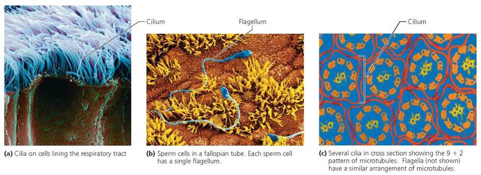
FIGURE 3.21. Microtubules are responsible for the movement of cilia and flagella.
Cilia and flagella differ in length, number per cell, and pattern of movement. Nevertheless, they have a similar arrangement of microtubules, called a 9 + 2 pattern (Figure 3.21c), at their core. This arrangement consists of nine pairs of microtubules arranged in a ring with two microtubules at the center.
Microfilaments are solid rods made of the protein actin. These fibers are best known for their role in muscle contraction, where they slide past thicker filaments made of the protein myosin. Finally, microfilaments play a role in cell division, forming a band that contracts and pinches the cell in two.
Intermediate filaments are a diverse group of ropelike fibers helping to maintain cell shape and anchoring certain organelles in place. Their protein composition varies from one type of cell to another.
Cellular Respiration and Fermentation in the Generation of ATP
Living requires work, and work requires energy. Logic tells us, therefore, that living requires energy.
We get our energy from the food we eat. Our digestive system (discussed in Chapter 15) breaks down complex macromolecules such as carbohydrates, proteins, and fats into their simpler components, such as glucose, amino acids, and fatty acids. These simpler molecules are then absorbed into the bloodstream and carried to our cells, where some of the energy stored in the molecules' chemical bonds is used to make ATP, the energy-rich molecule that our cells use to do their work. (Some energy is also given off as heat.) Although carbohydrates, proteins, and fats are all sources of cellular energy, we will focus on carbohydrates. Cells have two ways of breaking glucose molecules apart for energy: cellular respiration and fermentation. Cellular respiration requires oxygen; fermentation does not.
All the chemical reactions that take place in a cell constitute its metabolism. These chemical reactions are organized into metabolic pathways. Each pathway consists of a series of steps in which a starting molecule is modified, eventually resulting in a particular product. Specific enzymes speed up each step of the pathway. Cellular respiration and fermentation are examples of catabolic pathways—pathways in which complex molecules, such as carbohydrates, are broken down into simpler compounds, releasing energy. Anabolic pathways, on the other hand, build complex molecules from simpler ones and consume energy in the process.
Environmental Issue
The Deadly Interaction between Asbestos and Lysosomes
Asbestos is a fibrous silicate mineral, found in many forms in nature, that is strong, flexible, and resistant to heat and corrosion (Figure 3.A). Because of these properties, asbestos was used widely in construction—as an insulator on ceilings and pipes, for example, or to soundproof and fireproof the walls of schools.
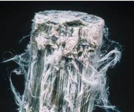
FIGURE 3.A. The deadly miracle material asbestos
The very properties that make asbestos an ideal building material—its fibrous nature and durability—also can make it deadly. For example, when fibers of asbestos insulation are dislodged, small, light particles become suspended in the air and can be inhaled into the lungs. There is some evidence that the different forms of asbestos differ in the time they persist in lung tissue. Nevertheless, particles of at least one form appear resistant to degradation and remain in the lungs for life (Figure 3.B).
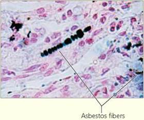
FIGURE 3.B. Asbestos fibers in lung tissue
Inhalation of asbestos particles can cause lung cancer and mesothelioma, a form of cancer specific to the lining of the lungs and chest cavity (pleura) and the lining of the abdomen (peritoneum). Asbestosis, a third condition, is the most common disease caused by exposure to asbestos. It results from the dangerous interaction between asbestos and lysosomes. Cells responsible for cleaning the respiratory passages engulf small particles of asbestos inhaled into the lungs; lysosomes inside the cleaning cells then fuse with the vesicles containing the asbestos particles. Unfortunately, the lysosomal enzymes cannot break down the asbestos particles. Instead, the particles destabilize the membranes of the lysosomes, causing massive release of enzymes, which destroy the cells of the respiratory tract. Irreversible scarring of lung tissue is the result, eventually interfering with the exchange of gases in the lungs. People with asbestos-damaged lungs experience chronic coughing and shortness of breath. These symptoms become more severe overtime and may cause death from impaired respiratory function.
At present, there is no effective treatment for asbestosis. The focus, therefore, has been on prevention. In the United States, the use of asbestos for insulation and fireproofing, or for any new purposes, is banned. However, asbestos is still present in many buildings constructed before the ban went into effect. In these buildings, it is generally recommended and often required that exposed asbestos be removed, enclosed by other building materials, or covered with a sealant. Experts should determine which method is best in any given situation. Moreover, the sealing or removal of asbestos must be done by experts, because the greatest risk of asbestos exposure occurs when asbestos is handled improperly. Finally, workers at risk of exposure to asbestos—plumbers, electricians, insulation workers, and carpenters, to name a few—should insist upon frequent testing of the air in their workplaces.
Questions to Consider
• Cigarette smoking worsens diseases caused by exposure to asbestos. If a worker who smokes is exposed over many years to asbestos in the workplace and subsequently develops lung cancer, then who is responsible for his developing cancer? Is the employer responsible or does the worker bear some personal responsibility?
• What information would you consider when assessing responsibility?
Cellular Respiration
Cellular respiration is the oxygen-requiring pathway by which cells break down glucose. It is an elaborate series of chemical reactions whose final products are carbon dioxide, water, and energy. In a laboratory beaker, glucose and oxygen can be combined to produce those products in a single step. However, under those circumstances, the glucose burns, and all the energy is lost as heat. The process used by cells, in which glucose is broken down in a series of steps, enables the cells to obtain much of the energy in a usable form—specifically, as a high-energy chemical bond in ATP. Recall from Chapter 2 that ATP is formed from ADP (adenosine diphosphate) and inorganic phosphate (Pi) in a process that requires energy.
Cellular respiration has four phases: (1) glycolysis, (2) the transition reaction, (3) the citric acid cycle, and (4) the electron transport chain. All four phases occur continuously within cells. Glycolysis takes place in the cytoplasm of the cell. The transition reaction, the citric acid cycle, and the electron transport chain take place in mitochondria. You will see that some of these phases consist of a series of reactions in which the products from one reaction become the substrates (raw materials) for the next reaction. You will also see that the transfer of electrons from one atom or molecule to another is a key feature of the process our cells use to capture energy from fuel. As the electrons are passed along a chain of intermediate compounds, their energy is used to make ATP.
Glycolysis. The first phase of cellular respiration, called glycolysis (glyco, sugar; lysis, splitting), begins with glucose, a six-carbon sugar, being split into 2 three-carbon sugars. These three-carbon sugars are then converted into two molecules of pyruvate (Figure 3.22), another three-carbon compound. Glycolysis occurs in several steps, each requiring a different, specific enzyme. During the first steps, two molecules of ATP are consumed because energy is needed to prepare glucose for splitting. During the remaining steps, four molecules of ATP are produced, for a net gain of two ATP. Glycolysis also produces two molecules of nicotine adenine dinucleotide (NADH), which are generated when electrons are donated to the coenzyme NAD+. Glycolysis does not require oxygen and releases only a small amount of the chemical energy stored in glucose. Most of the energy remains in the two molecules of pyruvate. The pyruvate molecules move from the cytoplasm into the inner compartment of the mitochondrion.
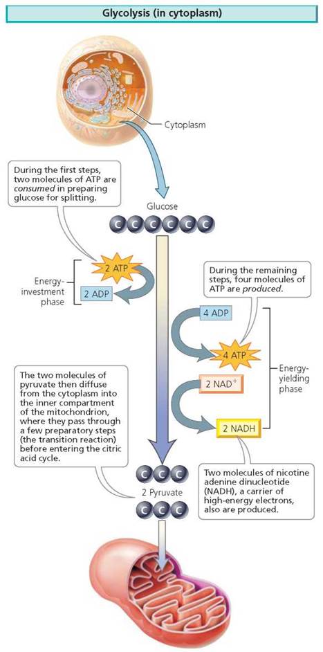
FIGURE 3.22. Glycolysis is a several-step sequence of reactions in the cytoplasm. Glucose, a six-carbon sugar, is split into 2 three-carbon molecules of pyruvate.
Transition reaction. Once inside the inner compartment of the mitochondrion, pyruvate reacts with a substance called coenzyme A (CoA) in a reaction called the transition reaction. The transition reaction results in the removal of one carbon (in the form of carbon dioxide, CO2) from each pyruvate (Figure 3.23). The resulting two-carbon molecule, called an acetyl group, then binds to CoA to form acetyl CoA. A molecule of NADH is also produced from each pyruvate.
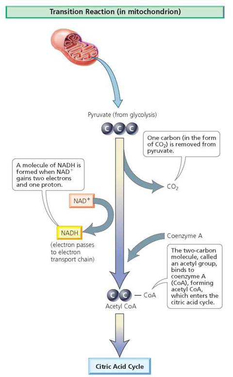
FIGURE 3.23. The transition reaction takes place inside the mitochondrion and is the link between glycolysis and the citric acid cycle.
Citric acid cycle. Still in the inner compartment of the mitochondrion, acetyl CoA reacts with a four-carbon compound in the first of a cyclic series of eight chemical reactions known as the citric acid cycle, named after the first product (citric acid, or citrate) formed along its route (Figure 3.24). The cycle is also called the Krebs cycle—after the scientist Hans Krebs, who described many of the reactions. Rather than considering each of the chemical reactions in the citric acid cycle, we will simply say that it completes the loss of electrons from glucose and yields two molecules of ATP (one from each acetyl CoA that enters the cycle) and several molecules of NADH and FADH2 (flavin adenine dinucleotide). NADH and FADH2 are carriers of high-energy electrons. The NADH and FADH2 produced in glycolysis, in the transition reaction, and in the citric acid cycle enter the electron transport chain, the final phase of cellular respiration. The citric acid cycle also produces CO2 as waste.

FIGURE 3.24. The citric acid cycle is a cyclic series of eight chemical reactions that occurs inside the mitochondrion and yields two molecules of ATP and several molecules of NADH and FADH2 per molecule of glucose.
Electron transport chain. During the final phase of cellular respiration, the molecules of NADH and FADH2 produced by earlier phases pass their electrons to a series of carrier proteins embedded in the inner membrane of the mitochondrion. These proteins are known as the electron transport chain (Figure 3.25). (Recall that the inner membrane of the mitochondrion is highly folded, providing space for thousands of sets of carrier proteins.) During the transfer of electrons from one protein to the next, energy is released and used to make ATP. Eventually, the electrons are passed to oxygen, the final electron acceptor, which then combines with two hydrogen ions to form water. Oxygen has a critical role in cellular respiration. When oxygen is absent, electrons accumulate in the carrier proteins, halting the citric acid cycle and cellular respiration. But when oxygen is present, and accepts the electrons, respiration continues. The electron transport chain produces 32 molecules of ATP per molecule of glucose. In the Health Issue essay, Mitochondrial Diseases Cause an Energy Shortage in Our Bodies, we describe what happens when there is a deficiency of proteins in the electron transport chain.
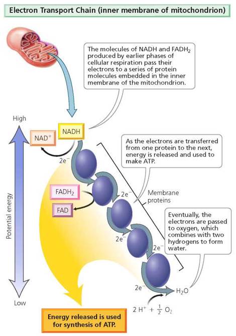
FIGURE 3.25. The electron transport chain is the final phase of cellular respiration. This phase yields 32 ATP molecules per molecule of glucose.
Altogether, cellular respiration generally produces 36 molecules of ATP per molecule of glucose: 2 ATP from glycolysis, 2 ATP from the citric acid cycle, and 32 ATP from the electron transport chain. The results of cellular respiration are summarized in Figure 3.26. Basic descriptions of each phase can be found in Table 3.4.
TABLE 3.4. Review of Cellular Respiration
Phase |
Location |
Description |
Main Products |
Glycolysis |
Cytoplasm |
Several-step process by which glucose is split into 2 pyruvate |
2 pyruvate 2 ATP 2 NADH |
Transition reaction |
Mitochondria |
One CO2 is removed from each pyruvate; the resulting molecules bind to CoA, forming 2 acetyl CoA |
2 acetyl CoA 2 NADH |
Citric acid cycle |
Mitochondria |
Cyclic series of eight chemical reactions by which acetyl CoA is broken down |
2 ATP 2 FADH2 6 NADH |
Electron transport chain |
Mitochondria |
Electrons from NADH and FADH2 are passed from one protein to the next, releasing energy for ATP synthesis |
32 ATP H2O |
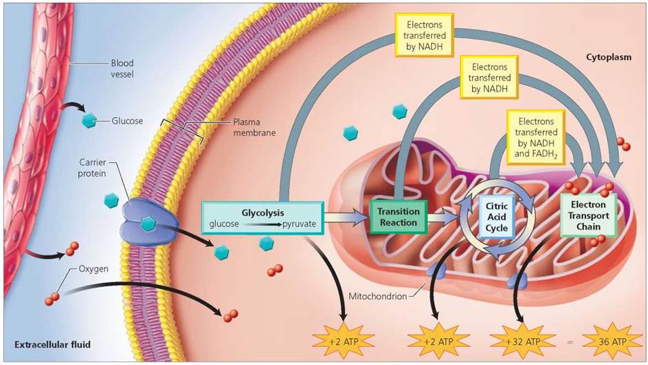
FIGURE 3.26. Summary of cellular respiration. Cellular respiration produces 36 ATP per molecule of glucose (2 ATP from glycolysis, 2 ATP from the citric acid cycle, and 32 ATP from the electron transport chain).
Fermentation
As noted earlier, cellular respiration depends on oxygen as the final electron acceptor in the electron transport chain. Without oxygen, the transport chain comes to a halt, blocking the citric acid cycle and stopping cellular respiration. Is there a way for cells to harvest energy when molecules of oxygen are scarce? The answer is yes, and the pathway is fermentation.
Fermentation is the breakdown of glucose without oxygen. It begins with glycolysis, which as you recall occurs in the cytoplasm and does not require oxygen. From one molecule of glucose, glycolysis produces two molecules each of pyruvate, the electron carrier NADH, and ATP. The remaining fermentation reactions also take place in the cytoplasm, transferring electrons from NADH to pyruvate or a derivative of pyruvate. This transfer of electrons is critical because it regenerates NAD+, which is essential for the production of ATP through glycolysis. Recall that in cellular respiration, oxygen is the final electron acceptor in the electron transport chain, whereas in fermentation it is pyruvate or a pyruvate derivative. Fermentation therefore nets only 2 molecules of ATP compared with the 36 molecules of ATP produced by cellular respiration (refer, again, to Figure 3.26). In short, fermentation is a very inefficient way for cells to harvest energy.
Lactic acid fermentation occurs in the human body. During strenuous exercise, the oxygen supply in our muscle cells runs low. Under these conditions, the cells increase lactic acid fermentation to ensure the continued production of ATP. The muscle pain we often experience after intense exercise is caused partly by the accumulation of lactic acid, a waste product of this type of fermentation. In time, the soreness disappears as the lactic acid moves into the bloodstream and is carried to the liver, where it is converted back to pyruvate.
Mitochondrial Diseases Cause an Energy Shortage in Our Bodies
Outages in the electrical power grid can cause many services—air and rail transport, communications, water purification, heating and cooling—to slow and sometimes fail. A similar situation can happen in our bodies when mitochondria, our energy-processing organelles, fail. Recall that mitochondria convert energy from food molecules into ATP for cells to use, and that several steps of cellular respiration occur in mitochondria and require particular proteins to proceed. More than 40 illnesses, collectively known as mitochondrial diseases, can result from deficiencies in the proteins that function in energy metabolism within mitochondria, including proteins that are part of the electron transport chain. When mitochondria fail to function properly, less energy is generated, cell function is compromised, and cell death can result. This, in turn, can affect tissues, organs, and organ systems. Mitochondrial diseases cause the most damage to parts of the body that need the most energy, such as the brain, heart, lungs, kidneys, endocrine glands, and skeletal muscles.
Each year in the United States, an estimated 1000 to 4000 babies are born with mitochondrial diseases. Many physicians believe that mitochondrial diseases are underdiagnosed and frequently misdiagnosed, so estimates may be low. These diseases are caused by mutations (changes) in DNA that encodes for proteins critical in energy metabolism within mitochondria. Some of these proteins are encoded by mitochondrial DNA, but others are encoded by nuclear DNA and then imported into mitochondria. The mutations may be spontaneous or inherited. Children inherit nuclear DNA from their mother and father, but they inherit their mitochondrial DNA only from their mother. Environmental factors, such as infections and certain drugs, can also damage mitochondria.
People with a mitochondrial disease may experience fatigue and fail to gain weight. Symptoms of specific mitochondrial diseases are diverse because they reflect which cells and organs have compromised mitochondria. If cells in the brain are affected, then symptoms may include seizures, developmental delays, and dementia. When sensory organs, such as the eyes or ears are affected, then a decline or complete loss of vision or hearing may occur. Skeletal muscle cells with defective mitochondria can result in muscle weakness, cramps, and exercise intolerance. Sometimes multiple organ systems are affected. Additionally, cells typically contain hundreds of mitochondria, so a single cell can have some mitochondria that are normal and others that are defective (i.e., carry a mutation). The health of cells (and the severity of disease symptoms) will depend on the relative proportions of normal and defective mitochondria. Symptoms may vary dramatically even among members of the same family with an inherited mutation in mitochondrial DNA. Because some eggs of a woman may have large proportions of defective mitochondria while other eggs have mostly healthy mitochondria, the severity of symptoms in the children of this woman will depend on the proportion of defective mitochondria passed to them by their mother. Finally, depending on the particular disorder, the onset of symptoms can occur in infancy, childhood, or adulthood. Adult onset may occur in a person who was born with the genetic mutation, but whose symptoms did not appear until an environmental trigger, such as a severe illness, brought them on.
Mitochondrial diseases are difficult to diagnose, so most physicians refer patients to an appropriate research center. Evaluation at such a center might include an assessment of family history, a complete physical exam to characterize affected organ systems (for example, imaging of the brain and heart, and tests for vision and hearing), and metabolic screening of blood samples for certain enzymes and products of cell metabolism, such as lactate and pyruvate. A muscle biopsy might be taken to evaluate the condition of mitochondria in muscle cells and to determine whether certain proteins are present.
Given variation in the number of organ systems affected and in the severity of symptoms, it is difficult to predict the course of a mitochondrial disease. Indeed, the quality of life experienced by some patients may be relatively good, while others have severe symptoms and do not survive childhood. There are no known cures for mitochondrial diseases, so medical professionals focus on alleviating symptoms, and slowing disease progression. For example, a dietician may help patients to maintain or gain weight, and physical therapy might benefit patients whose skeletal muscles are affected. Patients are also advised to avoid energetically stressful situations, such as fasting or extreme cold. Mitochondrial dysfunction also may be linked to autism, aging, and many chronic conditions of adulthood. Clearly, much remains to be learned about the roles that mitochondria play in human health.
Questions to Consider
•Consider a woman who has an inherited, adult onset mitochondrial disease that was diagnosed only after she had two sons and a daughter. Her symptoms are relatively mild and her disease has been traced to a mutation in her mitochondrial DNA. Which, if any, of her children will inherit the mutation?
• What factors determine the extent of symptoms in any afflicted child? If all of her children reach adulthood and have families of their own, which of her children will pass on the mutation?
Looking ahead
In Chapter 3 we learned about the basic structure of cells. In Chapter 4 we describe how specialized cells form tissues, organs, and organ systems.
Highlighting the Concepts
Eukaryotic Cells Compared with Prokaryotic Cells (pp. 45-47)
• There are two main types of cells. Prokaryotic cells, unique to bacteria and Archaea, lack membrane-bound organelles. Eukaryotic cells, found in all other organisms, have membrane-bound organelles.
Cell Size and Microscopy (p. 47)
• As a cell grows, its volume increases more than its surface area; therefore, a cell that is too large will encounter problems caused by inadequate surface area. For that reason, most cells are very small.
• Because most cells are very small, they can be seen only with a microscope. Whereas light microscopes use beams of light to image specimens, electron microscopes use beams of electrons. Electron microscopes can reveal finer details because the wavelength of an electron beam is smaller than the wavelengths of visible light.
Cell Structure and Function (pp. 47-48)
• All eukaryotic cells have certain features in common, including a plasma membrane and membrane-bound organelles. Structural differences between eukaryotic cells often reflect differences in function. For example, a sperm has a tail and streamlined shape so that it can swim to the egg. The egg, in contrast, is large, immobile, and packed with materials needed to initiate development.
Plasma Membrane (pp. 48-53)
• The plasma membrane is made of phospholipids arranged in a bilayer with proteins and molecules of cholesterol interspersed throughout and carbohydrates attached to the outer surface.
The structure of the plasma membrane is often described as a fluid mosaic.
• The plasma membrane maintains the cell's integrity, regulates movement of substances into and out of the cell, functions in cell-cell recognition, promotes communication between cells, and binds cells together to form tissues and organs.
• Substances cross the plasma membrane in several ways. Some cross by random movement from higher to lower concentration (simple diffusion); others need help from a carrier or channel protein (facilitated diffusion). Water also moves across the plasma membrane from higher to lower concentration (osmosis). Substances being concentrated by cells cross the plasma membrane from lower to higher concentration with the help of ATP and a carrier protein (active transport). Finally, cells may engulf outside materials by surrounding them with plasma membrane (endocytosis) or may release substances to the external environment by fusing internal vesicles with the plasma membrane and spilling their contents to the outside (exocytosis).
Organelles (pp. 53-58)
• Within a eukaryotic cell, membranes delineate specialized compartments within which specific processes occur. The nucleus contains almost all the genetic information and thus holds the code for the cell's structure and many of its functions. The nucleolus is a region within the nucleus that makes ribosomal RNA. Ribosomes are the sites where protein synthesis begins. Endoplasmic reticulum (ER) functions in membrane production and may be studded with ribosomes (rough endoplasmic reticulum, RER) or free of ribosomes (smooth endoplasmic reticulum, SER). The Golgi complex sorts, modifies, and packages products of the RER. Lysosomes digest bacteria engulfed by cells and break down old or defective components within cells. Mitochondria process energy for cells.
Cytoskeleton (pp. 58-59)
• The cytoskeleton, a complex network of fibers throughout the cytoplasm of the cell, consists of three categories of fibers, all made of proteins: microtubules, microfilaments, and intermediate filaments. Microtubules are hollow rods of tubulin that function in cell movement (cilia and flagella), support, and the movement within cells of chromosomes, organelles, and vesicles. Microfilaments are rods of actin that function in muscle contraction and cell division. Intermediate filaments, which are different kinds of ropelike fibers, maintain cell shape and anchor organelles.
Cellular Respiration and Fermentation in the Generation of ATP (pp. 59-64)
• Cells require energy to work. We get this energy from the food we eat. Carbohydrates, fats, and proteins that we consume are each broken down by our digestive system into smaller units such as simple sugars, fatty acids, and amino acids. These simpler substances are absorbed into the bloodstream and carried to our cells, where the energy stored in the substances' chemical bonds is transferred for the cell's use to the chemical bonds of ATP. (Some energy is also given off as heat.)
• Cells use two catabolic pathways—cellular respiration and fermentation—to break down the carbohydrate glucose and store its energy as ATP. Cellular respiration requires oxygen and usually yields 36 molecules of ATP per molecule of glucose.
• Cellular respiration has four phases—glycolysis, the transition reaction, the citric acid cycle, and the electron transport chain—all occurring continuously within cells. Glycolysis occurs in the cytoplasm and splits glucose into pyruvate while producing NADH (a carrier of high-energy electrons) and a net gain of 2 ATP. The molecules of pyruvate move from the cytoplasm into the inner compartment of the mitochondrion, where they pass through a few preparatory steps, known as the transition reaction, before entering the citric acid cycle. The citric acid cycle is an eight-step cycle that completes the breakdown of glucose into carbon dioxide. The citric acid cycle also yields 2 ATP and carriers of high-energy electrons (NADH and FADH2). In the electron transport chain, the carriers of high- energy electrons produced during glycolysis, the transition reaction, and the citric acid cycle pass their electrons to a series of proteins embedded in the inner membrane of the mitochondrion; oxygen is the final electron acceptor. Energy released during the transfer of electrons yields 32 ATP.
• Fermentation is the breakdown of glucose without oxygen. It occurs in the cytoplasm and begins with glycolysis, the splitting of glucose into pyruvate. The remaining chemical reactions involve the transfer of electrons from NADH to pyruvate or a derivative of pyruvate. Compared with cellular respiration, fermentation is an inefficient way for cells to harvest energy because it yields only 2 ATP as compared with the 36 ATP of cellular respiration.
Reviewing the Concepts
1. How do prokaryotic and eukaryotic cells differ? pp. 45-47
2. Describe the structure of the plasma membrane. pp. 48-49
3. List five functions of the plasma membrane. p. 49
4. What is the difference between simple and facilitated diffusion? Give an example of each. p. 50
5. Describe and differentiate endocytosis and exocytosis. pp. 52-53
6. Where are chromosomes found in the cell? p. 54
7. List two functions of lysosomes. pp. 54, 56
8. What are lysosomal storage diseases? p. 56
9. Name the three types of fibers that make up the cytoskeleton. Describe their structures and functions. p. 58
10. What is the basic function of cellular respiration? p. 59
11. How do cellular respiration and fermentation differ with respect to the number of ATP molecules produced and the requirement for oxygen? p. 63-64
12. Prokaryotic cells
a. have internal membrane-bound organelles.
b. are usually larger than eukaryotic cells.
c. lack internal membrane-enclosed organelles.
d. have linear strands of DNA within a nucleus.
13. The plasma membrane
a. is selectively permeable.
b. contains lipids that function in cell-cell recognition.
c. has cell adhesion molecules that prevent cells from sticking together.
d. is made of nucleic acids.
14. Facilitated diffusion is
a. the random movement of a substance from a region of higher concentration to a region of lower concentration.
b. the movement of water across the plasma membrane.
c. the movement of molecules across the plasma membrane against a concentration gradient with the aid of a carrier protein and energy supplied by the cell.
d. the movement of a substance from a region of higher concentration to a region of lower concentration with the aid of a membrane protein.
15. Almost all the genetic information of a cell is found in the
a. endoplasmic reticulum.
b. Golgi complex.
c. nucleus.
d. mitochondria.
16. Ribosomes
a. are found on smooth endoplasmic reticulum.
b. are sites where protein synthesis begins.
c. process and modify proteins.
d. break down foreign invaders and old organelles.
17. Mitochondria
a. process energy for cells.
b. lack ribosomes and DNA.
c. are bounded by a single membrane.
d. function in cell digestion.
18. Microtubules
a. are found in eukaryotic cilia and flagella.
b. are made of the protein actin.
c. play a role in muscle contraction.
d. pinch a cell in two during cell division.
19. Glycolysis
a. occurs in the mitochondria.
b. requires oxygen.
c. splits glucose into pyruvate.
d. nets 32 molecules of ATP per molecule of glucose.
20. _____ is the jellylike solution within a cell that contains everything between the nucleus and the plasma membrane.
21. _____ is called "cell drinking."
22. The _____ is a specialized region within the nucleus that is involved in the production of rRNA.
23. _____ is the final electron acceptor in the electron transport chain.
24. Our muscle cells may switch from cellular respiration to _____ when oxygen is low.
Applying the Concepts
1. Given what you know about the composition of the plasma membrane, would you expect an anesthetic to be soluble or insoluble in lipids? Explain your answer.
2. Would you expect to find more mitochondria in muscle cells or bone cells? Explain your answer.
3. During cell division, the nucleus and cytoplasm of a cell split into two cells. Cancer is characterized by uncontrolled cell division and is often treated with chemotherapy. Some drugs used in chemotherapy halt cell division by affecting the cyto- skeleton. Which of the cytoskeletal elements (microtubules, microfilaments, intermediate filaments) might be affected?
Becoming Information Literate
Using at least three reliable sources, prepare a brochure on Tay-Sachs disease. Be sure to include the following sections: (1) Symptoms; (2) Causes; (3) Transmission; (4) Diagnosis (before and after birth); (5) Treatment, if any; and (6) Prognosis. List each source you considered, and explain why you chose the sources you used.