THE LIVING WORLD
Unit Four. The Evolution and Diversity of Life
17. Protists: Advent of the Eukaryotes
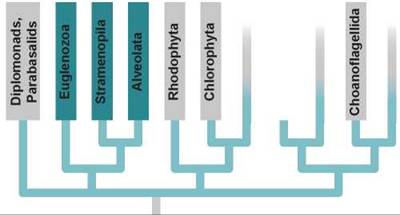
As we have mentioned, molecular comparisons allow us to place 12 of the 17 protist phyla with some confidence on the protist phylogenetic tree (we will get to the other five later). The largest branch of the protist phylogenetic tree contains seven phyla clustered within three monophyletic groups.
Euglenozoa Are Free-living Protists with Anterior Flagella
Euglenozoa are freshwater protists with the majority having two flagella. There are two major groups in the phylum Eugle- nozoa, the euglenoids and the trypanosomes.
Euglenoids like Euglena are euglenozoans with two flagella. As shown in figure 17.9, the flagella are attached at the base of a flask-shaped opening called the reservoir located at the anterior end of the cell. One of the flagella is long and has a row of very fine, short, hairlike projections along one side. A second, shorter flagellum is located within the reservoir but does not emerge from it. Contractile vacuoles collect excess water from all parts of the organism and empty it into the reservoir, which apparently helps regulate the osmotic pressure within the organism. The stigma is light-sensitive and helps these photosynthetic organisms move toward light. Reproduction is by mitotic cell division; no sexual reproduction is known in this group.
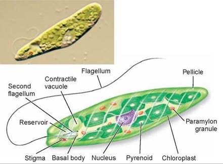
Figure 17.9. Euglena.
In euglenoids such as Euglena, starch forms around pyrenoids; other food reserves are stored in paramylon granules.
Euglenoids clearly illustrate the folly of attempting to classify protists as tiny animals or plants. About one-third of the 1,000 known species have chloroplasts and are photosynthetic; the others lack chloroplasts, ingest their food, and are heterotrophic. In the dark, many photosynthetic euglenoids reduce the size of their chloroplasts (they may appear to disappear!) and become heterotrophs until they return to the light.
Trypanosomes are euglenozoans with a unique single mitochondrion that contains two circles of DNA. Trypanosomes are important pathogens of human beings, responsible for many serious diseases. Perhaps the most devastating is African sleeping sickness, in which infected individuals experience extreme lethargy and fatigue. The trypanosomes spend some of their life cycle in the blood and saliva of a carrier, such as the tsetse fly shown in figure 17.10a. When the fly feeds on a human, the trypanosomes are spread to the human host, where they circulate in its blood. You can see a trypanosome in figure 17.10b, the wormlike shape among the circular red blood cells.
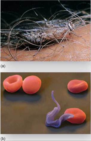
Figure 17.10. Trypanosomes cause sleeping sickness.
(a) A tsetse fly is sucking blood from a human arm in Tanzania, East Africa. The fly's saliva can transmit the trypanosomes that cause sleeping sickness to the human it bites. (b) In this photograph, the undulating, changeable shape of Trypanosoma is visible among human red blood cells.
Leishmaniasis is a trypanosome infection transmitted by sand flies that causes severe skin sores. If the protists reach internal organs, death can result. About 1.5 million new cases are reported each year.
Stramenopila Are Protists with Fine Hairs
Another major group on the protist phylogenetic tree contains three phyla that possess fine hairs on their flagella. These flagellated cells may only appear at certain times in the life cycle, or may have been lost completely, as in the diatoms.
Brown algae, members of the phylum Phaeophyta, contain the longest, fastest-growing, and most photosynthetically productive living organisms, the giant kelp. Kelp form underwater forests with individuals over 100 meters long. The 1,500 species of brown algae are all multicellular and almost all marine. They are the most conspicuous seaweeds in the ocean. The larger brown algae, like the one in figure 17.11a, have flattened blades, stalks, and anchoring bases and often contain complex internal plumbing like that of plants. The life cycle has alternating (a) generations, the large individuals being the sporophyte (diploid) generation.
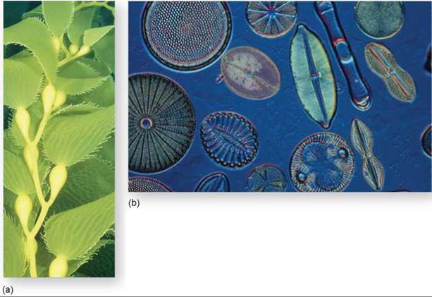
Figure 17.11. Two of the more common stramenopila.
(a) Massive "groves" of giant kelp, a kind of brown algae, contain some of the largest organisms on earth. (b) These diatoms have unique silica, two-part shells.
Diatoms, members of the phylum Chrysophyta, are photosynthetic unicellular protists with a unique double shell of silica. Like tiny oysters, the shells resemble small boxes with lids, one fitting inside the other.
Diatoms are abundant in both oceans and lakes. There are over 11,500 species, of two sorts: some with radial symmetry that look like tiny wheels and others with bilateral (twosided) symmetry (figure 17.11b). The shells of fossil diatoms form thick deposits that are mined commercially as “diato- maceous earth,” used as an abrasive or to make paint sparkle. Diatoms move in a complex manner that is still being investigated. Their movement involves protoplasmic streaming along grooves in their shell and the waving of tiny vibrating fibrils within the grooves.
Water molds, phylum Oomycota, are the downy mildews that are often seen in moist environments. There are 580 named species, all of which either parasitize living organisms or feed on dead organic matter. Many oomycetes are important plant pathogens, including Phytophthora infestans, which causes late blight in potatoes. This mold was responsible for the Irish potato famine of 1845-47, during which about 400,000 Irish people starved.
Alveolata Are Protists with Submembrane Vesicles
Another main “branch” on the protist phylogenetic tree contains three phyla, all of which have a layer of flattened vesicles called alveoli beneath their plasma membrane. The alveoli are thought to function in the transport of materials out of the cell, similar to Golgi bodies.
Ciliates, members of the phylum Ciliophora, are very complex and unusual unicellular heterotrophs with large numbers of cilia (tiny beating hairs) covering the outside of the body, and two nuclei per cell (the micronucleus and macronucleus). Ciliates are so different from other eukaryotes (they even use the genetic code differently!) that many taxonomists argue they should be placed in a separate kingdom of their own. About 8,000 species have been named.
Ciliates have a pellicle, a protein scaffold inside the plasma membrane that can change shape, which makes the body wall tough but flexible. The body interior is extremely complex, inspiring some biologists to consider ciliates multicellular organisms without cell boundaries rather than unicellular. The Paramecium in figure 17.12 is a typical ciliate. It has a complex digestive process, with a gullet (“mouth”) that serves as an intake channel for bacteria and food particles. Once ingested, they are then enclosed in membrane bubbles called food vacuoles and digested by enzymes. Reproduction is usually by fission, with the body splitting in half, but ciliates also undergo a form of sexual reproduction called conjugation (see page 362, figure 17.3b), in which haploid nuclei that have arisen by meiosis are exchanged.
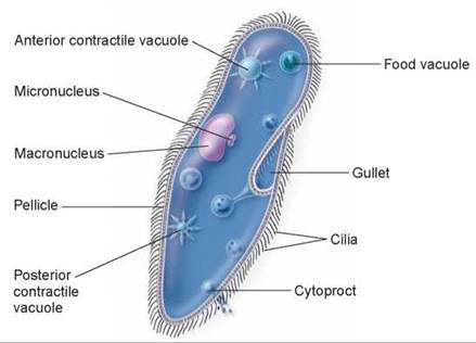
Figure 17.12. A ciliate.
The main features of the familiar ciliate Paramecium are shown.
Dinoflagellates, members of the phylum Pyrrhophyta, are photosynthetic unicellular protists, most with two flagella of unequal length. There are about 1,000 species. Some occur in freshwater, but most are marine. Bioluminescent dinofla-gellates produce the twinkling light sometimes seen in marine waters at night. Most dinoflagellates have a stiff coat of cellulose, often encrusted with silica, giving them unusual shapes. The four genera of dinoflagellates in figure 17.13 show how their flagella are unique, unlike those of any other phylum. One beats in a groove circling the body like a belt, the other in a groove perpendicular to it. Their beating rotates the body like a top.
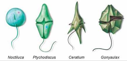
Figure 17.13. Dinoflagellates.
Noctiluca, which lacks the heavy cellulosic armor characteristic of most dinoflagellates, is one of the bioluminescent organisms that causes the waves to sparkle in warm seas at certain times of the year. In the other three genera, the shorter encircling flagellum may be seen in its groove, and the longer one projects away.
A few dinoflagellates produce powerful toxins such as the poisonous “red tides,” which are population explosions of such dinoflagellates. You can see in figure 17.14 why they are called red tides, coloring the water a reddish color. The toxins can affect humans when they eat seafood taken from red tide contaminated waters. Dinoflagellates reproduce by splitting in half.
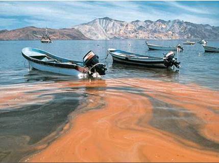
Figure 17.14 Red tide.
Red tides are caused by population explosions of dinoflagellates. The pigments in the dinoflagellates, or in some cases other organisms, color the water.
Sporozoans are spore-forming unicellular parasites of animals, all members of the phylum Apicomplexa. They are named after a unique arrangement of microtubules and other cell organelles at one end of the cell called an apical complex. This complex is used to facilitate invading a host cell.
Sporozoans are responsible for many diseases in humans and domestic animals. Sporozoans infect animals with small spores that are transmitted from host to host.
Sporozoans have complex life cycles that involve both asexual and sexual phases, as illustrated in figure 17.15. Sporozoans of the genus Plasmodium cause malaria and are spread among humans by mosquitoes of the genus Anopheles. When a mosquito inserts its proboscis into a human, it injects about a thousand sporozoites into the blood 1. They travel to the liver within a few minutes. If even one sporozoite reaches the liver, it will multiply rapidly there and still cause malaria 2. The sporozoites transform inside the liver and spread to the blood where they progress through several more stages 3, some of which develop into gametocytes 4. The gametocytes are ingested by a mosquito 5, where fertilization takes place forming sporozoites 6, and the cycle begins anew.
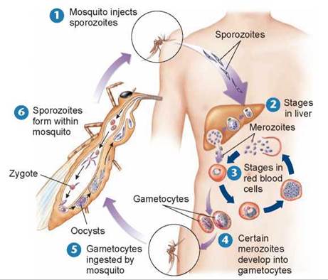
Figure 17.15. A sporozoan life cycle.
Plasmodium is the sporozoan that causes malaria. Plasmodium has a complex life cycle that alternates between mosquitoes and mammals.
Malaria is one of the most serious diseases in the world. About 500 million people are affected by it at any one time, and approximately 2 million of them, mostly children, die each year.
Key Learning Outcome 17.6. Seven phyla clustered in the largest branch of the protist phylogenetic tree exhibit an amazing diversity of form and function.