THE LIVING WORLD
Unit Five. Evolution of Animal Life
19. Evolution of the Animal Phyla
19.6. Solid Worms: Bilateral Symmetry
All eumetazoans other than cnidarians and ctenophores are bilaterally symmetrical—that is, they have a right half and a left half that are mirror images of each other. This is apparent when you compare the radially symmetrical sea anemone in figure 19.8 with the bilaterally symmetrical squirrel. Any of the three planes that cut the sea anemone in half produce mirror images, but only one plane, the green sagittal plane, produces mirror images of the squirrel. In looking at a bilaterally symmetrical animal, you refer to the top half of the animal as dorsal and the bottom half as ventral. The front is called anterior and the back posterior. Bilateral symmetry was a major evolutionary advance among the animals because it allows different parts of the body to become specialized in different ways. For example, most bilaterally symmetrical animals have evolved a definite head end, a process called cephalization. Animals that have heads are often active and mobile, moving through their environment headfirst, with sensory organs concentrated in front so the animal can test for food, danger, and mates, as it enters new surroundings.
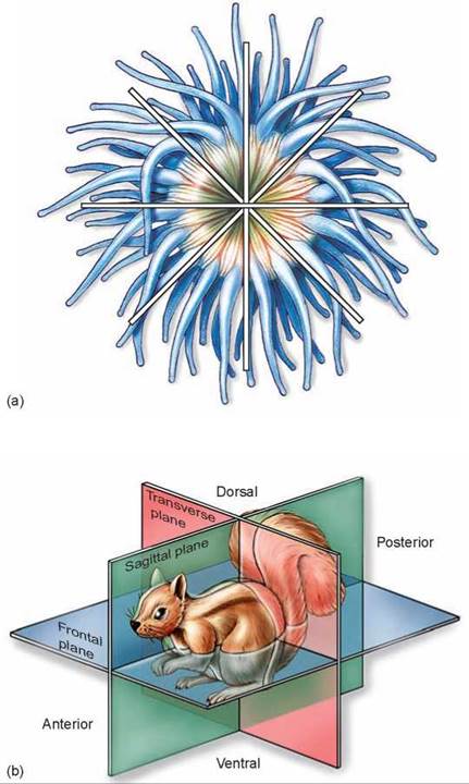
Figure 19.8. How radial and bilateral symmetry differ.
(a) Radial symmetry is the regular arrangement of parts around a central axis. (b) Bilateral symmetry is reflected in a body form that has a left and right half.
The bilaterally symmetrical eumetazoans produce three embryonic layers that develop into the tissues of the body: an outer ectoderm (colored blue in the drawing of a flatworm in figure 19.9), an inner endoderm (colored yellow), and a third layer, the mesoderm (colored red), between the ectoderm and endoderm. In general, the outer coverings of the body and the nervous system develop from the ectoderm, the digestive organs and intestines develop from the endoderm, and the skeleton and muscles develop from the mesoderm.
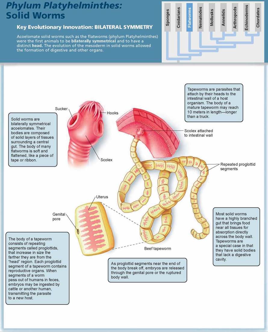
The simplest of all bilaterally symmetrical animals are the solid worms. By far the largest phylum of these, with about 20,000 species, is Platyhelminthes (pronounced plat-ee-hel-MIN-theeze), which includes the flatworms. Flatworms are the simplest animals in which organs occur. An organ is a collection of different tissues that function as a unit. The testes and uterus of flatworms are reproductive organs, for example. The dark spots on the head are eyespots that can detect light, although they cannot focus an image like your eyes can.
Solid worms lack any internal cavity other than the digestive tract. Flatworms are soft-bodied animals flattened from top to bottom, like a piece of tape or ribbon. If you were to cut a flatworm in half across its body, as in figure 19.9, you would see that the gut is completely surrounded by tissues and organs. This solid body construction is called acoelomate, meaning “without a body cavity.”
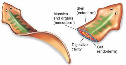
Figure 19.9. Body plan of a solid worm.
All bilaterally symmetrical eumetazoans produce three layers during embryonic development: an outer ectoderm, a middle mesoderm, and an inner endoderm. These layers differentiate to form the skin, muscles and organs, and gut, respectively, in the adult animal.
Flatworms
Although flatworms have a simple body design, they do have a definite head at the anterior end and they do possess organs. Flatworms range in size from a millimeter or less to many meters long, as in some tapeworms. Most species of flatworms are parasitic, occurring within the bodies of many other kinds of animals. Other flatworms are free-living, occurring in a wide variety of marine and freshwater habitats, as well as moist places on land (figure 19.10). Free-living flatworms are carnivores and scavengers; they eat various small animals and bits of organic debris. They move from place to place by means of ciliated epithelial cells concentrated on their ventral surfaces.
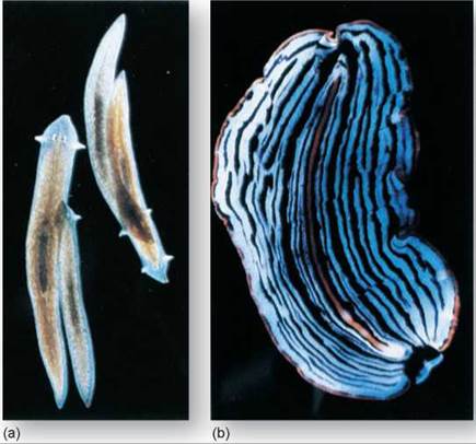
Figure 19.10. Flatworms.
(a) A common flatworm, Planaria. (b) A marine free-living flatworm.
There are two classes of parasitic flatworms, which live within the bodies of other animals: flukes and tapeworms. Both groups of worms have epithelial layers resistant to the digestive enzymes and immune defenses produced by their hosts—an important feature in their parasitic way of life. Some parasitic flatworms require only one host, but many flukes require two or more hosts to complete their life cycles. The liver fluke shown in figure 19.11 requires two hosts besides humans (or some other mammal). The eggs are released from the mammal 1 and ingested by a snail where the fluke develops into a tadpolelike larva 2 that is released into the water. The larvae bore into the muscle of a fish where they form cysts. Mammals become infected when they eat raw, infected fish 3. The parasitic lifestyle has resulted in the eventual loss of features not used or needed by the parasite. Parasitic flatworms lack certain features of the free-living flatworms, such as cilia in the adult stage, eyespots, and other sensory organs that lack adaptive significance for an organism that lives within the body of another animal, a loss sometimes dubbed “degenerative evolution.” The tapeworm described in the Phylum Facts illustration is a classic example of degenerative evolution. As you read the characteristics in the blue boxes, notice that the tapeworm’s body has been reduced to two functions, eating and reproducing.
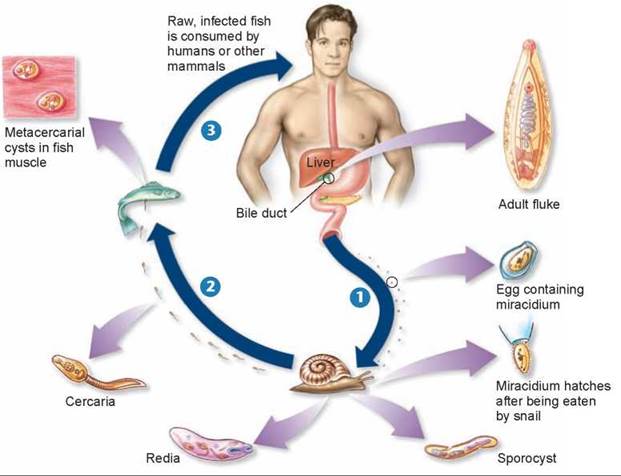
Figure 19.11. Life cycle of the human liver fluke, Clonorchis sinensis.
Adult flukes are 1-2 centimeters long and live in the bile passages of the liver. Eggs containing a complete, first-stage larva, or miracidium, are passed into water from feces and may be ingested by a snail 1. Within the snail, the egg transforms into a sporocyst, which produces larvae called rediae. These grow into tadpolelike larvae called cercariae. Cercariae escape into the water 2 where they bore into the muscles of certain fish (members of the goldfish and carp family) forming cysts. Mammals eating raw fish consume the cysts 3. The flukes emerge from the cyst and travel to the bile duct where they mature and infest the liver, causing liver damage.
Parasitic flatworms, like the human liver fluke Clonorchis sinensis, have had a significant impact on humans. Other very important flukes are the blood flukes of the genus Schistosoma. They afflict more than 200 million people throughout tropical Asia, Africa, Latin America, and the Middle East, about 1 in 20 of the world’s population. Three species of Schistosoma cause the disease called schistosomiasis, or bilharzia. Over 20,000 people die each year from this disease.
Characteristics of Flatworms
Those flatworms that have a digestive cavity have an incomplete gut, one with only one opening. As a result, they cannot eat, digest, and eliminate undigested particles of food simultaneously. Thus, flatworms cannot feed continuously, as more advanced animals can. The gut is branched and extends throughout the body (the gut is the green structure in the Planaria in figure 19.12), functioning in both digestion and transport of food. Cells that line the gut engulf most of the food particles by phagocytosis and digest them; but, as in the cnidar- ians, some of these particles are partly digested extracellularly. Tapeworms, which are parasitic flatworms, lack digestive systems. They absorb their food directly through their body walls.
Unlike cnidarians, flatworms have an excretory system, which consists of a network of fine tubules (little tubes) that runs throughout the body. Cilia line the hollow centers of bulblike flame cells (shown in the enlarged view in figure 19.12), which are located on the side branches of the tubules. Cilia in the flame cells move water and excretory substances into the tubules and then out through exit pores located between the epidermal cells. Flame cells were named because of the flickering movements of the tuft of cilia within them. They primarily regulate the water balance of the organism. The excretory function of flame cells appears to be a secondary one. A large proportion of the metabolic wastes excreted by flatworms probably diffuses directly into the gut and is eliminated through the mouth.
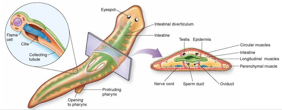
Figure 19.12 Diagram of flatworm anatomy.
The organism shown is Dugesia, the familiar freshwater "planaria" used in many biology laboratories.
Like sponges, cnidarians, and ctenophorans, flatworms lack a circulatory system, which is a network of vessels that carries fluids, oxygen, and food molecules to parts of the body. Consequently, all flatworm cells must be within diffusion distance of oxygen and food. Flatworms have thin bodies and highly branched digestive cavities that make such a relationship possible.
The nervous system of flatworms is very simple. Some primitive flatworms have only a loosely organized nerve net. However, most members of this phylum have longitudinal nerve cords (blue structures on the ventral side in the cross section above) that constitute a simple central nervous system. Between the longitudinal cords are cross connections, so that the flatworm nervous system resembles a ladder extending the length of the body.
Free-living flatworms use sensory pits or tentacles along the sides of their heads to detect food, chemicals, or movements of the fluid in which they are living. Free-living members of this phylum also have eyespots on their heads. These are inverted, pigmented cups containing light-sensitive cells connected to the nervous system. These eye-spots enable the worms to distinguish light from dark. Flat-worms are far more active than cnidarians or ctenophores. Such activity is characteristic of bilaterally symmetrical animals. In flatworms, this activity seems to be related to the greater concentration of sensory organs and, to some degree, the nervous system elements in the heads of these animals.
The reproductive systems of flatworms are complex. Most flatworms are hermaphroditic, with each individual containing both male and female sexual structures. In some parasitic flatworms, there is a complex succession of distinct larval forms. Some genera of flatworms are also capable of asexual regeneration; when a single individual is divided into two or more parts, each part can regenerate an entirely new flatworm.
Key Learning Outcome 19.6. Flatworms have internal organs, bilateral symmetry, and a distinct head. They do not have a body cavity.