THE LIVING WORLD
Unit Six. Animal Life
25. The Path of Food Through the Animal Body
25.5. The Esophagus and Stomach
Structure and Function of the Esophagus
Swallowed food enters a muscular tube called the esophagus, which connects the pharynx to the stomach. In adult humans, the esophagus is about 25 centimeters long; the upper third is enveloped in skeletal muscle, for voluntary control of swallowing, while the lower two-thirds is surrounded by involuntary smooth muscle. The swallowing center stimulates successive waves of contraction in these muscles that move food along the esophagus to the stomach. The muscles relax ahead of the food, allowing it to pass freely, and contract behind the food to push it along, as shown in figure 25.11. These rhythmic waves of muscular contraction are called peristalsis; they enable humans and other vertebrates to swallow even if they are upside down.
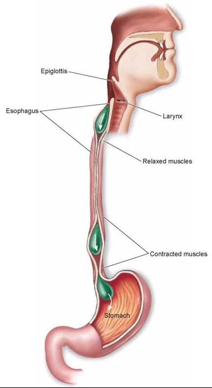
Figure 25.11. The esophagus and peristalsis.
In many vertebrates, the movement of food from the esophagus into the stomach is controlled by a ring of circular smooth muscle, the sphincter, that opens in response to the pressure exerted by the food. Contraction of this sphincter prevents food in the stomach from moving back into the esophagus. Rodents and horses have a true sphincter at this site, and thus stomach contents cannot move back out. Humans lack a true sphincter, and stomach contents can be brought back out during vomiting, when the sphincter between the stomach and esophagus is relaxed and the contents of the stomach are forcefully expelled through the mouth. The relaxing of this sphincter can also result in the movement of stomach acid into the esophagus, causing an irritation called heartburn. Chronic and severe heartburn is a condition known as acid reflux.
Structure and Function of the Stomach
The stomach is a saclike portion of the digestive tract. Its inner surface is highly convoluted, enabling it to fold up when empty and open out like an expanding balloon as it fills with food. Thus, while the human stomach has a volume of only about 50 milliliters when empty, it may expand to contain 2 to 4 liters of food when full.
The stomach contains an extra layer of smooth muscle for churning food and mixing it with gastric juice, an acidic secretion of the tubular gastric glands of the mucosa. The gastric glands lie at the bottom of deep depressions, the gastric pits shown in the enlargement in figure 25.12. These exocrine glands contain two kinds of secretory cells: parietal cells, which secrete hydrochloric acid (HCl); and chief cells, which secrete pepsinogen, a weak protease (protein-digesting enzyme) that requires a very low pH to be active. This low pH is provided by the HCl. Activated pepsinogen molecules then cleave each other at specific sites, producing a much more active protease, pepsin. This process of secreting a relatively inactive enzyme that is then converted into a more active enzyme outside the cell prevents the chief cells from digesting themselves. It should be noted that only proteins are partially digested in the stomach—there is no significant digestion of carbohydrates or fats.
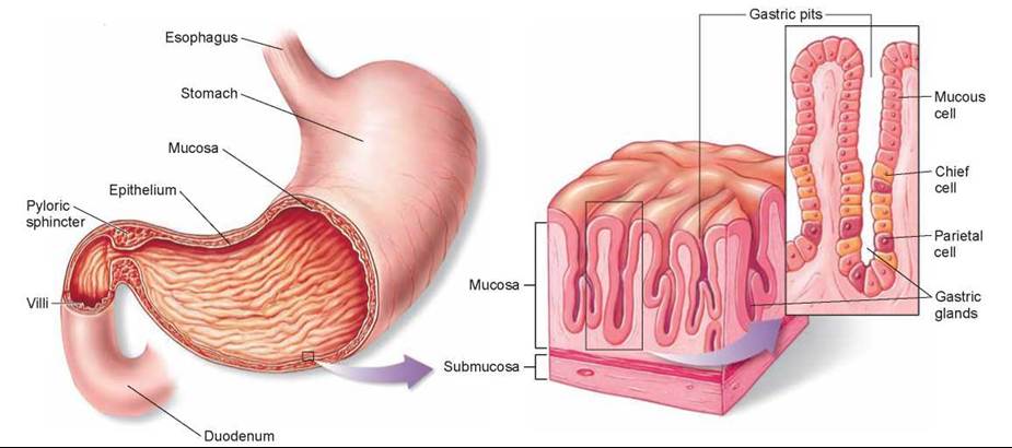
Figure 25.12. The stomach and gastric glands.
Food enters the stomach from the esophagus. The epithelial walls of the stomach are dotted with gastric pits, which contain glands that secrete hydrochloric acid (HCl) and the enzyme pepsinogen. The gastric glands consist of mucous cells, chief cells that secrete pepsinogen, and parietal cells that secrete HCl. Gastric pits are the openings of the gastric glands.
Action of Acid
The human stomach produces about 2 liters of HCl and other gastric secretions every day, creating a very acidic solution inside the stomach. The concentration of HCl in this solution is about 10 millimolar, corresponding to a pH of 2. Thus, gastric juice is about 250,000 times more acidic than blood, whose normal pH is 7.4. The low pH in the stomach helps denature food proteins, making them easier to digest, and keeps pepsin maximally active. Active pepsin hydrolyzes food proteins into shorter chains of polypeptides that are not fully digested until the mixture enters the small intestine. The mixture of partially digested food and gastric juice is called chyme.
The acidic solution within the stomach also kills most of the bacteria that are ingested with the food. The few bacteria that survive the stomach and enter the intestine intact are able to grow and multiply there, particularly in the large intestine. In fact, most vertebrates harbor thriving colonies of bacteria within their intestines, and bacteria are a major component of feces. As we discuss later, bacteria that live within the digestive tract of cows and other ruminants play a key role in the ability of these mammals to digest cellulose.
Ulcers
It is important that the stomach not produce too much acid. If it did, the body could not neutralize the acid later in the small intestine, a step essential for the final stage of digestion. Production of acid is controlled by hormones. These hormones are produced by endocrine cells scattered within the walls of the stomach. The hormone gastrin regulates the synthesis of HCl by the parietal cells of the gastric pits, permitting HCl to be made only when the pH of the stomach is higher than about 1.5.
Overproduction of gastric acid can occasionally eat a hole through the wall of the stomach. Such gastric ulcers are rare, however, because epithelial cells in the mucosa of the stomach are protected somewhat by a layer of alkaline mucus, and because those cells are rapidly replaced by cell division if they become damaged (gastric epithelial cells are replaced every two to three days). Over 90% of gastrointestinal ulcers are duodenal ulcers, which are ulcers of the small intestine. These may be produced when excessive amounts of acidic chyme are delivered into the duodenum, so that the acid cannot be properly neutralized through the action of alkaline pancreatic juice (described later). Susceptibility to ulcers is increased when the mucosal barriers to self-digestion are weakened by an infection of the bacterium Helicobacter pylori. Modern antibiotic treatments can reduce symptoms and often cure the ulcer.
In addition to producing HCl, the parietal cells of the stomach also secrete intrinsic factor, a polypeptide needed for the intestinal absorption of vitamin B12. Because this vitamin is required for the production of red blood cells, persons who lack sufficient intrinsic factor develop a type of anemia (low red blood cell count) called pernicious anemia.
Leaving the Stomach
Chyme leaves the stomach through the pyloric sphincter, shown at the base of the stomach in figure 25.12, to enter the small intestine. This is where all terminal digestion of carbohydrates, fats, and proteins occurs, and where the products of digestion—amino acids, glucose, and fatty acids—are absorbed into the blood. Only water from chyme and a few substances such as aspirin and alcohol are absorbed through the wall of the stomach.
Key Learning Outcome 25.5. Peristaltic waves of contraction propel food along the esophagus to the stomach. Gastric juice contains strong hydrochloric acid and the protein-digesting enzyme pepsin, which begins the digestion of proteins into shorter polypeptides. The acidic chyme is then transferred through the pyloric sphincter to the small intestine.