THE LIVING WORLD
Unit Six. Animal Life
27. How the Animal Body Defends Itself
27.2. Cellular Counterattack: The Second Line of Defense
When the body’s interior is invaded, a host of cellular and chemical defenses swing into action. Four are of particular importance: (1) cells that kill invading microbes; (2) proteins that kill invading microbes; (3) the inflammatory response, which speeds defending cells to the point of infection; and (4) the temperature response, which elevates body temperature to slow the growth of invading bacteria.
Although these cells and proteins roam throughout the body, there is a central location for their storage and distribution; it is called the lymphatic system. The lymphatic system consists of the structures shown in figure 27.3: lymph nodes, lymphatic organs, and a network of lymphatic capillaries that drain into lymphatic vessels. Although the lymphatic system has other functions involved with circulation (see chapter 23), it is also involved in the immune response.
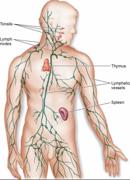
Figure 27.3. The lymphatic system.
The major lymphatic vessels, organs, and nodes are shown.
Cells That Kill Invading Microbes
The most important counterattack to infection is mounted by white blood cells, which attack invading microbes. These cells patrol the bloodstream and await invaders within the tissues. The three basic kinds of killing cells are macrophages and neutrophils, which are phagocytes, and natural killer cells. These three kinds of cells can distinguish the body’s cells (self) from foreign cells (nonself) because the body’s cells contain self-identifying MHC proteins (discussed in more detail later in this chapter). Each type of killing cell uses a different tactic to kill invading microbes.
Macrophages. White blood cells called macrophages (Greek, “big eaters”) kill bacteria by ingesting them, much as an amoeba ingests a food particle. The macrophage in figure 27.4 is sending out long, sticky cytoplasmic extensions that catch the sausage-shaped bacteria and draw them back to the cell where they are engulfed. Although some macrophages are anchored within particular organs, particularly the spleen, most patrol the byways of the body, circulating as precursor cells called monocytes in the blood, lymph, and fluid between cells. Macrophages are among the most actively mobile cells of the body.
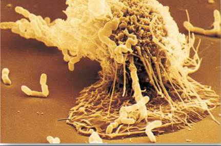
Figure 27.4. A macrophage in action.
In this scanning electron micrograph, a macrophage is "fishing" for rod-shaped bacteria.
Neutrophils. Other white blood cells called neutrophils act like kamikazes. In addition to ingesting microbes, they release chemicals (identical to household bleach) to “neutralize” the entire area, killing any bacteria in the neighborhood—and themselves in the process. A neutrophil is like a grenade tossed into an infection. It kills everything in the vicinity. Macrophages, by contrast, kill only one invading cell at a time but live to keep on doing it.
Natural Killer. Cells A third kind of white blood cell, called a natural killer cell, does not attack invading microbes but rather the body cells that are infected by them. Natural killer cells puncture the membrane of the infected target cell with a molecule called perforin. The natural killer cell in figure 27.5a is releasing several perforin molecules that insert in the membrane of the infected cell, like boards on a picket fence, forming a pore that allows water to rush in, causing the cell to swell and burst. Natural killer cells are particularly effective at detecting and attacking body cells that have been infected with viruses. They are also one of the body’s most potent defenses against cancer. The cancer cell shown in the photo in figure 27.5b was killed before it had a chance to develop into a tumor.
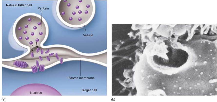
Figure 27.5. Natural killer cells attack target cells.
(a) The initial event is the tight binding of the natural killer cell to the target cell. Binding initiates a chain of events within the natural killer cell in which vesicles loaded with perforin molecules move to the outer plasma membrane and expel their contents into the intercellular space over the target. The perforin molecules insert into the membrane, forming a pore that admits water and ruptures the cell. (b) A natural killer cell has attacked this cancer cell, punching a hole in its plasma membrane. Water has rushed in, making it balloon out. Soon it will burst.
Proteins That Kill Invading Microbes
The cellular defenses of vertebrates are complemented by a very effective chemical defense called the complement system. This system consists of approximately 20 different proteins that circulate freely in the blood plasma in an inactive state. Their defensive activity is triggered when they encounter the cell walls of bacteria or fungi. The complement proteins then aggregate to form a membrane attack complex that inserts itself into the foreign cell’s plasma membrane, forming a pore like that produced by natural killer cells. Like the perforin pore, the membrane attack complex in figure 27.6 allows water to enter the foreign cell causing the cell to swell and burst. The difference between the two is that perforin attacks host cells that have become infected, while complement attacks the foreign cell directly. Aggregation of the complement proteins is also triggered by the binding of antibodies to invading microbes, as we’ll see in a later section.
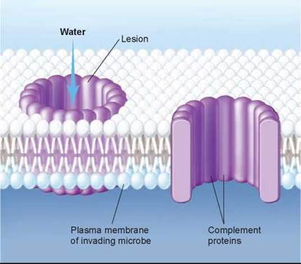
Figure 27.6. Complement proteins attack invaders.
Complement proteins form a transmembrane channel resembling the perforin-lined lesion made by natural killer cells, but complement proteins are free-floating, and they attach to the invading microbe directly, while perforin molecules insert into infected body cells.
The proteins of the complement system can augment the effects of other body defenses. Some amplify the inflammatory response (discussed next) by stimulating histamine release; others attract phagocytes (monocytes and neutrophils) to the area of infection; and still others coat invading microbes, roughening the microbes’ surfaces so that phagocytes may attach to them more readily.
Another class of proteins that play a key role in body defense are interferons. There are three major categories of interferons: alpha, beta, and gamma. These polypeptides act as messengers that protect other cells in the vicinity from viral infection. The viruses are still able to penetrate the other cells, but the ability of the viruses to replicate and assemble new virus particles is inhibited.
The Inflammatory Response
The aggressive cellular and chemical counterattacks to infection are made more effective by the inflammatory response. The inflammatory response can be broken down into three stages as shown in figure 27.7, where rod-shaped bacteria are entering the body through a wound:
1. In the first panel, infected or injured cells release chemical alarm signals, most notably histamine and prostaglandins.
2. These chemical alarm signals cause blood vessels to expand, both increasing the flow of blood to the site of infection or injury and, by stretching their thin walls, making the capillaries more permeable. This produces the redness and swelling so often associated with infection.
3. In the second panel, the increased blood flow through larger, leakier capillaries promotes the migration of phagocytes from the blood to the site of infection, squeezing between cells in the capillary walls. Neutrophils arrive first, spilling out chemicals that kill the microbes (as well as tissue cells in the vicinity and themselves). Monocytes follow and become macrophages, which engulf the pathogens as well as the remains of all the dead cells, as shown in the third panel. This counterattack takes a considerable toll; the pus associated with infections is a mixture of dead or dying neutrophils, tissue cells, and pathogens.
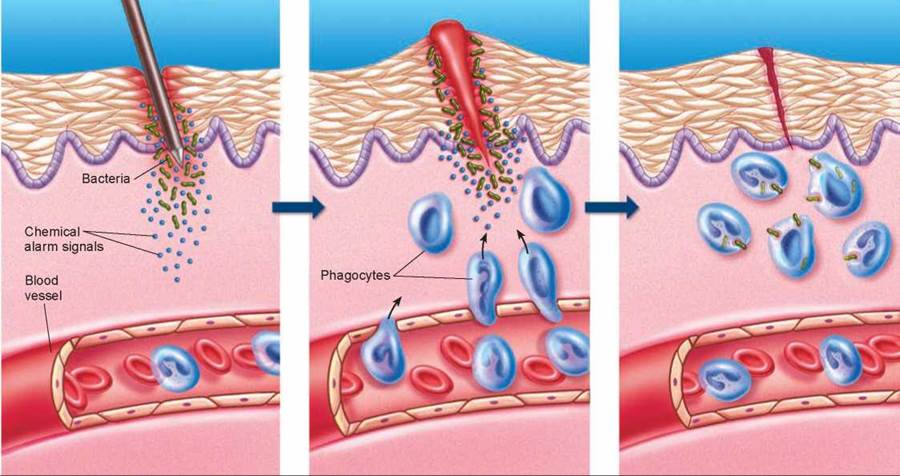
Figure 27.7. The events in a local inflammation.
When an invading microbe has penetrated the skin, chemicals, such as histamine and prostaglandins, act as alarm signals that cause nearby blood vessels to dilate. Increased blood flow brings a wave of phagocytic cells that attack and engulf invading bacteria.
Human pathogenic bacteria do not grow well at high temperatures. Thus, when macrophages initiate their counterattack, they increase the odds in their favor by sending a message to the brain to raise the body’s temperature. The cluster of brain cells that serves as the body’s thermostat responds to the chemical signal by boosting the body temperature several degrees above the normal value of 37°C (98.6°F). The higher-than-normal temperature that results is called a fever. Although fever is quite effective at inhibiting microbial growth, very high fevers are dangerous because excessive heat can inactivate critical cellular enzymes. In general, temperatures greater than 39.4°C (103°F) are considered dangerous; those greater than 40.6°C (105°F) are often fatal.
The second line of defense, with both chemical and cellular weapons, provides a sophisticated defense against microbial infection. Only occasionally do bacteria or viruses overwhelm this defense. When this happens, they face yet a third line of defense, more difficult to evade than any they have encountered. It is the specific immune response, the most elaborate of the body’s defenses.
Key Learning Outcome 27.2. Vertebrates respond to infection with a battery of cellular and chemical weapons, including cells and proteins that kill invading microbes, and inflammatory and temperature responses.