THE LIVING WORLD
Unit Six. Animal Life
30. Chemical Signaling Within the Animal Body
30.3. The Hypothalamus and the Pituitary
The hypothalamus, the “control center” of the neuroendocrine system, exerts its control by releasing hormones that influence the nearby pituitary gland, located in a bony recess in the brain just below the hypothalamus. The pituitary in turn produces hormones that influence the body’s other endocrine glands. Hormones produced by the back portion of the pituitary, or posterior lobe, regulate water conservation, milk letdown and uterine contraction in women; hormones produced by the front portion, or anterior lobe, regulate the other endocrine glands.
The Posterior Pituitary
The posterior pituitary contains axons that originate in cells within the hypothalamus (see figure 30.5). The hormones released from the posterior pituitary are actually produced by neuron cell bodies located in the hypothalamus. The hormones are transported to the posterior pituitary through axon tracts and are stored and released from the posterior pituitary.
The role of the posterior pituitary first became evident in 1912, when a remarkable medical case was reported: A man who had been shot in the head developed a surprising disorder—he began to urinate every 30 minutes, unceasingly. The bullet had lodged in his pituitary gland, and subsequent research demonstrated that surgical removal of the pituitary also produces these unusual symptoms. Pituitary extracts were shown to contain a substance that makes the kidneys conserve water, and in the early 1950s the peptide hormone vasopressin (also called antidiuretic hormone, ADH) was isolated. As you learned in chapter 26, ADH regulates the kidney’s retention of water. When ADH is missing, the kidneys cannot retain water, which is why the bullet caused excessive urination. Excessive alcohol and caffeine consumption, which inhibit ADH secretion, have a similar effect.
The posterior pituitary also releases a second hormone, oxytocin, of very similar structure—both are short peptides composed of nine amino acids— but very different function. Oxytocin initiates uterine contraction during childbirth and milk release in mothers. Here is how milk release works: Sensory receptors in the mother’s nipples, when stimulated by sucking, send messages to the hypothalamus, causing the hypothalamus to stimulate the release of oxytocin from the posterior pituitary. The oxytocin travels in the bloodstream to the breasts where it stimulates contraction of the muscles around the ducts into which the mammary glands secrete milk. Both oxytocin and ADH are produced in the cell bodies of the hypothalamus but stored and released from the posterior pituitary.
The Anterior Pituitary
The anterior pituitary gland produces seven major peptide hormones (blue in figure 30.4), each controlled by a particular releasing signal secreted from the hypothalamus:
1. Thyroid-stimulating hormone (TSH). TSH stimulates the thyroid gland to produce the thyroid hormone thyroxine, which in turn stimulates oxidative respiration.
2. Adrenocorticotropic hormone (ACTH). ACTH stimulates the adrenal gland to produce a variety of steroid hormones. Some regulate the production of glucose from fat; others regulate the balance of sodium and potassium ions in the blood.
3. Growth hormone (GH). GH stimulates the growth of muscle and bone throughout the body.
4. Follicle-stimulating hormone (FSH). FSH is significant in the female menstrual cycle by triggering the maturation of egg cells and stimulating the release of estrogen. In males, it stimulates cells in the testes, regulating development of the sperm.
5. Luteinizing hormone (LH). LH plays an important role in the female menstrual cycle by triggering ovulation, the release of a mature egg. It also stimulates the male gonads to produce testosterone, which initiates and maintains the development of male secondary sexual characteristics not involved directly in reproduction.
6. Prolactin (PRL). Prolactin stimulates the breasts to produce milk, which is released in response to oxytocin.
7. Melanocyte-stimulating hormone (MSH). In reptiles and amphibians, MSH stimulates color changes in the epidermis. The function of this hormone in humans is still poorly understood.
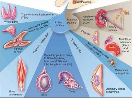
Figure 30.4 .The role of the pituitary.
How the Hypothalamus Controls the Anterior Pituitary
As noted earlier, the hypothalamus controls production and secretion of the anterior pituitary hormones by means of a family of special hormones. Neurons in the hypothalamus secrete these releasing and inhibiting hormones into blood capillaries at the base of the hypothalamus. Figure 30.5 shows the relationship of the two groups of neurons in the hypothalamus. As discussed earlier, some neurons (colored blue in the figure) extend into the posterior pituitary where axons deliver the hormones for storage and release. Other neurons of the hypothalamus (colored yellow in the figure) produce releasing and inhibiting hormones and release them into capillaries. These capillaries drain into small veins that run within the stalk of the pituitary to a second bed of capillaries in the anterior pituitary. This unusual system of vessels is known as the hypothalamohypophyseal portal system. It is called a portal system because it has a second capillary bed downstream from the first; the only other body location with a similar system is the liver.
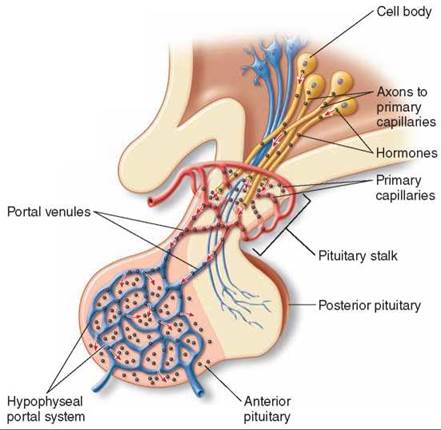
Figure 30.5. Hormonal control of the anterior pituitary gland by the hypothalamus.
Neurons in the hypothalamus secrete hormones that are carried by short blood vessels to the anterior pituitary gland, where they either stimulate or inhibit the secretion of anterior pituitary hormones.
Each releasing hormone delivered to the anterior pituitary by this portal system regulates the secretion of a specific anterior pituitary hormone. For example, thyrotropin-releasing hormone (TRH) stimulates the release of TSH; corticotropinreleasing hormone (CRH) stimulates the release of ACTH; gonadotropin-releasing hormone (GnRH) stimulates the release of FSH and LH; growth-hormone-releasing hormone (GHRH) stimulates the release of GH; and prolactin-releasing factor (PRF) stimulates the release of prolactin—however, this factor has not yet been chemically characterized and may actually be a chemical similar to thyrotropin-releasing hormone.
The hypothalamus also secretes hormones that inhibit the release of certain anterior pituitary hormones. To date, three such hormones have been discovered: Somatostatin inhibits the secretion of GH; prolactin-inhibiting hormone (PIH), possibly dopamine, i nhibits the secretion of prolactin; and melanotropin-inhibiting hormone (MIH) inhibits the secretion of MSH.
Because hypothalamic hormones control the secretions of the anterior pituitary gland, and the anterior pituitary hormones control the secretions of some other endocrine glands, it may seem that the hypothalamus functions as a “master gland,” in charge of hormonal secretion in the body. This idea is not generally valid, however, for two reasons. First, a number of endocrine organs, such as the adrenal medulla and the pancreas, are not directly regulated by this control system. Second, the hypothalamus and the anterior pituitary gland are themselves controlled by the very hormones whose secretion they stimulate! In most cases this is an inhibitory control. Figure 30.6 shows how the target gland hormones inhibit the hypothalamus and anterior pituitary. When enough of the target gland hormone has been produced, the hormone then feeds back and inhibits the release of the stimulating hormones from the hypothalamus and anterior pituitary, indicated by the dashed lines. This type of control system is an example of negative feedback (or feedback inhibition), which was also discussed in chapter 26.
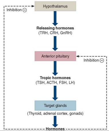
Figure 30.6. Negative feedback.
The hormones secreted by some endocrine glands feed back to inhibit the secretion of hypothalamic-releasing hormones and anterior pituitary tropic hormones.
Key Learning Outcome 30.3. The posterior pituitary gland contains axons originating from neurons in the hypothalamus that produce hormones. The anterior pituitary responds to hormonal signals from the hypothalamus and produces a family of pituitary hormones that are carried to distant glands that produce specific hormones.