THE LIVING WORLD
Unit Three. The Continuity of Life
11.3. Discovering the Structure of DNA
As it became clear that DNA was the molecule that stored the hereditary information, researchers began to question how this nucleic acid could carry out the complex function of inheritance. At the time, investigators did not know what the DNA molecule looked like.
We now know that DNA is a long, chainlike molecule made up of subunits called nucleotides. As you can see in figure 11.3, each nucleotide has three parts: a central sugar called deoxyribose, a phosphate (PO4) group, and an organic base. The sugar (the lavender pentagon structure) and the phosphate group (the yellow-circled structure) are the same in every nucleotide of DNA. However, there are four different kinds of bases: two large ones with double-ring structures, and two small ones with single rings. The large bases, called purines, are A (adenine) and G (guanine). The small bases, called pyrimidines, are C (cytosine) and T (thymine). A key observation, made by Erwin Chargaff, was that DNA molecules always had equal amounts of purines and pyrimidines. In fact, with slight variations due to imprecision of measurement, the amount of A always equals the amount of T, and the amount of G always equals the amount of C. This observation (A = T, G = C), known as Chargaff’s rule, suggested that DNA had a regular structure.
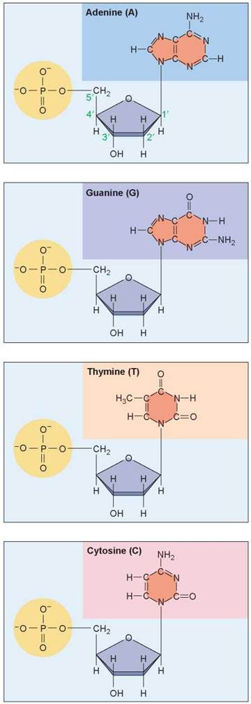
Figure 11.3. The four nucleotide subunits that make up DNA.
The nucleotide subunits of DNA are composed of three parts: a central five-carbon sugar called deoxyribose, a phosphate group, and an organic, nitrogen-containing base.
The significance of Chargaff’s rule became clear in 1953 when the British chemist Rosalind Franklin carried out an X-ray diffraction experiment. In these experiments, DNA molecules are bombarded with X-ray beams, and when individual rays encounter atoms, their paths are bent or diffracted like a thrown ball bounces off or around an object. Each atomic encounter creates a pattern on photographic film, shown in figure 11.4a, that looks like the ripples created by tossing a rock into a smooth lake. Franklin’s results suggested that the DNA molecule had the shape of a coiled spring or a corkscrew, a form called a helix, with the image in the photo from the viewpoint of looking down the center of the molecule.
Franklin’s work was shared with two researchers at Cambridge University, Francis Crick and James Watson, before it was published. Using Tinkertoy-like models of the bases, Watson and Crick deduced the true structure of DNA (figure 11.46): The DNA molecule is a double helix, a winding staircase of two strands whose bases face one another (figure 11.4c). Chargaff’s rule is a direct reflection of this structure—every bulky purine on one strand is paired with a slender pyrimidine on the other strand. Specifically, A (the blue bases) pairs with T (the orange bases), and G (the purple bases) pairs with C (the pink bases). Because hydrogen bonds, shown as dotted lines, can form between the base pairs, the molecule keeps a constant thickness.
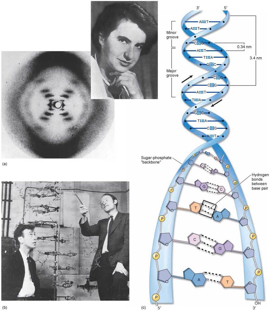
Figure 11.4. The DNA double helix.
(a) This X-ray diffraction photograph was made in 1953 by Rosalind Franklin (inset) in the laboratory of Maurice Wilkins. It suggested to Watson and Crick that the DNA molecule was a helix, like a winding staircase. (b) In 1953 Watson and Crick deduced the structure of DNA. James Watson (seated and peering up at their homemade model of the DNA molecule) was a young American postdoctoral student, and Francis Crick (pointing) was an English scientist. (c) The dimensions of the double helix were suggested by the X-ray diffraction studies. In a DNA duplex molecule, only two base pairs are possible: adenine (A) with thymine (T) and guanine (G) with cytosine (C). A G-C base pair has three hydrogen bonds; an A-T base pair has only two.
Key Learning Outcome 11.3. The DNA molecule consists of two strands of nucleotides held together by hydrogen bonds between bases. The two strands wind into a double helix.