MCAT Biology Review
Chapter 3: Embryogenesis and Development
3.3 Fetal Circulation
Recall that the placenta, shown in Figure 3.10, is the organ where nutrient, gas, and waste exchanges occur. It is crucial that maternal and fetal blood do not mix because they may have different blood types. The simplest method to move nutrients and waste products is by diffusion, and this is the preferred method for water, glucose, amino acids, and inorganic salts. Diffusion requires a gradient, so this implies that there is a higher partial pressure of oxygen in maternal blood than fetal blood. In addition to an oxygen gradient, fetal blood cells contain fetal hemoglobin ( HbF ), which exhibits a greater affinity for oxygen than does maternal (adult) hemoglobin (primarily HbA). This also assists with the transfer (and retention) of oxygen into the fetal circulatory system. Waste material and carbon dioxide move in the opposite direction.
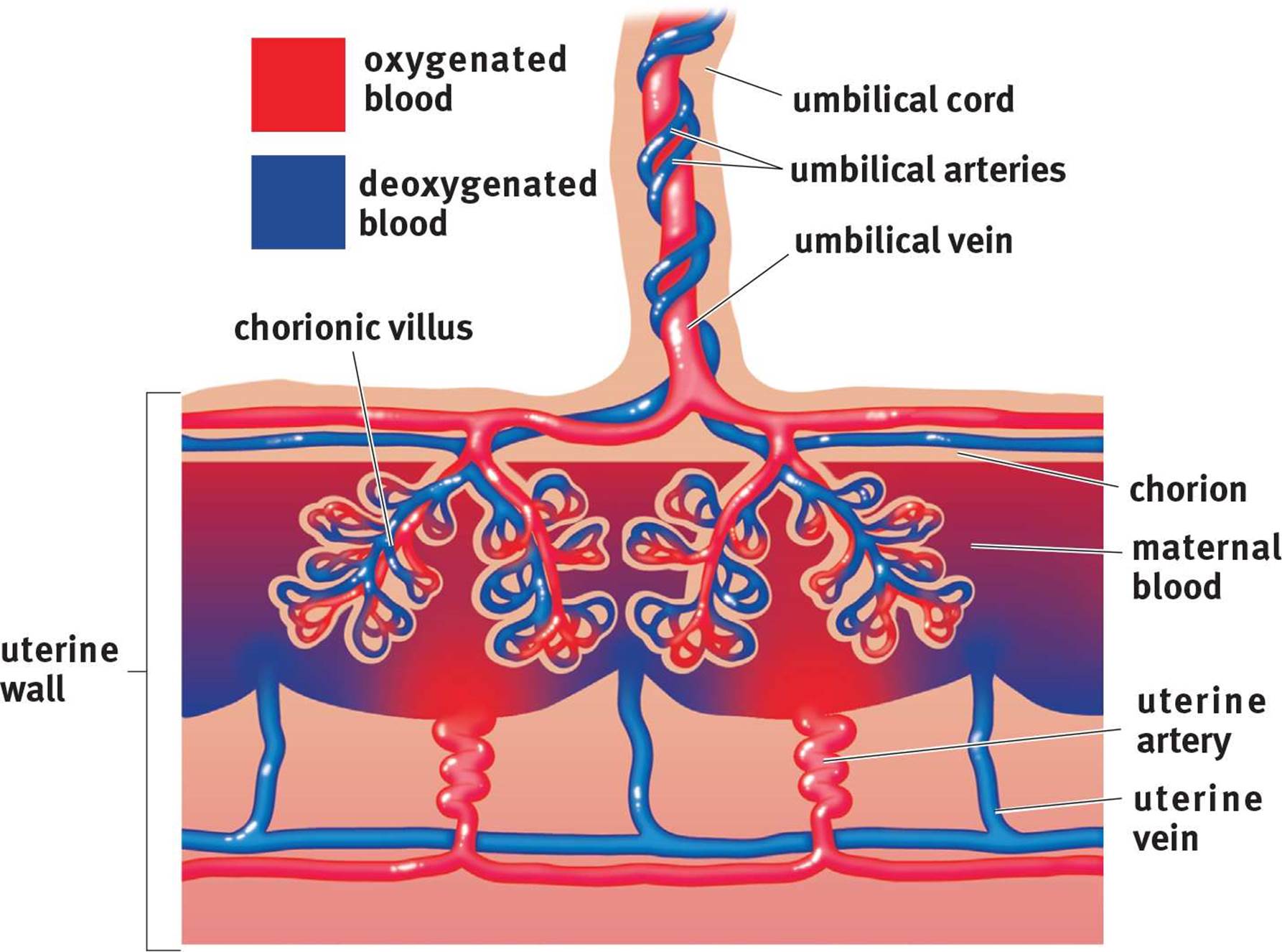 Figure 3.10. Placental Structure
KEY CONCEPT
Although the embryo obtains its nutrients and oxygen from the mother, there is no actual mixing of the blood. Instead, the placenta allows for the close proximity of the embryonic and maternal bloodstreams so that diffusion can occur between them.
KEY CONCEPT
Remember, gas exchange in the fetus occurs across the placenta. Fetal lungs do not function until birth.
The placental barrier also serves another function: immune protection. The fetus is immunologically naïve because it has not yet been exposed to any pathogens; however, accidental exposure can happen in utero. Thus, the crossing of antibodies across the placental membrane serves a protective function. The placenta also qualifies as an endocrine organ because it produces progesterone, estrogen, and human chorionic gonadotropin (hCG), all of which are essential for maintaining pregnancy.
REAL WORLD
Many pathogens are too large to cross the placental barrier by diffusion, but a set of pathogens called TORCHES infections can cross this barrier and cause significant birth defects. Therefore, screening for (and sometimes immunization against) these infections is recommended in pregnancy. TORCHES stands for TOxoplasma gondii, Rubella, Cytomegalovirus, HErpes or HIV, and Syphilis.
The umbilical vessels are commonly tested on the MCAT because they demonstrate the need to understand the proper biological definitions of artery and vein. Like all other arteries that carry blood away from the heart, the umbilical arteries carry blood away from the fetus toward the placenta. And, like all of the other veins that carry blood toward the heart, the umbilical vein carries blood toward the fetus from the placenta. Remember that oxygenation occurs at the placenta, rather than in the fetal lungs. Therefore, the umbilical arteries carry deoxygenated blood and the umbilical vein carries oxygenated blood.
KEY CONCEPT
Unlike most other arteries, the umbilical arteries carry deoxygenated blood with waste products. Unlike most other veins, the umbilical vein carries oxygenated blood with nutrients.
There are several key differences between fetal and adult circulation that demonstrate important characteristics of the developing organism. The lungs and liver both do not serve significant functions prior to birth. Gas exchange does not occur at the lungs, but rather at the placenta. Detoxification and metabolism are primarily controlled by the mother’s liver, and nutrient and waste exchange occurs at the placenta, as well. Thus, the fetus needs not depend on its own lungs and liver. Notably, these two organs are both underdeveloped and sensitive to the high blood pressures they will receive in postnatal life; thus, the developing child’s body constructs three shunts to actively direct blood away from these organs, as shown in Figure 3.11.
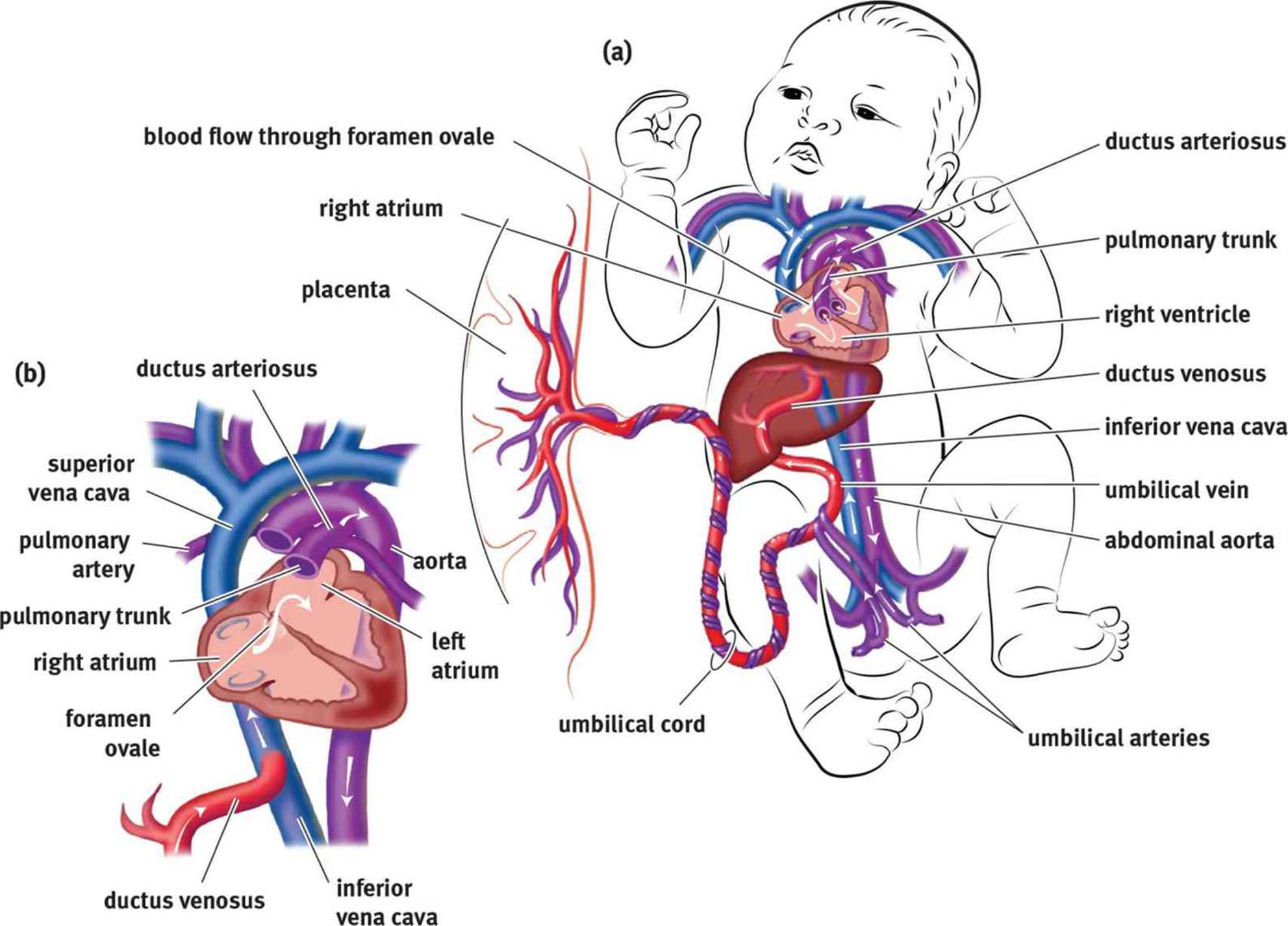 Figure 3.11. Fetal Circulation (a) Systemic fetal circulation; (b) Enlarged view of fetal circulation highlighting the three fetal shunts.
Two different shunts are used to reroute blood from the lungs. The first, called the foramen ovale, is a one-way valve that connects the right atrium to the left atrium. This allows blood entering the right atrium from the inferior vena cava to flow into the left atrium, instead of the right ventricle, and thereby be pumped through the aorta into systemic circulation directly. Unlike in adult circulation, the right side of the heart is at a higher pressure in the developing fetus than the left side, which pushes blood through the opening. After birth, this pressure differential reverses, shutting the foramen ovale. Second, the ductus arteriosus shunts leftover blood from the pulmonary artery to the aorta. Again, the pressure differential between the right and left sides of the heart pushes blood through this opening and into systemic circulation.
The liver is bypassed using the ductus venosus, which shunts blood returning from the placenta via the umbilical vein directly to the inferior vena cava. The liver still receives some blood supply from smaller hepatic arteries in the systemic circulation.
MCAT Concept Check 3.3:
Before you move on, assess your understanding of the material with these questions.
1. What is the oxygenation status of the blood in the umbilical arteries? In the umbilical vein?
· Umbilical arteries:
· Umbilical vein:
2. What are the three fetal shunts? What vessels or heart chambers do they connect? What organ does each shunt bypass?
|
Shunt |
Connected Vessels or Chambers |
Organ Bypassed |