MCAT Biology Review
Chapter 4: The Nervous System
Introduction
For generations, the indigenous peoples of South America used blow darts laced with a paralytic plant extract to hunt their prey. In the 1800s, English physicians who interacted with these indigenous South Americans recognized the possible uses of this paralytic agent, now known astubocurarine, as an anesthetic agent for surgeries. Physicians noticed that animals under the influence of tubocurarine would become temporarily immobilized but would recover after a period of paralysis. According to these physicians, this discovery would revolutionize surgery as an anesthetic agent. So confident were they in their discovery that one of the physicians volunteered to undergo surgery under the influence of tubocurarine to demonstrate its effectiveness. Unfortunately, he failed to realize that, although the drug was an effective paralyzing agent, it did not have any effect on the sensory receptors of the body, so he felt every cut of the surgery without being able to move or do anything about it.
It is through the nervous system that organisms sense pain, temperature, and all aspects of their environment. It also serves to coordinate this sensory information and respond to stimuli. Specifically, the nervous system is responsible for the control of muscular movement, neuromuscular reflexes, and glandular secretions (such as salivation and lacrimation). In addition, the nervous system is responsible for higher-level thinking and mental function.
Despite all of its complex functions, the nervous system operates through basic electrical and chemical signals. Biomedical scientists have discovered so much about the nervous system: its anatomical and functional divisions, the nature of the action potential, and its histological features under the microscope. However, there is so much more that we do not know. It is an inspirational challenge for future physicians to realize that the brain continues to be a vast frontier for human exploration and discovery.
4.1 Cells of the Nervous System
Neurons are specialized cells capable of transmitting electrical impulses and then translating those electrical impulses to chemical signals. In this section, we will consider the structure of the neuron as well as how neurons communicate with other parts of the nervous system.
NEURONS
Each neuron has a shape that matches its function, dictated by the other cells with which that neuron interacts. There are a variety of different types of neurons in the body, but they all share some specific features.
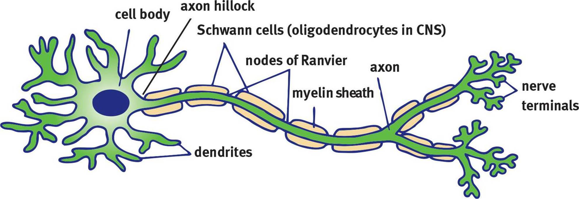 Figure 4.1. Structure of a Neuron
The anatomy of a neuron is shown in Figure 4.1. Like all other cells (besides mature red blood cells), neurons have nuclei. The nucleus is located in the cell body, also called the soma. The soma is also the location of the endoplasmic reticulum and ribosomes. The cell has many appendages emanating directly from the soma called dendrites, which receive incoming messages from other cells. The information received from the dendrites is transmitted through the cell body before it reaches the axon hillock, which integrates the incoming signals. The axon hillock plays an important role in action potentials, or the transmission of electrical impulses down the axon. Signals arriving from the dendrites can be either excitatory or inhibitory; the axon hillock sums these signals, and if the result is excitatory enough (reaching threshold, as discussed later in this chapter), it will initiate an action potential. The axon is a long appendage that terminates in close proximity to a target structure (a muscle, a gland, or another neuron). Most mammalian nerve fibers are insulated by myelin to prevent signal loss or crossing of signals. Just like insulation prevents wires next to each other from accidentally discharging each other, the myelin sheath maintains the electric signal within one neuron. In addition, myelin increases the speed of conduction in the axon. Myelin is produced by oligodendrocytes in the central nervous system andSchwann cells in the peripheral nervous system. At certain intervals along the axon, there are small breaks in the myelin sheath with exposed areas of axon membrane called nodes of Ranvier. As will be explored in the discussion of action potentials to follow, nodes of Ranvier are critical for rapid signal conduction. Finally, at the end of the axon is the nerve terminal or synaptic bouton (knob). This structure is enlarged and flattened to maximize neurotransmission to the next neuron and ensure proper release of neurotransmitters, the chemicals that transmit information between neurons.
REAL WORLD
Sometimes the body mounts an immune response against its own myelin, leading to the destruction of this insulating substance (demyelination). Because myelin speeds the conduction of impulses along a neuron, the absence of myelin results in the slowing of information transfer. A common demyelinating disorder is multiple sclerosis (MS). In MS, the myelin of the brain and spinal cord is selectively targeted. Because so many different kinds of neurons are demyelinated, MS patients experience a wide variety of symptoms including weakness, lack of balance, vision problems, and incontinence.
MNEMONIC
Axons carry neural signals away from the soma; dendrites carry signals toward the soma.
Neurons are not physically connected to each other. Between the neurons, there is a small space into which the terminal portion of the axon releases neurotransmitters, which bind to the dendrites of the postsynaptic neuron. This space is known as the synaptic cleft; together, the nerve terminal, synaptic cleft, and postsynaptic membrane are known as a synapse. Neurotransmitters released from the axon terminal traverse the synaptic cleft and bind to receptors on the postsynaptic neuron.
Multiple neurons may be bundled together to form a nerve in the peripheral nervous system. These nerves may be sensory, motor, or mixed, which refers to the type(s) of information they carry; mixed nerves carry both sensory and motor information. The cell bodies of neurons of the same type are clustered together into ganglia.
In the central nervous system, axons may be bundled together to form tracts. Unlike nerves, tracts only carry one type of information. The cell bodies of neurons in the same tract are grouped into nuclei.
OTHER CELLS IN THE NERVOUS SYSTEM
Neurons are not the only cells in the nervous system. Neurons must be supported and myelinated by other cells. These cells are often called glial cells, or neuroglia. Glial cells play both structural and supportive roles, as shown in Figure 4.2.
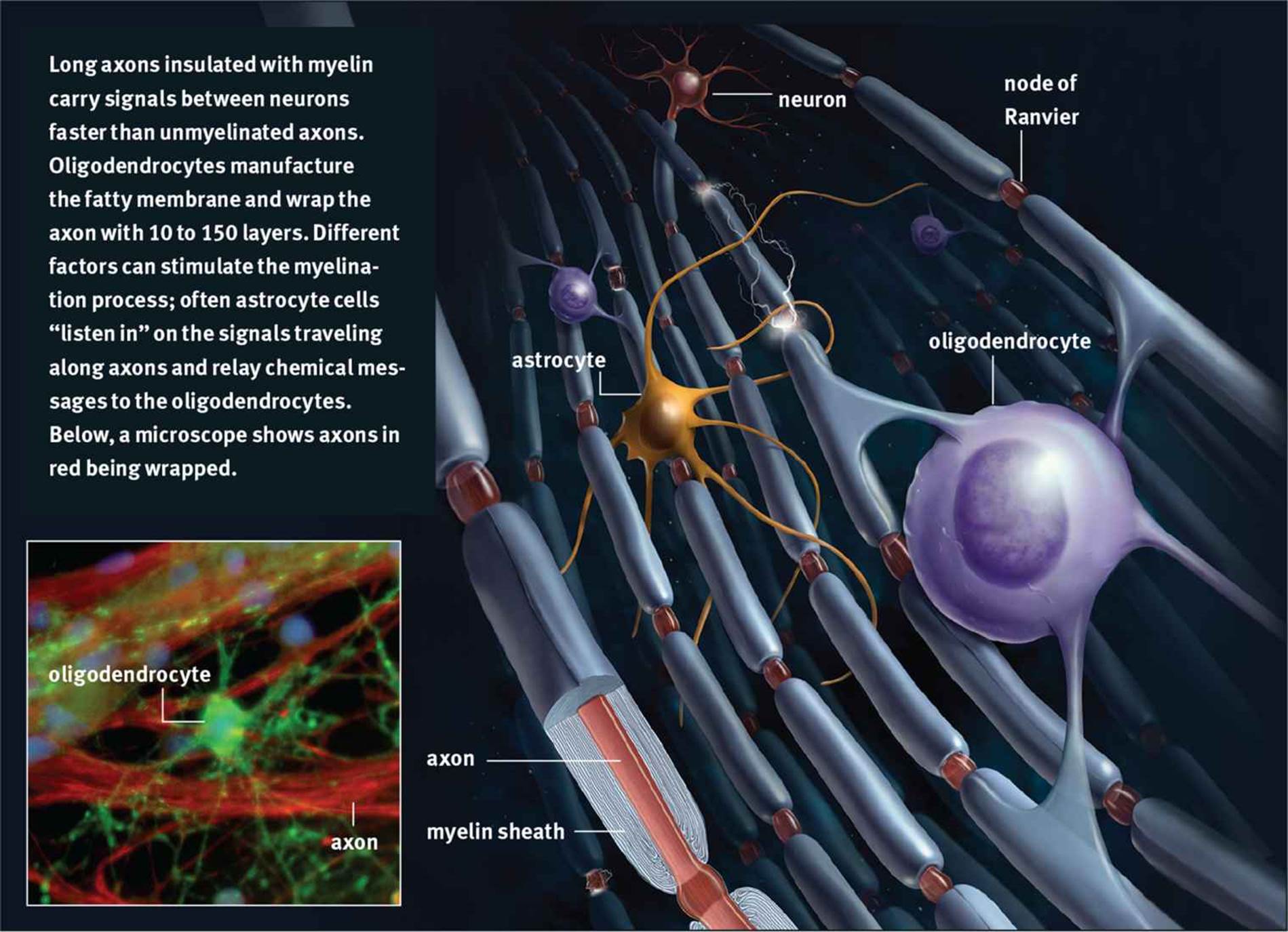 Figure 4.2. Glial Cells: Astrocytes and Oligodendrocytes
A detailed knowledge of these cell types is not necessary for the MCAT, so a familiarity with their basic functions will suffice:
· Astrocytes nourish neurons and form the blood–brain barrier, which controls the transmission of solutes from the bloodstream into nervous tissue.
· Ependymal cells line the ventricles of the brain and produce cerebrospinal fluid, which physically supports the brain and serves as a shock absorber.
· Microglia are phagocytic cells that ingest and break down waste products and pathogens in the central nervous system.
· Oligodendrocytes (CNS) and Schwann cells (PNS) produce myelin around axons.
MCAT Concept Check 4.1:
Before you move on, assess your understanding of the material with these questions.
1. For each of the following neuron structures, provide a brief description of its purpose:
· Axon:
· Axon hillock:
· Dendrite:
· Myelin sheath:
· Soma:
· Synaptic bouton:
2. What is a collection of cell bodies called in the CNS? In the PNS?
· CNS:
· PNS:
3. For each of the following glial cells, provide a brief description of its purpose:
· Astrocyte:
· Ependymal cell:
· Microglia:
· Oligodendrocyte:
· Schwann cell: