MCAT Biology Review
Chapter 2: Reproduction
Introduction
All mammals share certain characteristics: milk-producing mammary glands, three bones in the middle ear and one in the lower jaw, fur or hair, heterodont dentition (different kinds of teeth), and both sebaceous (oil-producing) and sudoriferous (sweat) glands. What about placenta formation during embryonic development? This is a characteristic of humans, as we’ll explore in Chapter 3 of MCAT Biology Review, but there are two groups of mammals that birth their young a bit differently: prototherians and metatherians.
Prototherians (monotremes), which include the duckbilled platypus and echidna (spiny anteater), encase their developing embryos within hard-shelled amniotic eggs and lay them to be hatched, like reptiles. This method of development is referred to as oviparity. Metatherians (marsupials) include koalas and kangaroos. A typical metatherian fetus (joey) undergoes some development in its mother’s uterus and then climbs its way out of the birth canal and into her marsupium, or pouch. It might seem a bit strange that something as essential as reproduction could be so different between mammalian species, but the truth is that there is a wide variety of reproductive mechanisms in nature. Many organisms reproduce without a sexual partner. Others can reproduce sexually or asexually depending on environmental conditions. In the Chapter 1 of MCAT Biology Review, we explored how bacteria and viruses reproduce. In this chapter, we’ll explore how eukaryotic cells reproduce, as well as the male and female reproductive systems.
2.1 The Cell Cycle and Mitosis
In animals, autosomal cells are said to be diploid (2n), which means that they contain two copies of each chromosome. Germ cells, on the other hand, are haploid (n), containing only one copy of each chromosome. In humans, these numbers are 46 and 23, respectively; we inherit 23 chromosomes from each parent. Eukaryotic cells replicate through the cell cycle, a specific series of phases during which a cell grows, synthesizes DNA, and divides. Derangements of the cell cycle can lead to unchecked cell division and may be responsible for the formation of cancer.
THE CELL CYCLE
The cell cycle, shown in Figure 2.1, is a perennial MCAT favorite. For actively dividing cells, the cell cycle consists of four stages: G1, S, G2, and M. The first three stages (G1, S, and G2) are known collectively as interphase. Interphase is the longest part of the cell cycle; even actively dividing cells spend about 90 percent of their time in interphase. Cells that do not divide spend all of their time in an offshoot of G1 called G0. During the G0 stage, the cell is simply living and serving its function, without any preparation for division.
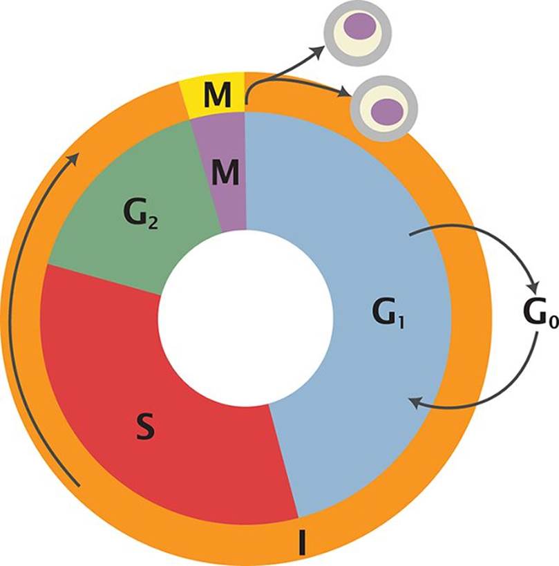 Figure 2.1. The Cell Cycle
During interphase, individual chromosomes are not visible with light microscopy. Rather, they are in a less condensed form known as chromatin. This is because the DNA must be available to RNA polymerase so that genes can be transcribed. During mitosis, however, it is preferable to condense the DNA into tightly coiled chromosomes to avoid losing any genetic material during cell division.
G1 Stage: Presynthetic Gap
During the G1 stage, cells create organelles for energy and protein production (mitochondria, ribosomes, and endoplasmic reticulum), while also increasing their size. In addition, passage into the S (synthesis) stage is governed by a restriction point. Certain criteria, such as containing the proper complement of DNA, must be met for the cell to pass the restriction point and enter the synthesis stage.
S Stage: Synthesis of DNA
During the S stage, the cell replicates its genetic material so that each daughter cell will have identical copies. After replication, each chromosome consists of two identical chromatids that are bound together at a specialized region known as the centromere, as shown in Figure 2.2. Note that the ploidy of the cell does not change even though the number of chromatids has doubled. In other words, humans in this stage still only have 46 chromosomes, even though 92 chromatids are present. Cells entering G2 have twice as much DNA as cells in G1.
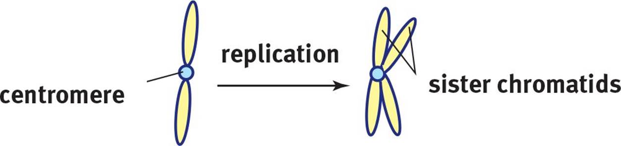 Figure 2.2. Chromosome Replication A single chromatid replicates to form two sister chromatids.
KEY CONCEPT
Each chromatid is composed of a complete, double-stranded molecule of DNA. Sister chromatids are identical copies of each other. The term chromosome may be used to refer to either a single chromatid before S phase or the pair of chromatids attached at the centromere after S phase.
G2 Stage: Postsynthetic Gap
During the G2 stage, the cell passes through another quality control checkpoint. DNA has already been duplicated, and the cell checks to ensure that there are enough organelles and cytoplasm to divide between two daughter cells. Furthermore, the cell checks to make sure that DNA replication proceeded correctly to avoid passing on an error to daughter cells that may further replicate the error in their progeny.
M Stage: Mitosis
The M stage consists of mitosis itself along with cytokinesis. Mitosis is divided into four phases: prophase, metaphase, anaphase, and telophase. The features of each phase will be discussed in the next section. Cytokinesis is the splitting of the cytoplasm and organelles into two daughter cells.
KEY CONCEPT
In autosomal cells, division results in two genetically identical daughter cells. In germ cells, the daughter cells are not equivalent.
CONTROL OF THE CELL CYCLE
The cell cycle is controlled by checkpoints, most notably between the G1 and S phase, and the G2 and M phase. At the G1/S checkpoint, the cell determines if the DNA is in good enough condition for synthesis. As mentioned above, this checkpoint is also known as the restriction point. If there has been damage to the DNA, the cell cycle goes into arrest until the DNA has been repaired. The main protein in control of this is known as p53.
At the G2/M checkpoint, the cell is mainly concerned with ensuring that the cell has achieved adequate size and the organelles have been properly replicated to support two daughter cells. p53 also plays a role in the G2/M checkpoint.
The molecules responsible for the cell cycle are known as cyclins and cyclin-dependent kinases (CDK). In order to be activated, CDKs require the presence of the right cyclins. During the cell cycle, concentrations of the various cyclins increase and decrease during specific stages. These cyclins bind to CDKs, creating an activated CDK–cyclin complex. This complex can then phosphorylate transcription factors. Transcription factors then promote transcription of genes required for the next stage of the cell cycle.
Cancer
Cell cycle control is essential to ensure that cells that are damaged or inadequately sized do not divide. When cell cycle control becomes deranged, and damaged cells are allowed to undergo mitosis, cancer may result. One of the most common mutations found in cancer is mutation of the gene that produces p53, called TP53. When this gene is mutated, the cell cycle is not stopped to repair damaged DNA. This allows for mutations to accumulate, eventually resulting in a cancerous cell that divides continuously and without regard to the quality or quantity of the new cells produced. Often, cancer cells undergo rapid cell division, creating tumors. Eventually, if the cell begins to produce the right factors (such as proteases that can digest basement membranes or factors that encourage blood vessel formation), the damaged cells are then able to reach other tissues. This may include both local invasion as well as distant spread of cancerous cells through the bloodstream or lymphatic systems. This latter result is known as metastasis.
BRIDGE
Cancer-causing genes can often be classified into oncogenes (genes that, when mutated, actively promote cell division) and tumor suppressor genes (genes that, when mutated, lose their ability to regulate or pause the cell cycle). Different cancer types are often associated with specific mutations in either oncogenes or tumor suppressor genes, or both. The biochemistry of these genes is discussed in Chapter 6 of MCAT Biochemistry Review.
MITOSIS
Mitosis, shown in Figure 2.3, is the process by which two identical daughter cells are created from a single cell. Mitosis consists of four distinct phases—prophase, metaphase, anaphase, and telophase—and occurs in somatic cells, or cells that are not involved in sexual reproduction.
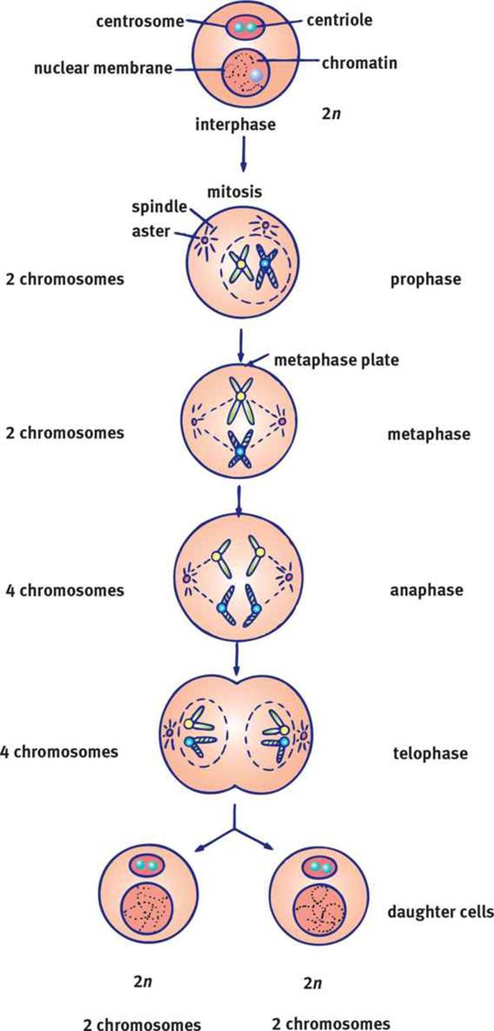 Figure 2.3. Mitosis Mitosis results in two identical daughter cells.
Prophase
Prophase is the first phase in mitosis. The first step in prophase involves condensation of the chromatin into chromosomes. Also, the centriole pairs separate and move toward opposite poles of the cell. These paired cylindrical organelles, shown in Figure 2.4, are located outside the nucleus in a region known as the centrosome and are responsible for the correct division of DNA. Once the centrioles migrate to opposite poles of the cell, they begin to form spindle fibers, which are made of microtubules. Each of the fibers radiates outward from the centrioles. Some microtubules form asters that anchor the centrioles to the cell membrane. Others extend toward the middle of the cell. The nuclear membrane dissolves during prophase, allowing these spindle fibers to contact the chromosomes. The nucleoli become less distinct, and may disappear completely.Kinetochores appear at the centromere. Kinetochores are protein structures located on the centromeres that serve as attachment points for specific fibers of the spindle apparatus appropriately called kinetochore fibers.
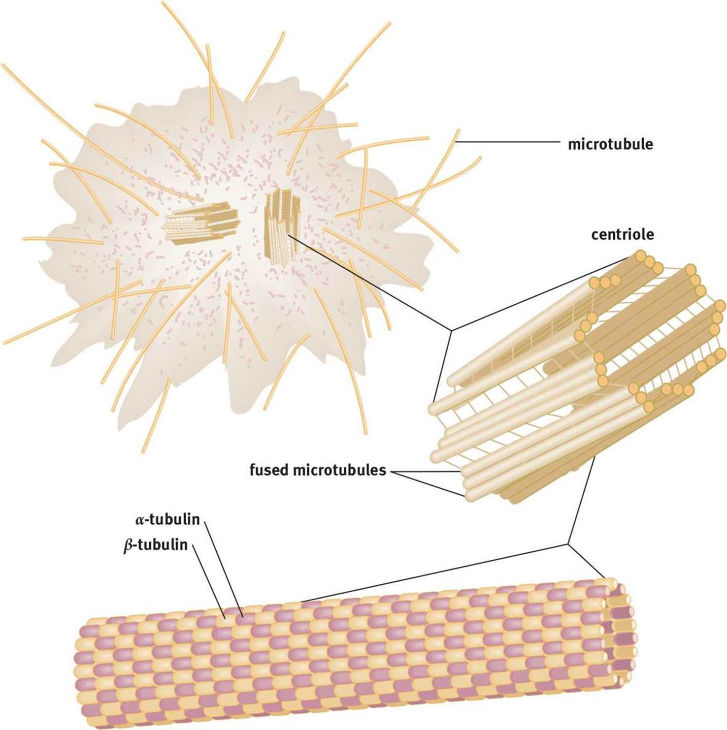 Figure 2.4. The Centrosome Each centrosome contains two tubulin-based centrioles responsible for proper movement of the chromosomes during mitosis.
KEY CONCEPT
The phases of mitosis:
· Prophase—chromosomes condense, spindle forms
· Metaphase—chromosomes align
· Anaphase—sister chromatids separate
· Telophase—new nuclear membranes form
Metaphase
In metaphase, the centriole pairs are now at opposite ends of the cell. The kinetochore fibers interact with the fibers of the spindle apparatus to align the chromosomes at the metaphase plate (equatorial plate), which is equidistant between the two poles of the cell.
Anaphase
During anaphase, the centromeres split so that each chromatid has its own distinct centromere, thus allowing the sister chromatids to separate. The sister chromatids are pulled toward the opposite poles of the cell by the shortening of the kinetochore fibers.
Telophase and Cytokinesis
Telophase is essentially the reverse of prophase. The spindle apparatus disappears. A nuclear membrane reforms around each set of chromosomes, and the nucleoli reappear. The chromosomes uncoil, resuming their interphase form. Each of the two new nuclei has received a complete copy of the genome identical to the original genome and to each other.
At the end of telophase, cytokinesis is the separation of the cytoplasm and organelles so that each daughter cell has sufficient supplies to survive on its own. Each cell undergoes a finite number of divisions before programmed death; for human somatic cells, this is usually between 20 and 50. After that, the cell can no longer divide continuously.
MCAT Concept Check 2.1:
Before you move on, assess your understanding of the material with these questions.
1. What are the five stages of the cell cycle? What happens in each stage?
|
Cell Cycle Stage |
Features |
2. What are the four phases of mitosis? What happens in each phase?
|
Mitotic Phase |
Features |