Case Files Biochemistry, 3rd Edition (2015)
SECTION II. Clinical Cases
CASE 6
A 21-year-old woman presents to the physician’s office with complaints of very painful vulvar ulcers. The patient states she has had the symptoms for approximately 3 days, and they are worsening. She says that prior to the ulcerations, there was a burning and tingling sensation of the skin in the same area. She has noted similar symptoms like this before and has been told it is sexually transmitted. On examination you see multiple extremely tender, vesicular (blister-like) lesions on an erythematous (red) base on both labia majora of the vulva. She has a moderate amount of tender inguinal lymph nodes bilaterally. The physician uses a swab to sample the ulcer and send it off for diagnostic analysis.
![]() What is the most likely diagnosis?
What is the most likely diagnosis?
![]() If the test is for DNA, then what method would be used to amplify the fragments of DNA sampled?
If the test is for DNA, then what method would be used to amplify the fragments of DNA sampled?
ANSWERS TO CASE 6:
Herpes Simplex Virus/Polymerase Chain Reaction
Summary: A 21-year-old woman presents with recurrent episodes of painful ulcerations on vulva. The physician uses a swab to sample the ulcer for diagnosis. She also has some neurologic symptoms at the vulva prior to appearance of lesions.
• Most likely diagnosis: Herpes simplex virus (HSV) type 2 outbreak.
• Biochemical Technique: Polymerase chain reaction (PCR) to amplify the small amount of HSV DNA. This is a very sensitive diagnostic technique that is able to detect small amounts of HSV DNA, and through in vitro techniques, it is able to rapidly produce large quantities of DNA.
CLINICAL CORRELATION
This 21-year-old woman has a recurrent episode of vulvar ulcers accompanied by burning and tingling in the same region. These symptoms are caused by the HSV affecting the afferent nerve. After a primary infection, the herpes virus lays dormant in the dorsal ganglia of the nerve root. Then, in times of stress or for unknown reasons, the virus becomes active and travels down the nerve to the skin. Thus, the patient often has neurologic symptoms even before the outbreak on the skin. Viral culture is an accurate method of diagnosis. Perhaps even better is PCR, which can also be used for detection of numerous infectious organisms, genetic mutations, and forensic testing.
APPROACH TO:
Polymerase Chain Reaction
OBJECTIVES
1. Describe the life cycle of HSVs.
2. Describe the process of PCR.
3. Know the definitions and purpose of restriction endonucleases and oligonucleotides.
4. Be familiar with how PCR may be used for identifying infections and mutations.
5. Cite the advantages of PCR over other biotechnology involving recombinant DNA.
DEFINITIONS
ANNEALING: The process of allowing single-stranded lengths of DNA to base pair to form double-stranded DNA. It is used most frequently when referring to the process of binding an oligonucleotide primer or probe to a longer DNA fragment.
DENATURATION: The process by which the secondary and tertiary structure of proteins and nucleic acids is broken down to form random chains. In the case of DNA, it specifically refers to the separation of double-stranded DNA into single strands by the breaking of the hydrogen bonds that form the complementary base pairing, usually by increasing the temperature.
POLYMERASE CHAIN REACTION (PCR): PCR is a method by which DNA or DNA fragments can be amplified through several steps of denaturation, annealing, and elongation. Through this process, the amount of target DNA can be increased by a factor of 106 to 109.
RESTRICTION ENZYMES: Endonucleases isolated from bacteria that will selectively cleave DNA having specific nucleotide sequences. The enzyme recognizes particular palindromic nucleotide sequences and will hydrolyze both strands within that sequence. Molecular biologists use these enzymes in various recombinant DNA techniques.
TAQ DNA POLYMERASE: A DNA polymerase isolated from the thermophilic bacteria Thermus aquaticus. It has the advantage that it remains active during the denaturation steps of PCR in which the temperature is increased to separate the DNA strands.
PALINDROME: Having the same sequence in the complimentary strand as the original strand of DNA or RNA when read from the 5′ to the 3′ direction.
ACYCLOVIR (ACYCLOGUANOSINE): Acycloguanosine, a prodrug that is activated to the active acycloguanosine triphosphate (acyclo-GTP), which inhibits viral DNA polymerase by acting as a DNA chain terminator.
DISCUSSION
HSVs are members of Herpesviridae, which also includes Epstein-Barr virus (infectious mononucleosis), varicella-zoster virus (chicken pox), and cytomegalovirus (CMV). Herpesviruses are large (150 nm in diameter) enveloped linear double-stranded DNA viruses. Because of the phospholipid envelope, herpesviruses are sensitive to acids, detergents, solvents, and drying. In general, herpesvirus replication begins with the attachment of the viral particle to the host cell surface, followed by the fusion of the viral envelope and cell membrane and release of the viral nucleocapsid into the cell cytoplasm. The nucleocapsid attaches to the nuclear envelope and then the viral genome is released into the cellular nucleus. The herpesvirus genome is transcribed and translated by host factors but is replicated by a viral encoded DNA polymerase. This HSV DNA polymerase is an important target for drug therapy. The viral nucleocapsids are formed in the nucleus, and the envelope is acquired from the nuclear or Golgi membrane. Mature viral particles are released from the host cell by exocytosis or cell lysis. Depending on the herpesvirus and type of host cell that is infected, a lytic, persistent (macrophages and lymphocytes), or latent (nerve cells) infection can be established.
HSV can infect many cell types (including macrophages, lymphocytes, and neurons), causing lytic, persistent, and latent infections. Because of nerve cell involvement in latent infections, a recurrence of the disease is often preceded by a prodrome (sensations such as burning or tingling). Initial HSV-1 and HSV-2 infections are usually established on mucous membranes. Although HSV-1 and HSV-2 are respectively referred to as oral herpes and genital herpes based on their typical site of infection, HSV-1 and HSV-2 can be found on both oral and genital tissue.
Diagnosis can be made by examining infected tissue or cells for characteristic cytopathologic effects, virus isolation and culture, or serology. Molecular methods (such as DNA in situ hybridization and PCR of vesicle fluid or scrapings) are also used for diagnosis. The molecular methods are gaining favor because they yield rapid results and identify the viral type or strain. Because of these diagnostic advantages, PCR is the preferred method for diagnosis of HSV encephalitis.
The PCR is an in vitro method for the exponential amplification of a specific DNA region. Today, PCR is one of the most important biochemical techniques and is applied in virtually all fields of modern molecular biology. The ingredients for PCR reactions are very simple: the target DNA to be amplified, a heat-stable DNA polymerase, the 4 deoxyribonucleotides, 2 oligonucleotide primers, and a reaction buffer. The entire PCR reaction is carried out in a single tube containing all these necessary components. The target DNA segment is amplified with a high degree of specificity by using 2 DNA oligonucleotide primers that are complementary to sequences on the 3′-flanking regions of opposite strands of the target segment. The PCR amplification occurs by repeated cycles of three temperature-dependent steps called denaturation, annealing, and elongation (Figure 6-1). Native DNA exists as a double helix; therefore, the first step, denaturation, of the process separates the 2 DNA chains by heating the reaction mixture to 194 to 203°F (90–95°C). In the second step, annealing, the reaction mixture is cooled to 113 to 140°F (45–60°C) so that the oligonucleotide primers can bind or anneal to the separated strands of the target DNA. In the final step of the process, elongation, the DNA polymerase adds nucleotides to the 3′ ends of the primers to complete a copy of the target DNA template. A heat-stable DNA polymerase obtained from the thermophilic bacterium T. aquaticus is used to synthesize the new strands in most PCRs. Since this Taq DNA polymerase works best at around 161.6°F (72°C), the temperature of the PCR mixture is raised to that temperature for elongation to proceed efficiently. At the end of a three-step cycle, each target region of DNA in the vial has been duplicated. This 3-step cycle is then repeated multiple times. Each new DNA strand can then act as a new template in the next cycle, yielding approximately 1 million copies of a target DNA after 20 cycles. Therefore, PCR is a method able to amplify any DNA sequence virtually without limit and allows the separation of the nucleic acid of interest from its context.
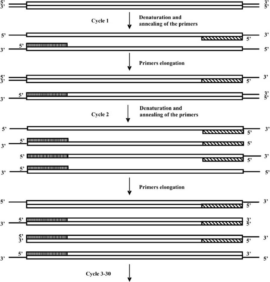
Figure 6-1. Polymerase chain reaction diagram.
PCR is also becoming the leading method for detection of the continuously increasing number of human pathogens. Examples include HSV, human papillomavirus, HIV, human T. lymphotropic virus types 1 and 2, CMV, Epstein-Barr virus, human herpesvirus-6 (HHV-6), hepatitis B virus, B16 parvovirus, JC and BK viruses, rubella virus, mycobacteria, Toxoplasmosis gondii, Trypanosoma cruzi, and malaria, with many more to follow. PCR can be applied as a detection method for virtually any pathogen for which even limited nucleotide sequence information is known and for which a specimen of infected tissue can be readily obtained. In most cases, PCR assays are more discriminative than conventional serology. For example, it is difficult to distinguish HSV-1 from HSV-2 or HIV-1 from HIV-2 by serology, yet such distinctions can be readily made on the basis of PCR amplification of type-specific genetic sequences. PCR yields rapid results, typically in 1 to 2 days in a clinical setting. It is applicable to a wide variety of clinical, pathologic, or forensic specimens, as well as to formalin-fixed tissue, inactivated bacterial cultures, and archaeological specimens. However, since PCR detects nucleic acids from both living and dead microbes, this must be taken into account if PCR is used to monitor response to therapy.
Prior to the PCR method for detection of HSV-1 and HSV-2, clinical specimens were analyzed using immunologic methods to confirm the virus identity following a viral culture. Such methods are time consuming (3-12 days) and expensive. In addition, cultivation of the virus is not always successful. Turnaround time for viral culture averaged 108 hours for positive results and 154 hours for negative results. In comparison, the present PCR method of HSV detection is specific, relatively fast and accurate. The method allows detection of one to ten virus particles in the presence of microgram quantities of cellular or heterogeneous DNA. The PCR assay offers increased sensitivity, specificity, and improved turnaround time (24-48 hours) when compared with traditional viral culture techniques. These methods have allowed a better understanding of the physiopathogeny of the disease. In particular, PCR has revealed the importance of asymptomatic viral shedding in infected patients. PCR also helps diagnosis in many situations where viral isolation by culture proves difficult or impossible, for example, in treated or atypical lesions, or in newborn central nervous system infections. PCR has demonstrated the existence of prolonged viremia in infected newborns. PCR helps sequencing of the viral genome for further epidemiologic studies or analysis of resistance to antiviral drugs. Recently, PCR-derived techniques have been developed to quantify viral load in real time, thus allowing a diagnosis in a few hours.
After amplification of the samples using PCR, the identification of the virus species can be achieved through restriction enzyme digestion, which yields a unique pattern of different fragment sizes characteristic for each herpesvirus (or other pathogen). Restriction endonucleases are enzymes able to cut double-stranded DNA at specific palindromic recognition sequences that are mostly 4 to 6 nucleotides in length. Furthermore, a well-designed restriction enzyme panel allows the discrimination between human herpes virus 6 variant A and variant B. In this way, this method can readily detect human herpesviruses, including occasional multiple infections, in a variety of clinical samples. When PCR assay was compared to isolation and electron microscopy for the detection of HSV in clinical samples, all specimens positive by conventional methods were also positive by PCR. However, in a number of clinical specimens in which HSV could not be detected by conventional methods, PCR was able to demonstrate the presence of the virus.
Treatment of HSV typically involves the use of nucleoside analog prodrugs that when activated specifically inhibit the viral DNA polymerase, thereby preventing replication of the viral genome. Acyclovir is a guanosine analog (Figure 6-2) that has been shown to be effective against HSV. It is first phosphorylated to the mono-phosphate derivative by viral thymidine kinase. Cellular kinases then convert it to the triphosphate. The now activated drug, acyclo-GTP, inhibits replication of the viral genome. The viral DNA polymerase will incorporate the acycloguanine nucleotide into the viral DNA in the replication process, but because there is no 3′-hydroxyl to form the next phosphodiester, the chain terminates and replication stops.
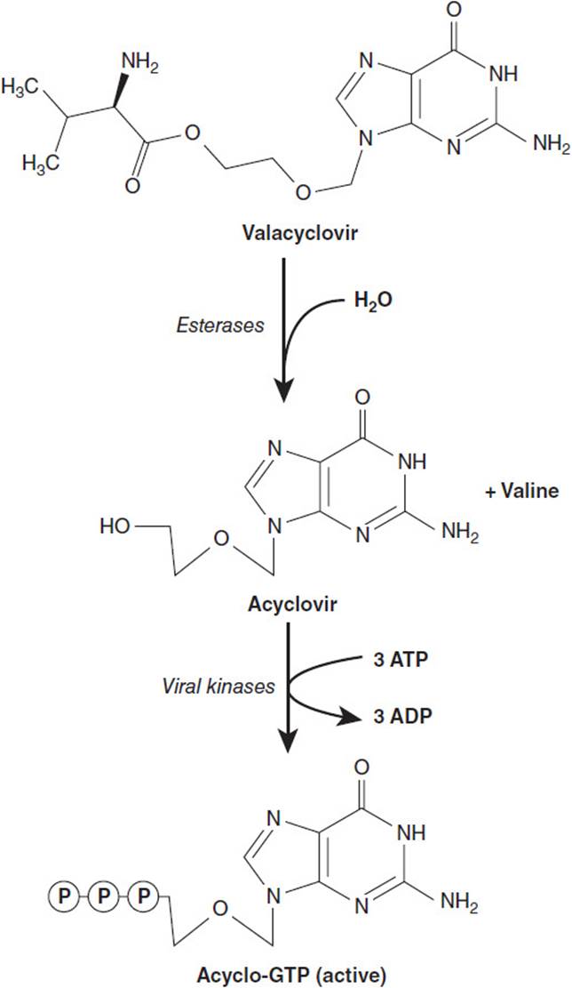
Figure 6-2. Conversion of the prodrug valacyclovir to the active acycloguanosine triphosphate.
Acyclovir has a relatively poor oral bioavailability. Valacyclovir is a modified version of acyclovir that has been esterified with the amino acid valine. Its oral bioavailability is about 3 to 5 times that of acyclovir. When it is absorbed, it is converted to acyclovir by cellular esterases and then further phosphorylated to the active acyclo-GTP (see Figure 6-2).
COMPREHENSION QUESTIONS
6.1 One of the steps in the PCR amplification of DNA fragments is the denaturation step in which the temperature is raised to break the hydrogen bonds that make up the base pairing. Which of the following DNA fragments would most likely require the greatest increase in temperature to cause complete denaturation?
A. 5′–C-A-A-T-G-T-A-A-T-T-G-C-A-T-3′
3′–G-T-T- A-C-A-T-T-A-A-C-G-T-A-5′
B. 5′–A-T-A-T-A-T-A-T-A-T-A-T-A-T-3′
3′–T- A-T- A-T-A-T-A-T- A-T-A-T-A-5′
C. 5′–A-A-C-C-G-G-A-C-C-G-C-G-A-T-3′
3′–T- T-G-G-C-C-T-G-G-C-G-C-T-A-5′
D. 5′–A-G-A-G-A-G-A-G-A-G-A-G-A-G-3′
3′–T-C-T-C-T-C-T-C-T- C-T-C-T-C-5′
E. 5′–G-A-C-T-G-T-A-A-T-A-C-G-A-T-3′
3′–C-T-G-A-C-A-T-T-A-T-G-C-T-A-5′
6.2 Restriction enzymes are used to cleave genomic DNA into smaller fragments. Which of the following single-strand DNA sequences has the best potential to be a site of action for a restriction endonuclease?
A. T–A–G–C–T–T
B. C–T–G–C–A–G
C. A–A–C–C–A–A
D. G–T–G–T–G–T
E. A–A–A–C–C–C
6.3 A 21-year-old woman was abducted when she went to the local convenience store. Her body was found the next morning in a wooded area behind the store. The autopsy revealed that she had been sexually assaulted and strangled. Crime scene investigators were able to collect a semen sample from vaginal fluid as well as tissue samples from underneath the victim’s fingernails. DNA samples were obtained from three suspects besides the victim. A variable number of tandem repeats (VNTR) analysis was performed on the DNA samples from the evidence collected, the victim, and the suspect, and the results were compared. Which of the following techniques is the most appropriately applied for this analysis?
A. Allele-specific oligonucleotide probes
B. DNA sequencing
C. Northern blot
D. Southern blot
E. Western blot
6.4 A 40-year-old man presents to his primary care physician due to a painful lesion on his scrotum. On examination, the physician observed what appeared to be herpetic vesicles on the left underside of the scrotum. He prescribed a course of valacyclovir. Valacyclovir treats outbreaks of HSV infection by which of the following?
A. Acting as a protease inhibitor
B. Inhibiting protein translation
C. Inhibiting reverse transcriptase
D. Inhibiting packaging of new viral particles
E. Inhibiting replication of the viral genome
ANSWERS
6.1 C. The temperature at which the DNA strands separate is dependent on the number of hydrogen bonds that make up the base pairing. Because G-C pairs have three hydrogen bonds while A-T pairs only have 2, then the strand that has the greatest number of G-C pairs will have the higher melting temperature, Tm, which is the temperature at which one-half of the base pairs are broken. Interestingly, the TATA box, the starting point of transcription in eukaryotes, has weaker bonding.
6.2 B. Most of the sequences recognized by restriction endonucleases are palindromes; that is, they have the same nucleotide sequence on both strands when read in the 5′ to 3′ direction. Because answer B has the only sequence that will be a palindrome when paired with its complementary strand, it is the most likely to be a site recognized by a restriction endonuclease (it is the site recognized by the restriction endonuclease Pst1).
6.3 D. A VNTR analysis examines the hypervariable regions of the human genome. These contain sequences that are repeated in tandem a variable number of times and the length is unique for each individual. Because it is DNA fragments that are being analyzed, the Southern blot is the most appropriate technique to use to separate and detect these regions. DNA sequencing is too time consuming to be practical for forensic analyses. The Northern blot is used to separate and detect RNA, whereas the Western blot is used for proteins. Allele-specific oligonucleotide probes are used when testing for the presence of a genetic mutation that either introduces or removes a restriction site.
6.4 E. Valacyclovir is a prodrug that is metabolized to an analog of GTP. The nucleotide analog is incorporated into viral DNA as it is replicating and terminates the replication process.
BIOCHEMISTRY PEARLS
![]() PCR is an in vitro method for the exponential amplification of a specific DNA region.
PCR is an in vitro method for the exponential amplification of a specific DNA region.
![]() PCR involves (1) denaturation, which separates the 2 DNA chains by heating the reaction mixture to 194 to 203°F (90-95°C), (2) annealing or cooling to 113 to 140°F (45-60°C) so that the oligonucleotide primers can bind or anneal to the separated strands of the target DNA, and (3) elongation with DNA polymerase (at 162°F [72°C] when Taq DNA polymerase used) adding nucleotides to the 3′ ends of the primers to a complete copy of the target DNA template.
PCR involves (1) denaturation, which separates the 2 DNA chains by heating the reaction mixture to 194 to 203°F (90-95°C), (2) annealing or cooling to 113 to 140°F (45-60°C) so that the oligonucleotide primers can bind or anneal to the separated strands of the target DNA, and (3) elongation with DNA polymerase (at 162°F [72°C] when Taq DNA polymerase used) adding nucleotides to the 3′ ends of the primers to a complete copy of the target DNA template.
![]() GC pairs have 3 hydrogen bonds, whereas AT pairs have only 2 hydrogen bonds and are therefore weaker.
GC pairs have 3 hydrogen bonds, whereas AT pairs have only 2 hydrogen bonds and are therefore weaker.
![]() A heat-stable DNA polymerase obtained from the thermophilic bacterium T. aquaticus is used to synthesize the new strands in most PCR reactions.
A heat-stable DNA polymerase obtained from the thermophilic bacterium T. aquaticus is used to synthesize the new strands in most PCR reactions.
![]() Endonucleases usually act at sites of palindromes.
Endonucleases usually act at sites of palindromes.
REFERENCES
Granner DK, Weil PA. Molecular genetics, recombinant DNA, & genomic technology. In: Murray RK, Granner DK, Mayes PA, et al, eds. Harper’s Illustrated Biochemistry. 26th ed. New York: Lange Medical Books/McGraw-Hill; 2003.
Johnson G, Nelson S, Petric M, et al. Comprehensive PCR-based assay for detection and species identification of human herpesviruses. J Clin Microbiol. 2000;38(9):3274-3279.