Harper’s Illustrated Biochemistry, 29th Edition (2012)
SECTION II. Bioenergetics & the Metabolism of Carbohydrates & Lipids
Chapter 18. Glycolysis & the Oxidation of Pyruvate
David A. Bender, PhD & Peter A. Mayes, PhD, DSc
OBJECTIVES
After studying this chapter, you should be able to:
![]() Describe the pathway of glycolysis and its control and explain how glycolysis can operate under anaerobic conditions.
Describe the pathway of glycolysis and its control and explain how glycolysis can operate under anaerobic conditions.
![]() Describe the reaction of pyruvate dehydrogenase and its regulation.
Describe the reaction of pyruvate dehydrogenase and its regulation.
![]() Explain how inhibition of pyruvate metabolism leads to lactic acidosis
Explain how inhibition of pyruvate metabolism leads to lactic acidosis
BIOMEDICAL IMPORTANCE
Most tissues have at least some requirement for glucose. In the brain, the requirement is substantial, and even in prolonged fasting the brain can meet no more than about 20% of its energy needs from ketone bodies. Glycolysis, the major pathway for glucose metabolism, occurs in the cytosol of all cells. It is unique, in that it can function either aerobically or anaerobically, depending on the availability of oxygen and the electron transport chain. Erythrocytes, which lack mitochondria, are completely reliant on glucose as their metabolic fuel, and metabolize it by anaerobic glycolysis. However, to oxidize glucose beyond pyruvate (the end product of glycolysis) requires both oxygen and mitochondrial enzyme systems: the pyruvate dehydrogenase complex, the citric acid cycle (Chapter 17), and the respiratory chain (Chapter 13).
Glycolysis is both the principal route for glucose metabolism and also the main pathway for the metabolism of fructose, galactose, and other dietary carbohydrates. The ability of glycolysis to provide ATP in the absence of oxygen is especially important, because this allows skeletal muscle to perform at very high levels of work output when oxygen supply is insufficient, and it allows tissues to survive anoxic episodes. However, heart muscle, which is adapted for aerobic performance, has relatively low glycolytic activity and poor survival under conditions of ischemia. Diseases in which enzymes of glycolysis (eg, pyruvate kinase) are deficient are mainly seen as hemolytic anemias or, if the defect affects skeletal muscle (eg, phosphofructokinase), as fatigue. In fast-growing cancer cells, glycolysis proceeds at a high rate, forming large amounts of pyruvate, which is reduced to lactate and exported. This produces a relatively acidic local environment in the tumor, which may have implications for cancer therapy. The lactate is used for gluconeogenesis in the liver (Chapter 20), an energy-expensive process, which is responsible for much of the hypermetabolism seen in cancer cachexia. Lactic acidosis results from various causes, including impaired activity of pyruvate dehydrogenase, especially in thiamin (vitamin B t) deficiency.
GLYCOLYSIS CAN FUNCTION UNDER ANAEROBIC CONDITIONS
Early in the investigations of glycolysis it was realized that fermentation in yeast was similar to the breakdown of glycogen in muscle. It was noted that when a muscle contracts in an anaerobic medium, that is, one from which oxygen is excluded, glycogen disappears and lactate appears. When oxygen is admitted, aerobic recovery takes place and lactate is no longer produced. However, if contraction occurs under aerobic conditions, lactate does not accumulate and pyruvate is the major end product of glycolysis. Pyruvate is oxidized further to CO2 and water (Figure 18–1). When oxygen is in short supply, mitochondrial reoxidation of NADH formed during glycolysis is impaired, and NADH is reoxidized by reducing pyruvate to lactate, so permitting glycolysis to proceed (Figure 18–1). While glycolysis can occur under anaerobic conditions, this has a price, for it limits the amount of ATP formed per mole of glucose oxidized, so that much more glucose must be metabolized under anaerobic than aerobic conditions. In yeast and some other microorganisms, pyruvate formed in anaerobic glycolysis is not reduced to lactate, but is decarboxylated and reduced to ethanol.
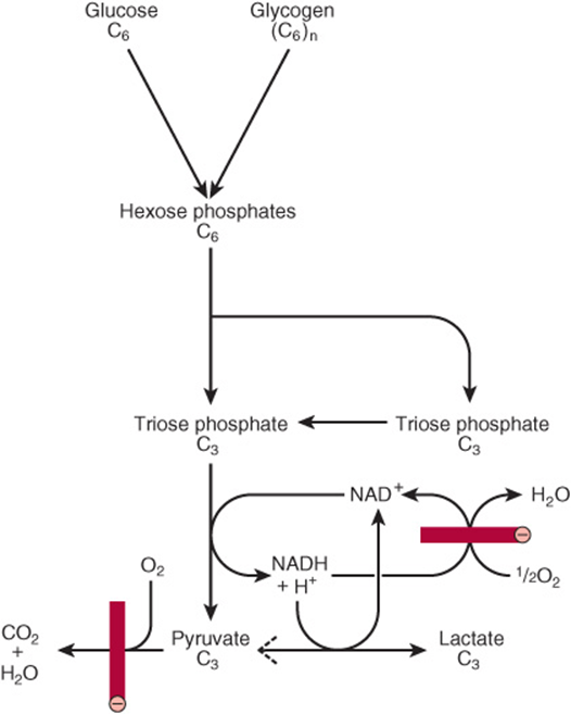
FIGURE 18–1 Summary of glycolysis. ![]() , blocked by anaerobic conditions or by absence of mitochondria containing key respiratory enzymes, as in erythrocytes.
, blocked by anaerobic conditions or by absence of mitochondria containing key respiratory enzymes, as in erythrocytes.
THE REACTIONS OF GLYCOLYSIS CONSTITUTE THE MAIN PATHWAY OF GLUCOSE UTILIZATION
The overall equation for glycolysis from glucose to lactate is as follows:
![]()
All of the enzymes of glycolysis (Figure 18–2) are found in the cytosol. Glucose enters glycolysis by phosphorylation to glucose 6-phosphate, catalyzed by hexokinase, using ATP as the phosphate donor. Under physiologic conditions, the phosphorylation of glucose to glucose 6-phosphate can be regarded as irreversible. Hexokinase is inhibited allosterically by its product, glucose 6-phosphate.
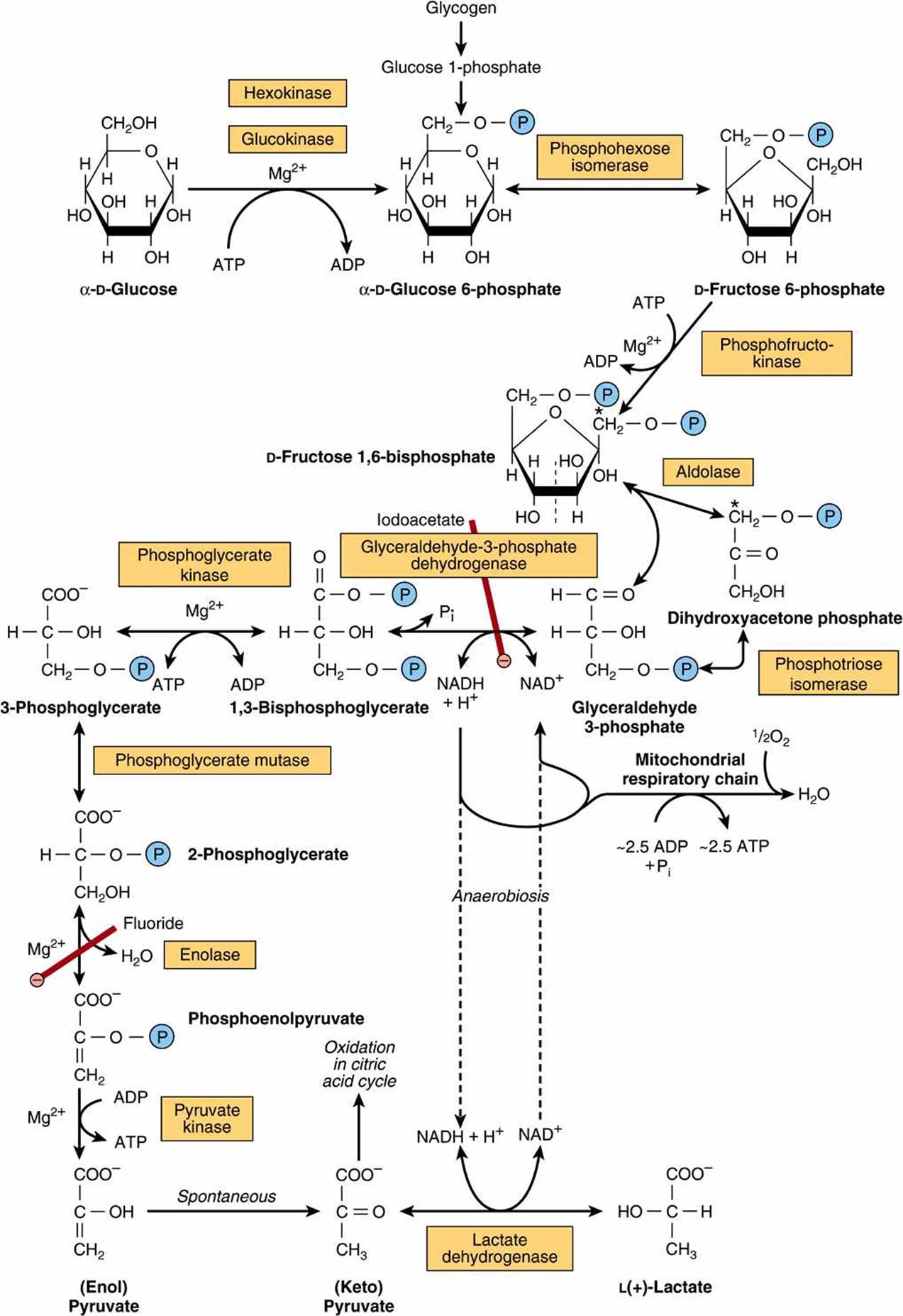
FIGURE 18–2 The pathway of glycolysis. (![]() ,
, ![]()
![]()
![]() , inhibition.) *Carbons 1-3 of fructose bisphosphate form dihydroxyacetone phosphate, and carbons 4-6 form glyceraldehyde 3-phosphate. The term “bis-,” as in bisphosphate, indicates that the phosphate groups are separated, whereas the term “di-,” as in adenosine diphosphate, indicates that they are joined.
, inhibition.) *Carbons 1-3 of fructose bisphosphate form dihydroxyacetone phosphate, and carbons 4-6 form glyceraldehyde 3-phosphate. The term “bis-,” as in bisphosphate, indicates that the phosphate groups are separated, whereas the term “di-,” as in adenosine diphosphate, indicates that they are joined.
In tissues other than the liver (and pancreatic β-islet cells), the availability of glucose for glycolysis (or glycogen synthesis in muscle, Chapter 19, and lipogenesis in adipose tissue, Chapter 23) is controlled by transport into the cell, which in turn is regulated by insulin. Hexokinase has a high affinity (low Km) for glucose, and in the liver it is saturated under normal conditions, and so acts at a constant rate to provide glucose 6-phosphate to meet the liver’s needs. Liver cells also contain an isoenzyme of hexokinase, glucokinase, which has a Km very much higher than the normal intracellular concentration of glucose. The function of glucokinase in the liver is to remove glucose from the blood following a meal, providing glucose 6-phosphate in excess of requirements for glycolysis, which is used for glycogen synthesis and lipogenesis. Glucokinase is also found in pancreatic β-islet cells, where it functions to detect high concentrations of glucose; onward metabolites of the glucose 6-phosphate formed stimulate the secretion of insulin.
Glucose 6-phosphate is an important compound at the junction of several metabolic pathways: glycolysis, gluconeogenesis, the pentose phosphate pathway, glycogenesis, and glycogenolysis. In glycolysis, it is converted to fructose 6-phosphate by phosphohexose isomerase, which involves an aldose-ketose isomerization. This reaction is followed by another phosphorylation catalyzed by the enzyme phosphofructokinase (phosphofructokinase-1) forming fructose 1,6-bisphosphate. The phosphofructokinase reaction may be considered to be functionally irreversible under physiologic conditions; it is both inducible and subject to allosteric regulation, and has a major role in regulating the rate of glycolysis. Fructose 1,6-bisphosphate is cleaved by aldolase (fructose 1,6-bisphosphate aldolase) into two triose phosphates, glyceraldehyde 3-phosphate and dihydroxyacetone phosphate, which are interconverted by the enzyme phosphotriose isomerase.
Glycolysis continues with the oxidation of glyceraldehyde 3-phosphate to 1,3-bisphosphoglycerate. The enzyme catalyzing this oxidation, glyceraldehyde 3-phosphate dehydrogenase, is NAD dependent. Structurally, it consists of four identical polypeptides (monomers) forming a tetramer. Four—SH groups are present on each polypeptide, derived from cysteine residues within the polypeptide chain. One of the—SH groups is found at the active site of the enzyme (Figure 18–3). The substrate initially combines with this—SH group, forming a thiohemiacetal that is oxidized to a thiol ester; the hydrogens removed in this oxidation are transferred to NAD+. The thiol ester then undergoes phosphorolysis; inorganic phosphate (Pi) is added, forming 1,3-bisphosphoglycerate, and the—SH group is reconstituted.
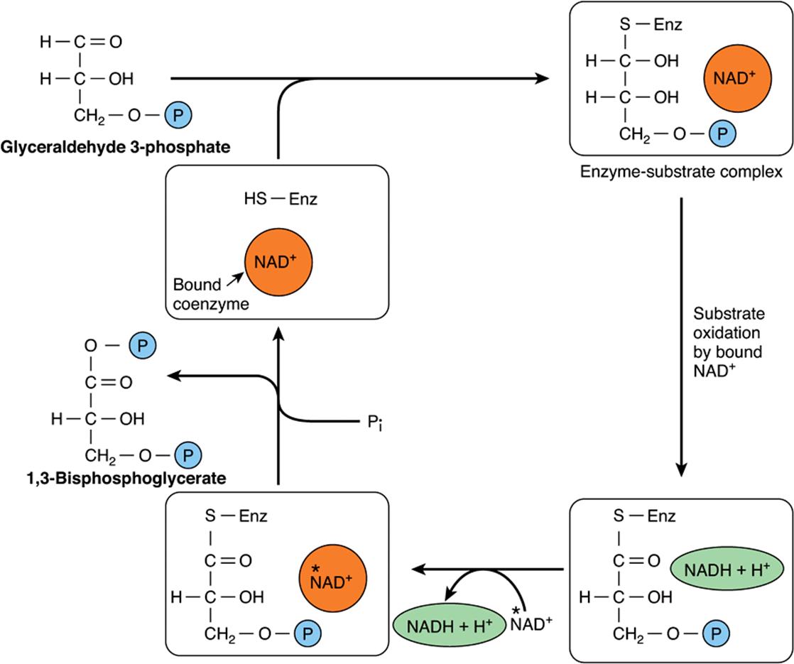
FIGURE 18–3 Mechanism of oxidation of glyceraldehyde 3-phosphate. (Enz, glyceraldehyde 3-phosphate dehydrogenase.) The enzyme is inhibited by the—SH poison iodoacetate, which is thus able to inhibit glycolysis. The NADH produced on the enzyme is not so firmly bound to the enzyme as is NAD+. Consequently, NADH is easily displaced by another molecule of NAD+.
In the next reaction, catalyzed by phosphoglycerate kinase, phosphate is transferred from 1,3-bisphosphoglycerate onto ADP, forming ATP (substrate-level phosphorylation) and 3-phosphoglycerate. Since two molecules of triose phosphate are formed per molecule of glucose undergoing glycolysis, two molecules of ATP are formed in this reaction per molecule of glucose undergoing glycolysis. The toxicity of arsenic is the result of competition of arsenate with inorganic phosphate (Pi) in this reaction to give 1-arseno-3-phosphoglycerate, which undergoes spontaneous hydrolysis to 3-phosphoglycerate without forming ATP. 3-Phosphoglycerate is isomerized to 2-phosphoglycerate by phosphoglycerate mutase. It is likely that 2,3-bisphosphoglycerate (diphosphoglycerate, DPG) is an intermediate in this reaction.
The subsequent step is catalyzed by enolase and involves a dehydration, forming phosphoenolpyruvate. Enolase is inhibited by fluoride, and when blood samples are taken for measurement of glucose, it is collected in tubes containing fluoride to inhibit glycolysis. The enzyme is also dependent on the presence of either Mg2+ or Mn2+. The phosphate of phosphoenolpyruvate is transferred to ADP by pyruvate kinase to form two molecules of ATP per molecule of glucose oxidized. The reaction of pyruvate kinase is essentially irreversible under physiological conditions, partly because of the large free energy change involved and partly because the immediate product of the enzyme-catalyzed reaction is enol-pyruvate, which undergoes spontaneous isomerization to pyruvate, so that the product of the reaction is not available to undergo the reverse reaction.
The redox state of the tissue now determines which of the two pathways is followed. Under anaerobic conditions, the NADH cannot be reoxidized through the respiratory chain to oxygen. Pyruvate is reduced by the NADH to lactate, catalyzed by) lactate dehydrogenase. There are different tissue-specific isoenzymes lactate dehydrogenases that have clinical significance (Chapter 7). The reoxidation of NADH via lactate formation allows glycolysis to proceed in the absence of oxygen by regenerating sufficient NAD+ for another cycle of the reaction catalyzed by glyceraldehyde 3-phosphate dehydrogenase. Under aerobic conditions, pyruvate is taken up into mitochondria, and after oxidative decarboxylation to acetyl-CoA is oxidized to CO2 by the citric acid cycle (Chapter 17). The reducing equivalents from the NADH formed in glycolysis are taken up into mitochondria for oxidation via one of the two shuttles described in Chapter 13.
Tissues That Function Under Hypoxic Conditions Produce Lactate
This is true of skeletal muscle, particularly the white fibers, where the rate of work output, and hence the need for ATP formation, may exceed the rate at which oxygen can be taken up and utilized. Glycolysis in erythrocytes always terminates in lactate, because the subsequent reactions of pyruvate oxidation are mitochondrial, and erythrocytes lack mitochondria. Other tissues that normally derive much of their energy from glycolysis and produce lactate include brain, gastrointestinal tract, renal medulla, retina, and skin. Lactate production is also increased in septic shock, and many cancers also produce lactate. The liver, kidneys, and heart usually take up lactate and oxidize it, but produce it under hypoxic conditions.
When lactate production is high, as in vigorous exercise, septic shock, and cancer cachexia, much is used in the liver for gluconeogenesis (Chapter 20), leading to an increase in metabolic rate to provide the ATP and GTP needed. The increase in oxygen consumption as a result of increased oxidation of metabolic fuels to provide the ATP and GTP needed for gluconeogenesis is seen as oxygen debt after vigorous exercise.
Under some conditions, lactate may be formed in the cytosol, but then enter the mitochondrion to be oxidized to pyruvate for onward metabolism. This provides a pathway for the transfer of reducing equivalents from the cytotol into the mitochondrion for the electron transport chain in addition to the glycerophosphate (Figure 13–12) and malate (Figure 13–13) shuttles.
GLYCOLYSIS IS REGULATED AT THREE STEPS INVOLVING NONEQUILIBRIUM REACTIONS
Although most of the reactions of glycolysis are reversible, three are markedly exergonic and must therefore be considered physiologically irreversible. These reactions, catalyzed by hexokinase (and glucokinase), phosphofructokinase, and pyruvate kinase, are the major sites of regulation of glycolysis. Phosphofructokinase is significantly inhibited at normal intracellular concentrations of ATP; as discussed in Chapter 20, this inhibition can be rapidly relieved by 5′ AMP that is formed as ADP begins to accumulate, signaling the need for an increased rate of glycolysis. Cells that are capable of gluconeogenesis (reversing the glycolytic pathway, Chapter 20) have different enzymes that catalyze reactions to reverse these irreversible steps; glucose 6-phosphatase, fructose 1,6-bisphosphatase and, to reverse the reaction of pyruvate kinase, pyruvate carboxylase, and phosphoenolpyruvate carboxykinase. Fructose enters glycolysis by phosphorylation to fructose 1-phosphate, and bypasses the main regulatory steps, so resulting in formation of more pyruvate (and acetyl-CoA) than is required for ATP formation (Chapter 21). In the liver and adipose tissue, this leads to increased lipogenesis, and a high intake of fructose may be a factor in the development of obesity.
In Erythrocytes, the First Site of ATP Formation in Glycolysis May Be Bypassed
In erythrocytes, the reaction catalyzed by phosphoglycerate kinase may be bypassed to some extent by the reaction of bisphosphoglycerate mutase, which catalyzes the conversion of 1,3-bisphosphoglycerate to 2,3-bisphosphoglycerate, followed by hydrolysis to 3-phosphoglycerate and Pi, catalyzed by 2,3-bisphosphoglycerate phosphatase (Figure 18–4). This alternative pathway involves no net yield of ATP from glycolysis. However, it does serve to provide 2,3-bisphosphoglycerate, which binds to hemoglobin, decreasing its affinity for oxygen, and so making oxygen more readily available to tissues (see Chapter 6).
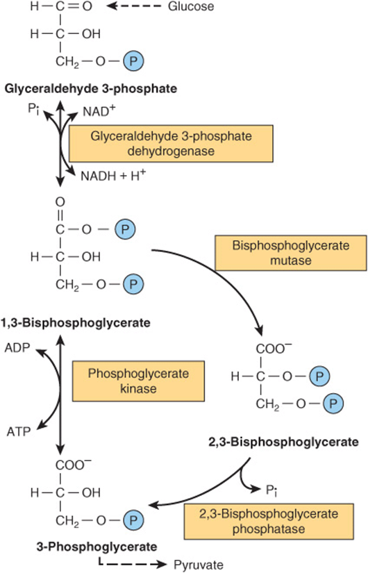
FIGURE 18–4 2,3-Bisphosphoglycerate pathway in erythrocytes.
THE OXIDATION OF PYRUVATE TO ACETYL CoA IS THE IRREVERSIBLE ROUTE FROM GLYCOLYSIS TO THE CITRIC ACID CYCLE
Pyruvate, formed in the cytosol, is transported into the mitochondrion by a proton symporter (Figure 13–10). Inside the mitochondrion, it is oxidatively decarboxylated to acetyl-CoA by a multienzyme complex that is associated with the inner mitochondrial membrane. This pyruvate dehydrogenase complex is analogous to the α-ketoglutarate dehydrogenase complex of the citric acid cycle (Figure 17–3). Pyruvate is decarboxylated by the pyruvate dehydrogenase component of the enzyme complex to a hydroxyethyl derivative of the thiazole ring of enzyme-bound thiamin diphosphate, which in turn reacts with oxidized lipoamide, the prosthetic group of dihydrolipoyl transacetylase, to form acetyl lipoamide (Figure 18–5). Thiamin is vitamin B1 (Chapter 44) and in deficiency, glucose metabolism is impaired, and there is significant (and potentially life-threatening) lactic and pyruvic acidosis. Acetyl lipoamide reacts with coenzyme A to form acetyl-CoA and reduced lipoamide. The reaction is completed when the reduced lipoamide is reoxidized by a flavoprotein, dihydrolipoyl dehydrogenase, containing FAD. Finally, the reduced flavoprotein is oxidized by NAD+, which in turn transfers reducing equivalents to the respiratory chain. The overall reaction is:
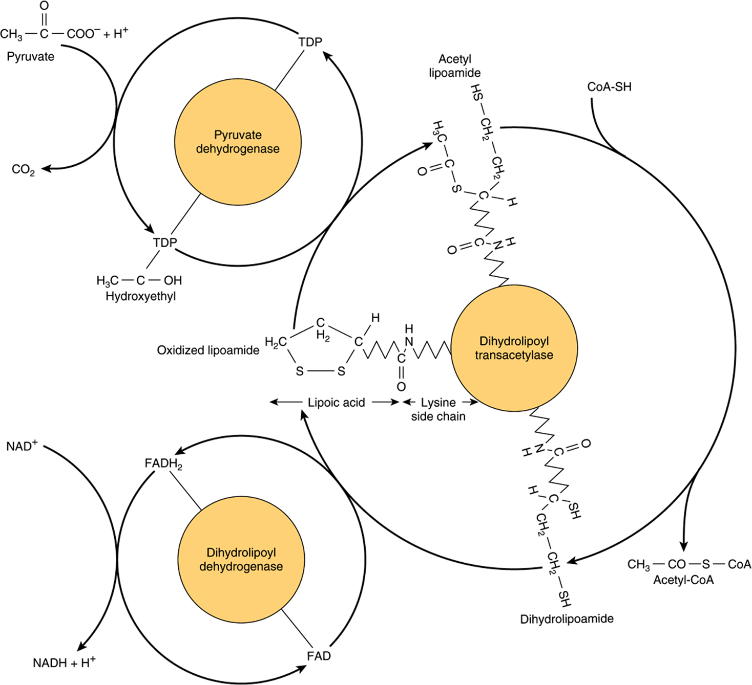
FIGURE 18–5 Oxidative decarboxylation of pyruvate by the pyruvate dehydrogenase complex. Lipoic acid is joined by an amide link to a lysine residue of the transacetylase component of the enzyme complex. It forms a long flexible arm, allowing the lipoic acid prosthetic group to rotate sequentially between the active sites of each of the enzymes of the complex. (FAD, flavin adenine dinucleotide; NAD+, nicotinamide adenine dinucleotide; TDP, thiamin diphosphate.)
![]()
The pyruvate dehydrogenase complex consists of a number of polypeptide chains of each of the three component enzymes, and the intermediates do not dissociate, but remain bound to the enzymes. Such a complex of enzymes, in which the substrates are channeled from one enzyme to the next, increases the rate of reaction and prevents side reactions, increasing overall efficiency.
Pyruvate Dehydrogenase Is Regulated by End-Product Inhibition & Covalent Modification
Pyruvate dehydrogenase is inhibited by its products, acetyl-CoA, and NADH (Figure 18–6). It is also regulated by phosphorylation (catalyzed by a kinase) of three serine residues on the pyruvate dehydrogenase component of the multienzyme complex, resulting in decreased activity and by dephosphorylation (catalyzed by a phosphatase) that causes an increase in activity. The kinase is activated by increases in the [ATP]/[ADP], [acetyl-CoA]/[CoA], and [NADH]/[NAD+] ratios. Thus, pyruvate dehydrogenase, and therefore glycolysis, is inhibited both when there is adequate ATP (and reduced coenzymes for ATP formation) available, and also when fatty acids are being oxidized. In fasting, when free fatty acid concentrations increase, there is a decrease in the proportion of the enzyme in the active form, leading to a sparing of carbohydrate. In adipose tissue, where glucose provides acetyl-CoA for lipogenesis, the enzyme is activated in response to insulin.
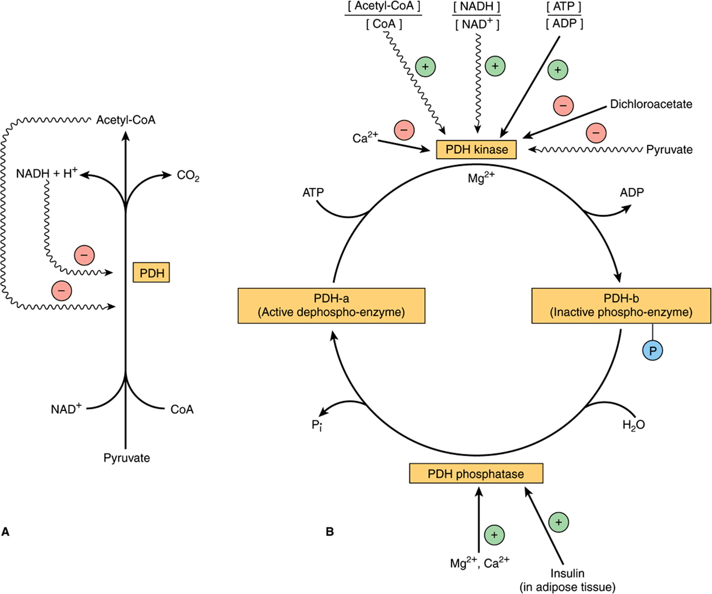
FIGURE 18–6 Regulation of pyruvatede hydrogenase (PDH). Arrows with wavy shafts indicate allosteric effects. (A) Regulation by end-product inhibition. (B) Regulation by interconversion of active and inactive forms.
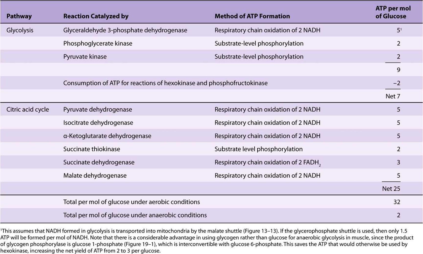
Oxidation of Glucose Yields Up to 32 Mol of ATP Under Aerobic Conditions, But Only 2 Mol When O2 Is Absent
When 1 mol of glucose is combusted in a calorimeter to CO2 and water, approximately 2870 kJ are liberated as heat. When oxidation occurs in the tissues, approximately 32 mol of ATP are generated per molecule of glucose oxidized to CO2 and water. In vivo, ΔG for the ATP synthase reaction has been calculated as approximately 51.6 kJ. It follows that the total energy captured in ATP per mole of glucose oxidized is 1651 kJ, or approximately 58% of the energy of combustion. Most of the ATP is formed by oxidative phosphorylation resulting from the reoxidation of reduced coenzymes by the respiratory chain. The remainder is formed by substrate level phosphorylation (Table 18-1).
TABLE 18–1 ATP Formation in the Catabolism of Glucose
CLINICAL ASPECTS
Inhibition of Pyruvate Metabolism Leads to Lactic Acidosis
Arsenite and mercuric ions react with the—SH groups of lipoic acid and inhibit pyruvate dehydrogenase, as does a dietary deficiency of thiamin (Chapter 44), allowing pyruvate to accumulate. Many alcoholics are thiamin deficient (both because of a poor diet and also because alcohol inhibits thiamin absorption), and may develop potentially fatal pyruvic and lactic acidosis. Patients with inherited pyruvate dehydrogenase deficiency, which can be the result of defects in one or more of the components of the enzyme complex, also present with lactic acidosis, particularly after a glucose load. Because of the dependence of the brain on glucose as a fuel, these metabolic defects commonly cause neurologic disturbances.
Inherited aldolase A deficiency and pyruvate kinase deficiency in erythrocytes cause hemolytic anemia. The exercise capacity of patients with muscle phosphofructokinase deficiency is low, particularly if they are on high-carbohydrate diets. By providing lipid as an alternative fuel, for example, during starvation, when blood-free fatty acid and ketone bodies are increased, work capacity is improved.
SUMMARY
![]() Glycolysis is the cytosolic pathway of all mammalian cells for the metabolism of glucose (or glycogen) to pyruvate and lactate.
Glycolysis is the cytosolic pathway of all mammalian cells for the metabolism of glucose (or glycogen) to pyruvate and lactate.
![]() It can function anaerobically by regenerating oxidized NAD+ (required in the glyceraldehyde-3-phosphate dehydrogenase reaction), by reducing pyruvate to lactate.
It can function anaerobically by regenerating oxidized NAD+ (required in the glyceraldehyde-3-phosphate dehydrogenase reaction), by reducing pyruvate to lactate.
![]() Lactate is the end product of glycolysis under anaerobic conditions (eg, in exercising muscle) or, in erythrocytes, when there are no mitochondria to permit the further oxidation of pyruvate.
Lactate is the end product of glycolysis under anaerobic conditions (eg, in exercising muscle) or, in erythrocytes, when there are no mitochondria to permit the further oxidation of pyruvate.
![]() Glycolysis is regulated by three enzymes catalyzing nonequilibrium reactions: hexokinase, phosphofructokinase, and pyruvate kinase.
Glycolysis is regulated by three enzymes catalyzing nonequilibrium reactions: hexokinase, phosphofructokinase, and pyruvate kinase.
![]() In erythrocytes, the first site in glycolysis for generation of ATP may be bypassed, leading to the formation of 2,3-bisphosphoglycerate, which is important in decreasing the affinity of hemoglobin for O2.
In erythrocytes, the first site in glycolysis for generation of ATP may be bypassed, leading to the formation of 2,3-bisphosphoglycerate, which is important in decreasing the affinity of hemoglobin for O2.
![]() Pyruvate is oxidized to acetyl-CoA by a multienzyme complex, pyruvate dehydrogenase, which is dependent on the vitaminderived cofactor thiamin diphosphate.
Pyruvate is oxidized to acetyl-CoA by a multienzyme complex, pyruvate dehydrogenase, which is dependent on the vitaminderived cofactor thiamin diphosphate.
![]() Conditions that involve an impairment of pyruvate metabolism frequently lead to lactic acidosis.
Conditions that involve an impairment of pyruvate metabolism frequently lead to lactic acidosis.
REFERENCES
Behal RH, Buxton DB, Robertson JG, Olson MS: Regulation of the pyruvate dehydrogenase multienzyme complex. Annu Rev Nutr 1993;13:497.
Boiteux A, Hess B: Design of glycolysis. Philos Trans R Soc Lond B Biol Sci 1981;293:5.
Fothergill-Gilmore LA: The evolution of the glycolytic pathway. Trends Biochem Sci 1986;11:47.
Gladden LB: Lactate metabolism: a new paradigm for the third millennium. J Physiol 2004;558:5.
Kim J-W, Dang CV: Multifaceted roles of glycolytic enzymes. Trends Biochem Sci 2005;30:142.
Levy B: Lactate and shock state: the metabolic view. Curr Opin Crit Care 2006;1:315.
Maj MC, Cameron JM, Robinson BH: Pyruvate dehydrogenase phosphatase deficiency: orphan disease or an under-diagnosed condition? Mol Cell Endocrinol 2006;249:1.
Martin E, Rosenthal RE, Fiskum G: Pyruvate dehydrogenase complex: metabolic link to ischemic brain injury and target of oxidative stress. J Neurosci Res 2005;79:240.
Patel MS, Korotchkina LG: Regulation of the pyruvate dehydrogenase complex. Biochem Soc Trans 2006;34:217.
Philp A, Macdonald AL, Watt PW: Lactate—a signal coordinating cell and systemic function. J Exp Biol 2005;208:4561.
Pumain R, Laschet J: A key glycolytic enzyme plays a dual role in GABAergic neurotransmission and in human epilepsy. Crit Rev Neurobiol 2006;18:197.
Rider MH, Bertrand L, Vertommen D, et al: 6-Phosphofructo-2-kinase/fructose-2,6-bisphosphatase: head-to-head with a bifunctional enzyme that controls glycolysis. Biochem J 2004;381:561.
Robergs RA, Ghiasvand F, Parker D: Biochemistry of exercise-induced metabolic acidosis. Am J Physiol 2004;287:R502.
Sugden MC, Holness MJ: Mechanisms underlying regulation of the expression and activities of the mammalian pyruvate dehydrogenase kinases. Arch Physiol Biochem 2006;112:139.
Wasserman DH: Regulation of glucose fluxes during exercise in the postabsorptive state. Annu Rev Physiol 1995;57:191.