CHEMICAL BIOLOGY
Oxygen-Activating Enzymes, Chemistry of
Corinna R. Hess*, Richard W. D. Welford* and Judith P. Klinman**, Departments of Chemistry and Molecular Cell Biology, University of California, Berkeley, California
doi: 10.1002/9780470048672.wecb431
Aerobic organisms derive the energy required for cellular processes from the conversion of dioxygen to water, which highlights the importance of dioxygen chemistry in biologic systems. The biochemistry of dioxygen is far from simple and has been the subject of intense study in a range of chemical and biologic disciplines. The reduction of dioxygen is energetically favorable; however, dioxygen is a ground state triplet, kinetically unreactive with singlet organic molecules. Nature has developed a diverse array of catalysts to overcome this kinetic barrier. These dioxygen-activating enzymes are divided into two classes: oxygenases and oxidases. Oxygenases incorporate directly at least one atom from dioxygen into the organic products of their reaction. Oxidases couple the reduction of dioxygen with the oxidation of substrate. Typically, enzymes that react with dioxygen contain transition metal ions and/or conjugated organic molecules as cofactors. The reaction with dioxygen is initiated by electron transfer from the cofactor to O2. The subsequent chemistry varies depending on both the nature of the cofactor and the protein scaffold. Here we review the fascinating chemistry of the dioxygen-activating enzymes and identify some of the common strategies and themes that have emerged from over half a century of research.
____________________
* These authors contributed equally.
** Corresponding author.
Biological Background
The introduction of an oxygen-rich atmosphere led to the evolution of enzymes capable of exploiting this diatomic gas for a variety of different chemistries. Viewing life today, we can conclude that the use of dioxygen ultimately turned out to be advantageous. Dioxygen is required by all aerobic forms of life for a variety of chemical transformations and biological processes, which are conducted by a rich and diverse set of protein catalysts (1). In addition to its crucial function in the respiratory chain, dioxygen also is involved in biosynthesis, signaling, xenobiotic metabolism, DNA repair, and biodegradation. However, a darker side exists to dioxygen biochemistry. The reactive oxygen species (ROS) formed by reduction of dioxygen species have been linked to several detrimental processes such as aging and cancer (2). These ROS can damage proteins, cell walls, and DNA. One key area of interest is how dioxygen-activating enzymes use ROS without damaging their own peptide backbone in the process.
Dioxygen Chemistry
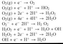
The full four-electron, four-proton reduction of O2 to two molecules of H2O is strongly exothermic (Table 1). However, most redox processes involve sequential one or two-electron transfer pathways. The first electron transfer to dioxygen is the most difficult step (Table 1) because of the loss in O-O bond strength associated with formation of superoxide (3). Most organic molecules do not possess enough reducing power to facilitate this initial reduction, and as a consequence, their one-electron oxidation by dioxygen is thermodynamically unfavorable. Moreover, dioxygen is a triplet in its ground state, whereas most organic molecules possess singlet ground states. Consequently, the direct reaction of dioxygen with organic molecules necessitates a spin conversion to the singlet excited state of O2, which has 22.5 kcal/mol more energy.
Enzymes have evolved to couple efficiently the reduction of dioxygen to the oxidation of biological compounds. Difficult C-H abstraction and O-atom insertion reactions are catalyzed by oxygen-activating enzymes under mild conditions and with high specificity.
Table 1. Reduction potentials of dioxygen and reduced oxygen species in water (3)
|
Redox reactions of O2, pH 7, 25° C |
E° (V vs. NHE) |
|
|
-0.33 -0.13 0.281 0.815 0.89 0.38 1.349 2.31 |
Cofactors used in Dioxygen Activation
Practically all known oxygen-activating enzymes employ a cofactor in the form of a metal center, a highly conjugated organic molecule, or both. Ultimately, the reactions involve the reduction of O2by two or four electrons to generate hydrogen peroxide or water. Metallocofactors and flavin molecules are powerful reductants, able to overcome the large potential associated with the formation of superoxide (Table 1), and with comparatively stable one-electron oxidized forms. The activated dioxygen products are highly reactive and readily accept additional electrons to yield the final enzymatic products.
Dioxygen activation by metalloenzymes is dominated by Fe and Cu, although a few examples of other transition metal active sites, such as Mn, have been documented. Metallocofactors bind dioxygen and its many reduced forms. The resultant metal-oxygen bonds compensate partly for the loss of bond strength upon reduction of O2. The rich spectroscopy associated with metal sites has advanced our understanding of the chemistry of dioxygen-activating enzymes. Methods such as electron paramagnetic resonance spectroscopy (EPR), X-ray absorbtion spectroscopy (XAS), and Mossbauer have been used to obtain structural information regarding the metallocofactors at various stages in the catalytic mechanisms (4).
Dioxygen-activating enzymes can be categorized according to the type of cofactor(s) employed. Although similar types of cofactors can catalyze different reactions, common structural features and reaction intermediates often exist for a given type of cofactor, as discussed below.
Iron-containing motifs for dioxygen activation
Iron-containing proteins are classified as heme, mononuclear non-heme, and binuclear non-heme enzymes. The Fe center in heme proteins is ligated by four nitrogens of a porphyrin molecule, in addition to one or two axial ligands (Fig. 1a). The reaction of FeII with O2 generates an FeIII-superoxide, in which the reduced O2 is coordinated at an axial position of the heme unit in a bent, end-on fashion.
Mononuclear non-heme iron centers with several different types of coordination environments have been identified (5-6-7). The most prevalent coordination is an octahedral or square pyramidal site with a 2His-1carboxylate ligand set coordinated to one face of an FeII center (Fig. 1b). In the resting state, the other positions are occupied by water molecules, which can be readily substituted during the reaction cycle by the substrate, the co-substrate, or O2. Another group of mononuclear non-heme iron enzymes activate their organic substrate for direct reaction with dioxygen. These enzymes contain an FeIII, in which the coordination environment differs by subfamily. For example, intradiol dioxygenases contain a trigonal bipyrimidal FeIII ligated by 2His, 2Tyr, and a hydroxide in the resting state (Fig. 1c).
The active sites of binuclear non-heme enzymes consist of two Fe atoms, separated by 3-4 A and bridged by O-atoms derived from hydroxide or carboxylate residues (Fig. 1d) (8). The iron centers can adopt 4-, 5-, or 6-coordinate geometries, with the bridging ligands bound via one or two O-atoms. The remaining coordination sites are occupied by His and Asp/Glu residues.
The reactions of iron-containing enzymes with O2 often involve high oxidation states of the metal. Generally, the initial reaction of dioxygen with both heme and mononuclear non-heme ferrous enzymes results in the formation of FeIII-superoxide intermediates. Highly reactive FeIV=O intermediates often are employed often for C-H activation. The mechanism of substrate oxidation by binuclear non-heme enzymes involves high valent, oxo-bridged species, with Fe in the +3 or +4 oxidation state.

Figure 1. X-ray crystal structures of representative motifs used in reactions with dioxygen. Amino acids and the heme cofactor are shown as sticks. Iron, copper, and water are shown as green, aquamarine, and red spheres, respectively. PDB codes are given in parentheses. (a) P450, the red stick represents a bound O2 molecule (1DO9); (b), non-heme iron, 2His-1 carboxylate (1RXF); (c) non-heme iron, intradiol dioxygenase (1DLM); (d) di-iron, methane monooxygenase, including a bound molecule of acetic acid (1MMO); (e) type 2 copper, copper amine oxidase (1A2V); (f) coupled binuclear copper, tyrosinase (1WX3); (g) reduced flavin.
Copper-containing motifs for dioxygen activation
The copper-containing enzymes can be classified as mononuclear, binuclear, or multicopper proteins (9). Unlike iron, copper cannot readily access high oxidation states and the formation of CuIII in biological systems remains controversial. Generally, CuI centers undergo one electron oxidations to activate dioxygen. Thus, the Cu-containing enzymes tend to employ multiple copper atoms or an additional cofactor for the final two or four-electron oxidation of their substrates.
Mononuclear copper enzymes capable of dioxygen activation contain a Type 2 copper center (Fig. 1e). Generally, Type 2 copper sites have a tetragonal geometry in the CuII state and can be identified by their EPR spectra (10). Reduction of the Cu site is accompanied by a loss of water molecules and a change in coordination geometry; trigonal or tetrahedral geometries are common for CuI(11). Dioxygen is thought to bind to the reduced copper site in an end-on or side-on fashion, to yield a CuII-superoxide intermediate.
A Type 3 active site, which consists of two antiferromagnetically coupled Cu atoms, ~ 3 A apart, is found in binuclear copper enzymes (10). Each copper is coordinated by three histidine residues and by one or two water molecules that serve as η2 bridging ligands (Fig. 1f). These enzymes all bind to O2 in an η2 fashion, where each O atom is bound by the two coppers. Each copper atom transfers one electron to dioxygen to yield a CuII2-peroxide intermediate (for example, “oxy state” in Fig. 3g). Proteins with Type 3 copper centers include dioxygen transport and dioxygen-activating enzymes.
The multicopper enzymes contain a trinuclear metal cluster that consists of a Type 2 copper site and a binuclear, Type 3 copper site (10). The Type 3 site of multicopper enzymes is distinct from the active site of the coupled binuclear copper enzymes described above. Although the Type 3 Cu centers are antiferromagnetically coupled, the centers are separated by ~ 5 A and are bridged by a hydroxide ligand. The multicopper oxidases also contain at least one mononuclear, Type 1, blue copper site in addition to the trinuclear Cu cluster. The Type 1 Cu site is ligated by two histidines, a cysteine, and often a weakly coordinating axial 4th ligand. Blue copper sites also are found commonly in electron transfer proteins and are defined by their intense Cys-to-metal chargetransfer transition at ~ 600 nm (11).
Dioxygen activation by flavins
A large group of enzymes react with O2 by using an organic flavin cofactor (Fig. 1g) (12, 13). The reactions can be divided into two half-reactions. First the flavin is reduced by substrate, and then the reduced flavin reacts with an electron acceptor, such as dioxygen. When O2 is the electron acceptor, the first step in the oxidative half-reaction is the rate-limiting electron transfer, which leads to the formation of a caged radical pair of superoxide anion and flavin semiquinone. The fate of the radical pair depends on whether the enzyme is an oxidase or an oxygenase and will be discussed below. The second-order reaction of a reduced free flavin with O2 in solution proceeds at a rate of 2.5 x 10² M-1 s-1. For an enzyme-bound flavin, the rate can vary between 2 M-1 s-1 and 106 M-1 s-1. It is not fully understood how enzymes with extremely similar active sites can access such a range of rates for reaction with O2.
The following sections describe specific examples of oxygenase and oxidase chemistry catalyzed by the cofactors described above. It is by no means an extensive list, but it should offer the reader a flavor of the diverse mechanisms of dioxygen activation by enzymes.
Oxygenases
The enzymes in this section can incorporate either one or two atoms from dioxygen into the organic product(s) of their reaction. It is of note that many oxygenases also can catalyze oxidase like reactions, such as desaturation and ring closure. Figure 2 shows illustrative examples of the reactions catalyzed by some of these enzymes, whereas Fig. 3 shows partial reaction mechanisms, including key intermediates.
Iron oxygenases
Heme-Fe: The cytochromes P450
The cytochromes P450 are some of the most well-studied oxygenase enzymes; their oxidation reactions, typically hydroxylations, are important for xenobiotic metabolism and biosynthesis (18, 19). Several notable differences exist between P450s, which catalyze oxygenations, and the O2 transport globins, which simply bind O2 reversibly. P450s have an axial cysteine thiolate iron ligand on the proximal side of the heme, whereas the transport globins have a histidine residue (20). Additionally, P450s have a conserved GX(E/D)T sequence motif on the distal side of the heme, which is thought to be involved in the proton donation required for cleavage of O2. In the resting state, the P450s contain a heme-ligated FeIII. Typically, the electrons for the reduction of O2 are supplied by NAD(P)H via protein redox partners. Extensive spectroscopic work has led to the observation of several of the peroxy intermediates involved in the conversion of the Fe-oxygen complex, Fem-O-O•, to the compound I + H2O (Fig. 3a). Most recently, elegant one-electron cryo reduction and EPR spectroscopy allowed the detection of the Fem-O-O2- and Fem-O-OH1- intermediates. The loss of water from the latter generates the highly oxidizing compound I, which is believed to be an FeIV=O species with radical character localized on either the porphyrin ring or thiol ligand. Compound I can abstract a hydrogen atom from an organic substrate. The substrate radical combines rapidly with the iron-ligated hydroxyl in a “radical rebound” mechanism (21). Several facets of P450 catalysis are underlying themes for some other oxygenase enzymes. The reduction of the ferric heme and the reaction with dioxygen occur only after binding of the organic substrate molecule. This “substrate-triggering” mechanism is of primary importance, as it prevents the formation of potentially damaging ROS in the absence of a suitable oxidizable substrate. Additionally, the substrate selectivity can be either broad or narrow, depending on the enzyme’s function.
Mononuclear non-heme iron oxygenases
The mononuclear, non-heme iron oxygenases can be classified into two groups: those that activate substrate for reaction with O2 and those in which the iron activates O2 directly (5, 7). Examples of enzymes that employ substrate activation mechanisms are the intradiol cleaving dioxygenases and lipoxygenases, which catalyze ring opening and hydroperoxidation, respectively (Figs. 2 and 3).
Intradiol dioxygenases prime their substrate for direct reaction with O2 by inducing radical character in the organic molecule. The iron is FeIII in the resting state, and spectroscopic studies have shown that its oxidation state does not change detectably throughout the catalytic cycle (Fig. 3b). The substrate-bound form is believed to have some semiquinone radical character and is thought to combine directly with O2. The transient peroxy substrate complex then breaks down by a Criegee-type alkyl migration to yield the products.
The FeIII center in lipoxygenase is coordinated by 3His/Asn, the protein’s C-terminal carboxylate and a hydroxide (Fig. 3c). The FeIII-OH abstracts a hydrogen atom from the substrate yielding a five-carbon delocalized radical intermediate (designated as R•) that combines directly with O2 to yield the peroxy product. Entry of O2 to the enzyme active site is believed to be controlled tightly, such that it can access only a single carbon of the substrate radical, giving strict regio- and stereospecificity (22).
The 2His-1carboxylate mononuclear, non-heme iron enzymes can be divided into subfamilies, which differ in the cosubstrate that provides the electrons for the reduction of O2 and the relative positions of the 2His-1carboxylate residues in the protein sequence (7). Even among members of the same subfamily, the only conserved residues are the 2His-1carboxylate, which indicates the use of this motif for activation of O2. Despite the conserved ligand set, the chemistry can vary greatly. In the reaction cycle of the α-ketogluturate (α-KG) dependent subfamily, it is the α-KG co-substrate that ligates the Fe, whereas the prime substrate occupies a second sphere position. The counter is observed for the extradiol catechol dioxygenases where the “prime” substrate ligates directly to the iron.
The largest subfamily in the 2His-1carboxylate group of non-heme FeII-dependent enzymes depends on α-KG as a cosubstrate (6). The substrate binding is ordered. First α-KG ligates the FeII, then the prime substrate binds adjacent to the metal site, and finally O2 binds to the metal (Fig. 3d). The ternary complex reacts intramolecularly to yield the oxidizing species. This was characterized recently as an FeIV=O by Mossbauer, Raman, and EXAFS spectroscopy, which is the first enzymatic example of a non-heme FeIV (14). As with P450s, the FeIV=O is thought to be the oxidizing intermediate in a wide variety of reactions, including hydroxylation, desaturation, and oxidative ring closure (6). The multifaceted, yet highly controlled, nature of this chemistry is exemplified brilliantly by the enzyme clavaminate acid synthase, which uses a single active site to catalyze a hydroxylation, a desaturation, and a ring closure at different stages in a single biosynthetic pathway. The α-KG-dependent enzymes, like other oxygenases, can hydroxylate aromatic amino acid side chains in the vicinity of the active site under certain conditions (7). This illustrates the enzymes’ need to control the oxidizing intermediate to minimize such deleterious side reactions. In fact, mammals are thought to require vitamin C for the reduction of FeIII, generated as a by-product of unproductive reaction cycles of α-KG enzymes.
The extradiol dioxygenases, similar to their intradiol counter parts, cleave aromatic compounds, but the position of ring opening differs (Fig. 2) (7). In the resting state, the extradiol dioxygenases contain a 2His-1carboxylate, five-coordinate FeII. Bidentate binding of the catechol to the Fe activates the metal for O2 binding. This process is analogous to the α-KG enzymes, except an additional substrate is not required to make the center five-coordinate. The FeIII superoxide formed on dioxygen binding can induce radical character in the catechol that leads eventually to ring opening.
Another subgroup of the 2His-1carboxylate family is dependent on a reduced pterin cofactor (5). They catalyze hydroxylations at the aromatic positions of amino acids in phenylalanine catabolism and hormone biosynthesis (Fig. 2). Unlike the α-KG-dependent enzymes, the pterin co-substrate does not ligate to the iron directly. In the reaction cycle, the pterin cosubstrate supplies two electrons for the heterolysis of O2 to give a yet to be characterized iron-oxygen hydroxylating species.
X-ray crystallography and magnetic circular dichroism spectroscopy have shown that, for the α-KG- and pterin-dependent mononuclear non-heme iron enzymes, the binding of the penultimate substrate promotes a change to a five-coordinate iron center. As with the P450s, this finding implies that dioxygen will not bind in the absence of an organic substrate, which prevents the build up of potentially damaging intermediates (23).

Figure 2. Some common reactions carried out by oxygenase enzymes. A * represents an oxygen atom derived from O2. The reactions listed are those thought to be the enzyme's biologically relevant reaction. Where appropriate the name of the enzyme catalyzing the example reaction is given.
Binuclear non-heme iron oxygenases
Soluble methane monooxygenase (sMMO) is the best studied binuclear non-heme iron oxygenase enzyme, largely due to its remarkable ability to hydroxylate the stable C-H (440 kJ /mol) of methane (15). sMMO is a three-component enzyme system, which consists of the di-iron hydroxylating protein, a flavin-Fe2S2 protein, and a third regulatory protein that does not contain a cofactor. The role of the flavin-Fe2S2 protein is to provide two electrons from NADPH to form the active FeII-FeIi form of the di-iron hydroxylating protein. Although incompletely understood, the regulatory protein seems to coordinate the interaction of the other two components such that uncoupled reaction cycles do not occur. The intermediates in the sMMO reaction cycle accumulate in the absence of methane, which is a feature that has allowed their spectroscopic characterization (14). Studies on the Mossbauer suggest that O2 binds initially to the binuclear Fe site forming a μ-1,2-peroxo-FeIII2 intermediate, which then decays to form the highly oxidizing intermediate termed Q (Fig. 3e). EXAFS and Mossbauer have indicated collectively that Q is a bis-(μ-oxo)-FeIV2 with a short Fe-Fe separation of 2.46 A. A strong consensus on how this intermediate compound hydroxylates methane has not yet been reached despite a wealth of studies using isotopically labeled and radical clock substrates (15). Currently, a radical rebound mechanism or a nonsynchronous concerted rearrangement remains possible.

Figure 3. Illustration of possible partial reaction cycles of some oxygenase enzymes. Water molecules and protein ligands have sometimes been omitted for clarity. (a) P450 (18); (b) intradiol dioxygenase (7); (c) lipoxygenase (7); (d) α-KG-dependent non-heme iron enzymes (14); (e) soluble methane monooxygenase (15); (f) uncoupled binuclear copper (16); (g) coupled binuclear copper; (h) flavin monooxygenases (17).
Copper oxygenases
Copper-containing oxygenases are less common than their iron counterparts, perhaps because of the greater difficulty in obtaining more reactive, higher oxidation states. Examples include dopamine β-monooxygenase (DβM), tyramine β-monooxygenase (TβM), peptidylglycine α-hydroxylating enzyme (PHM), tyrosinase, and a membrane bound form of MMO (16, 17, 24). The membrane bound form of MMO contains both a Type 3 binuclear Cu site and a mononuclear Type 2 Cu site (25). At the moment, its mechanism has not been studied in detail and will not be discussed additionally here.
Uncoupled binuclear copper oxygenases
DβM, TβM, and PHM all employ two uncoupled, Type 2 copper sites in the biosynthesis of neurotransmitters and of hormones (16). The reaction entails the hydroxylation of unactivated carbon centers, similar to the chemistry of the P450s and α-KG-dependent non-heme iron enzymes. The two metal sites of the uncoupled binuclear Cu monooxygenases are separated by more than 11 zA, which is a feature that distinguishes this family of enzymes from the binuclear Type 3 copper proteins. Each Cu site has a discrete coordination environment and serves a unique function in the hydroxylation reaction. The CuM site functions as the dioxygen activation site, whereas CuH serves as the electron transfer site, providing the additional electron required for substrate oxidation. Evidence suggests that the reaction of dioxygen with the reduced CuIM generates a CuIIM -superoxide intermediate (Fig. 3f), which subsequently abstracts a hydrogen atom from the substrate (26, 27). A methionine ligand to CuM is believed to stabilize the reduced enzyme form, such that oxygen activation is coupled strongly to the ensuing C-H activation step. The steps following C-H activation are still unresolved. A CuII-oxyl radical (CuII—O•) is a postulated, short-lived intermediate along the reaction pathway. This high energy intermediate is unprecedented in copper chemistry, but it could provide the driving force for the requisite electron transfer from CuIH to CuIIM.
Coupled binuclear copper oxygenases
A characteristic Type 3, binuclear Cu site is found in tyrosinase (10). Tyrosinase catalyzes the conversion of monophenols to orthoquinones, the formation of which is coupled to the two electron reduction of O2 and proceeds in two steps. The first step occurs via electrophilic attack on the phenol ring of the substrate and is followed by the oxidation of the di-hydroxybenzene to yield the quinone product. Thus, tyrosinase functions as both an oxygenase and an oxidase. Much of the structural information pertaining to the tyrosinase active site and various Cu-dioxygen intermediates was derived by comparison to several spectroscopic studies on model complexes (10). The resting form of tyrosinase assumes two forms: 15% of the enzyme exists in the oxy state (CuII2O22—), whereas 85% is present in the met state (both Cu sites are oxidized, but O2 is not bound). The reaction is initiated by the binding of phenolic substrate to one of the Cu atoms (CuA) in the oxy form (Fig. 3g). Substrate hydroxylation converts the active site to the met state, which binds diphenol in a bidentate fashion (as for catechol oxidase, Fig. 5c). The ensuing oxidation to yield the o -quinone generates the reduced, deoxy state (CuI2) of the enzyme. The oxy form is regenerated by the reaction with dioxygen and the catalytic cycle resumes. Alternative mechanisms, invoking the formation of CuIII intermediates or radicals, also have been postulated.
Flavin oxygenases
Several types of flavoprotein monooxygenases exist. One group catalyzes electrophilic aromatic substitution or heteroatom oxidation reactions, whereas the other group catalyzes Baeyer-Villiger-type oxidations of ketones (Fig. 2) (13, 17). In the well-studied, single-component flavoprotein monooxygenases, the flavin is reduced by a hydride delivered from a single molecule of NAD(P)H (Fig. 3h). The reduced flavin then reacts with O2 producing a peroxyflavin intermediate, which is in equilibrium with the hydroperoxyflavin form. For electrophilic substrates, as in the Baeyer-Villiger-type oxidation, it is the peroxyflavin intermediate that reacts with substrate. For hydroxylation of aromatic rings, the more electrophilic hydroperoxy intermediate is the reactive species. In the final step, the flavin is dehydrated to regenerate the resting state.
Single-component flavin monooxygenases employ different strategies to prevent the buildup of ROS. Extensive work on the p-hydroxybenzoate hydroxylase has demonstrated how protein dynamics compartmentalize different parts of the reaction (17). Reduction of the flavin and reaction with dioxygen occur in different conformations. Flavin reduction by NADPH only occurs in the presence of substrate, leading to the dioxygen reactive conformation. The Baeyer-Villiger monooxygenases and mammalian flavin monooxygenases use an alternative strategy. The flavin is reduced readily by NADPH in the absence of substrate, but the (hydro)peroxyflavin intermediate is stabilized by the bound NADP until a suitable substrate binds.
Members of the single-component flavin monooxygenase family have been shown to have wide substrate and reaction selectivities. In particular, one Baeyer-Villiger monooxygenase is known to react with over a 100 different substrates and is finding use as a tool for green biocatalysis in synthetic organic chemistry (28).
Oxidases
Oxidases couple the oxidation of an organic substrate to the two- or four-electron reduction of O2, producing H2O2 or two molecules of H2O, respectively. Oxygen atoms from dioxygen are not incorporated into the product, unlike reactions catalyzed by oxygenases. Oxidase reactions may proceed via inner-sphere or outer-sphere mechanisms.
Iron oxidases
Non-heme mononuclear iron oxidases
As mentioned, many non-heme iron enzymes also catalyze oxidase-type reactions, such as desaturation, in biological systems (7). Similar to the non-heme iron oxygenases, the reactions are thought to proceed through an FeIV=O intermediate. Two examples of enzymes that catalyze biologically interesting oxidase reactions are isopenicillin N-synthase (IPNS) and 1-aminocyclopropane-1-carboxylate oxidase (ACCO).
IPNS catalyzes a double ring closure in the formation of isopenicillin concomitant with the four-electron reduction of dioxygen to two molecules of water, without the use of any cofactors or cosubstrates other than the Fe (Fig. 4a) (7). Ligation of the substrate thiolate activates the Fe for reaction with dioxygen by converting the active site from a six-coordinate to a five-coordinate metal center. The first ring closure is believed to occur with heterolysis of FeIII-O-OH forming FeIV=O, which then closes the second ring.
ACCO breaks down 1-aminocyclopropane-1-carboxylate (ACC) in plants to form the growth hormone ethylene, hydrogen cyanide, and CO2 (Fig. 4b) (7). In the initial stages of catalysis, ACC ligates the iron, priming the system for reaction with dioxygen. Electrons for the reduction of O2 are derived from ascorbate, leading to formation of the reactive iron oxygen intermediate, possibly an FeIV=O. Although the later steps of the reaction are poorly characterized, it is thought that an FeIV=O may remove a hydrogen atom from ACC, generating a substrate radical, which breaks down to generate the gaseous product molecules.

Figure 4. Illustration of possible partial reaction cycles of some iron oxidase enzymes. (a) isopenicillin N-synthase (7); (b) 1-aminocyclopropane-1-carboxylate oxidase (7); (c) ribonucleotide reductase R2 (14).
Binuclear non-heme iron oxidases: Class I ribonucleotide reductas
Ribonucleotide reductases (RNRs) catalyze the conversion of nucleotides to deoxynucleotides and can be found in all organisms. RNRs are categorized into three classes and employ different cofactors for this process. The Class I RNRs contain a binuclear iron cofactor (29). The iron active site is located in the R2 subunit, one of two homodimeric subunits that compose the Class I RNRs, and is very similar structurally to the active site of sMMO (Fig. 1d). The binuclear iron site reacts with dioxygen, ultimately generating a tyrosyl radical, which transfers an electron across 35 A and oxidizes a cysteine residue in the R1 subunit. The resulting Cys radical abstracts a hydrogen atom from the nucleotide to initiate deoxynucleotide synthesis. The di-iron center is required by the enzyme for the initial activation of the tyrosine residue but is not necessary for catalytic activity, as electrons are shuttled back and forth between the Tyr• in R2 and the Cys• in R1. Spectroscopic methods such as EPR, XAS, and Mossbauer have been employed to characterize many of the short-lived di-iron intermediates formed on reaction of the metal site with dioxygen (14). The initial activation of dioxygen yields a peroxide-bound binuclear Fe111 center (Fig. 4c). This intermediate molecule may require protonation prior to reduction. An additional electron is transferred to the FeIII2-peroxide adduct, generating an oxo-bridged FeIII/FeIV center. The nature of the electron donor was revealed to be a tryptophan residue, which is oxidized to a cation radical in the electron transfer process. The ultimate formation of the tyrosyl radical by the Trp•/ + species has been shown to occur by several pathways. The iron active site in RNR-R2 shares several features and intermediates with sMMO. However, formation of an FeIV/FeIV species is unique to the latter enzyme and may dictate its monooxygenase ability.
Copper Oxidases
Mononuclear copper oxidases
Most copper-containing oxygenases and oxidases use multiple metal centers to conduct their biotransformations. Copper amine oxidases (CAO) and galactose oxidase (GalO), instead, employ posttranslationally derived amino acid side chains as cofactors to supply additional electrons (24).
CAOs use a quinone cofactor to catalyze the oxidation of primary amines to aldehydes (Fig. 5a) (30). The metal cofactor of CAO is a square pyramidal CuII center ligated by three histidines and by two water molecules (Fig. 1e). A distance of ~ 3 A separates the copper site and the organic cofactor. Although its catalytic function is as an oxidase, CAO also functions as an oxygenase in the self-processing mechanism of quinone cofactor biogenesis (31). The biogenesis reaction requires two equivalents of molecular dioxygen for the six-electron oxidation of an active site tyrosine to 2,4,5-trihydroxyphenylquinone (TPQ). The catalytic oxidase reaction of the CAOs can be separated into two half-reactions. In the reductive half-reaction, the amine substrate is oxidized to the corresponding aldehyde. Concurrently, TPQ is converted to the reduced aminoquinol form. Dioxygen subsequently reacts with the reduced aminoquinol, generating TPQ and one equivalent of hydrogen peroxide. Reduction of the copper center has not proven essential for oxidase activity. In selected CAOs dioxygen has been shown to be activated via direct electron transfer from the reduced TPQ (30). Kinetic studies suggest that the metal center in the oxidase reaction contributes primarily to charge stabilization of the activated dioxygen (Fig. 5a).
GalO contains a unique cofactor, which is composed of a cysteine-crosslinked tyrosine ligated to a copper center, derived from posttranslational crosslinking of the two amino acids (Fig. 5b) (32, 33). A second Tyr, two His residues, and water molecule comprise the remaining ligands of the square pyramidal CuII site. Cofactor formation is catalyzed by the enzyme itself, in a dioxygen and CuI-dependent reaction. The Tyr-Cys residue is oxidized during the posttranslational process to yield the active form of GalO, which is a CuII-Tyr-Cys• cation radical. The reaction catalyzed by GalO is the oxidation of primary alcohols to their corresponding aldehydes. The oxidation of substrate is coupled to the two-electron reduction of dioxygen to H2O2. Kinetic studies support the oxidation of alcohols via a ping-pong mechanism. In the reductive half-reaction, a hydrogen atom is abstracted from the C-a position of the metal bound alcohol to produce CuI and the reduced cysteinyl-tyrosine. In the oxidative half-reaction (Fig. 5b), dioxygen is believed to bind directly to the reduced Cu site, displacing the water molecule. The Tyr-Cys ligand supplies the additional electron that is required for the two-electron reduction of dioxygen to peroxide, and for the regeneration of the cofactor radical.

Figure 5. Illustration of possible partial reaction cycles of some copper- and flavin-dependent oxidase enzymes. (a) Copper amine oxidase 30, 31; (b) galactose oxidase (32); (c) catechol oxidase (10); (d) multicopper oxidases (10); (e) flavin oxidases (30); (f) cytochrome c oxidase (38).
Binuclear copper oxidases
The active site of the catechol oxidases is virtually identical to the Type 3 binuclear Cu site found in tyrosinases (vide ultra) (10, 34). In contrast to the tyrosinases, catechol oxidases do not exhibit monooxygenase activity and are capable only of the second reaction, the oxidation of diphenols to quinones. The resting form of catechol oxidases lies exclusively in the met state. According to the accepted mechanism, two molecules of catechol are oxidized on binding to either the reduced, deoxy form or the oxy form. Dioxygen reacts with the reduced form after product release to yield the CuII2-peroxide adduct and allow binding of the next substrate molecule (Fig. 5c). The difference in reactivity toward O2 for the Type 3 copper centers in tyrosinases, catechol oxidases, and hemocyanins has been attributed to the partial or the complete occlusion of the substrate binding site in the latter two enzyme families. The degree of flexibility around the copper active site also has been cited as a possible factor (35).
Multicopper oxidases
The multicopper oxidases couple the one-electron or two-electron oxidation of their substrates to the four-electron reduction of dioxygen to water (36). The reaction with substrate can proceed via an outer-sphere or an inner-sphere mechanism, and as a result, the substrate specificity varies substantially among the enzymes. The best-characterized enzymes are laccase, ascorbate oxidase, and ceruloplasmin. Radical phenol and amine species formed by laccase and ascorbate oxidase react further via polymerization reactions with other organic molecules, or disproportionate to generate the final biological products. The substrate for ceruloplasmin is FeII, which is oxidized to FeIII. Four substrate molecules transfer electrons sequentially to the Type 1 Cu site, which shuttles three electrons to the trinuclear cluster to generate the fully reduced enzyme form. Dioxygen is activated by the trinuclear copper cluster, which obtains two electrons from the Type 3 site (Fig. 5d). The Type 2 center is required for dioxygen activation but remains reduced at this stage in the reaction. The hydroperoxide is bound to the trinuclear cluster near the Type 3 binuclear Cu center, but both the Type 2 and the binuclear copper centers contribute significant electron density to the reduced oxygen molecule (37). The bound peroxide is reduced further by two electrons, one from the Type 2 Cu center and one from the distant Type 1 Cu site, forming the native intermediate. The native intermediate is believed to contain an oxide coordinated by the three Cu atoms of the trinuclear cluster. This species reacts with the substrate in the catalytic cycle.
Flavin Dependent Oxidases
Flavoprotein oxidases can conduct a variety of oxidation reactions, such as the conversion of alcohols and amines to aldehydes (12). Typically, the organic substrate provides an equivalent of hydride to reduce the flavin. As mentioned previously, the rate-limiting step in the reaction of the reduced flavin with dioxygen is the initial electron transfer to form a superioxide anion (30). Unlike the flavin oxygenases, a hydroperoxyflavin intermediate has never been detected for the flavin oxidases during catalysis. Instead, the mechanism is thought to proceed by two sequential one-electron transfers forming H2O2 (Fig. 5e). The lack of a solvent deuterium isotope effect in kcat/Km(O2) for glucose oxidase has provided evidence that proton transfer is not rate-limiting for the reduction of dioxygen.
Cytochrome c Oxidase
Cytochrome c oxidase is a vital enzyme in the respiratory pathway of most aerobic organisms (38, 39). The enzyme couples the four-electron reduction of dioxygen to the generation of a proton gradient and the resulting synthesis of ATP, which is the primary source of energy for all cellular processes. Cytochrome c oxidase contains two iron-porphyrin units (heme a and heme a3) and two mononuclear Cu sites (CuA and CuB). Heme a and CuA serve as electron transfer sites, whereas heme a 3 and CuB form a binuclear metal site that activates dioxygen. CuB is coordinated by three His residues and is located 5 A from the Fe center of heme a3. A tyrosine residue is bound covalently to one CuB His ligand and is believed to be critical to the four-electron reduction of O2. The first intermediate generated upon the reaction of dioxygen with the reduced CuIB-FeIIa3 center has been identified, based on resonance Raman spectroscopic studies, as an FenIIa3-O2, although a peroxide bridged FeIIIa3-O2 2--CuIIB species has not been ruled out entirely (Fig. 5f) (38). The heme-bound dioxygen molecule is reduced rapidly by four electrons, two of which are obtained from the iron center and one each obtained from CuB and the active site tyrosine. The resultant intermediate Pm consists of an FeIV=O, CuII, and a tyrosyl radical, as deduced from Raman and EPR spectroscopy (40, 41). A series of proton and electron transfer events regenerates the resting, fully oxidized form of cytochrome c oxidase. The electrons for this process are derived from cytochrome c and shuttled through the CuA and heme a sites, during which protons are pumped across the cell membrane. Thus, cytochrome c oxidase functions as an electron-coupled proton pump.
Conclusions
The mechanisms of the oxygenases and oxidases detailed here represent some of the numerous strategies employed by enzymes to overcome the kinetic barrier for reaction of organic molecules with dioxygen. This list is far from exhaustive; new reactions are discovered continually, and many intermediates in more established systems have not yet been characterized. A key feature of the dioxygen-activating enzymes is their ability to form highly ROS using carefully tuned cofactors. The enzymes prevent the release of ROS at a stage in the catalytic cycle when damage to the protein or the wider cellular environment could occur as a consequence. Efforts to mimic nature’s dioxygen chemistry with synthetic inorganic complexes often are only partially successful (7, 42). Many biomimetic complexes will react with dioxygen, but the resultant M-O bonds are either too stable to catalyze the subsequent chemistry or the products are too unstable and lead to undesirable side reactions.
Although great strides have been made to understand dioxygen activation by enzymes, many questions remain. The relationship between protein structure and enzyme catalysis is not well understood. Changes in the active site, beyond the primary coordination sphere, lead to altered cofactor redox properties. An identical cofactor often is employed by different enzymes to carry out dissimilar chemistry. The protein fold tunes the reactivity of these sites through electrostatic effects, control of solvent and substrate access, and by carefully organizing the orientation of substrates around the active site. The role of protein dynamics in tuning enzyme reactivity has become the focal point of recent studies, as well. These aspects of enzyme catalysis present the primary difficulties in the design of synthetic complexes that function as protein mimics.
References
1. Hamilton GA. Mechanisms of Biological Oxidations. In: Biological Oxidation Systems. 1990. Channa Reddy C, Hamilton GA, Madyastha KM, eds. Academic Press Inc., San Diego, CA.
2. Valko M, Izakovic M, Mazur M, Rhodes CJ, Telser J. Role of oxygen radicals in DNA damage and cancer incidence. Mol. Cell. Biochem. 2004; 266:37-56.
3. Sawyer DT. The chemistry and activation of dioxygen species in biology. In: Oxygen Complexes and Oxygen Activation by Transition Metals. 1988. Martell AE, Sayer DT, eds. Plenum, New York.
4. Solomon EI, Lever ABP. Inorganic Electronic Structure and Spectroscopy. 1999. Wiley-Interscience, New York.
5. Abu-Omar MM, Loaiza A, Hontzeas N. Reaction mechanisms of mononuclear non-heme iron oxygenases. Chem. Rev. 2005; 105:2227-2252.
6. Clifton IJ, McDonough MA, Ehrismann D, Kershaw NJ, Granatino N, Schofield CJ. Structural studies on 2-oxoglutarate oxygenases and related double-stranded beta-helix fold proteins. J. Inorg. Biochem. 2006; 100:644-669.
7. Costas M, Mehn MP, Jensen MP, Que L. Dioxygen activation at mononuclear nonheme iron active sites: enzymes, models, and intermediates. Chem. Rev. 2004; 104:939-986.
8. Kurtz DM. Structural similarity and functional diversity in diiron-oxo proteins. J. Biol. Inorg. Chem. 1997; 2:159-167.
9. Holm RH, Kennepohl P, Solomon EI. Structural and functional aspects of metal sites in biology. Chem. Rev. 1996; 96:2239-2314.
10. Solomon EI, Chen P, Metz M, Lee SK, Palmer AE. Oxygen binding, activation, and reduction to water by copper proteins. Angew. Chem. Int. Ed. 2001; 40:4570-4590.
11. Gray HB, Malmstrom BG, Williams RJP. Copper coordination in blue proteins. J. Biol. Inorg. Chem. 2000; 5:551-559.
12. Massey V. Activation of molecular-oxygen by flavins and flavoproteins. J. Biol. Chem. 1994; 269:22459-22462.
13. Mattevi A. To be or not to be an oxidase: challenging the oxygen reactivity of flavoenzymes. Trend. Biochem. Sci. 2006; 31:276-283.
14. Bollinger JM, Krebs C. Stalking intermediates in oxygen activation by iron enzymes: Motivation and method. J. Inorg. Biochem. 2006; 100:586-605.
15. Merkx M, Kopp DA, Sazinsky MH, Blazyk JL, Muller J, Lippard SJ. Dioxygen activation and methane hydroxylation by soluble methane monooxygenase: a tale of two irons and three proteins. Angew. Chem. Int. Ed. 2001; 40:2782-2807.
16. Klinman JP. The copper-enzyme family of dopamine β-mono-oxygenase and peptidylglycine α-hydroxylating monooxygenase: resolving the chemical pathway for substrate hydroxylation. J. Biol. Chem. 2006; 281:3013-3016.
17. Ballou DP, Entsch B, Cole LJ. Dynamics involved in catalysis by single-component and two-component flavin-dependent aromatic hydroxylases. Biochem. Biophys. Res. Comm. 2005; 338:590-598.
18. Denisov IG, Makris TM, Sligar SG, Schlichting I. Structure and chemistry of cytochrome P450. Chem. Rev. 2005; 105:2253-2277.
19. Sligar SG, Makris TM, Denisov IG. Thirty years of microbial P450 monooxygenase research: Peroxo-heme intermediates - the central bus station in heme oxygenase catalysis. Biochem. Biophys. Res. Comm. 2005; 338:346-354.
20. Green MT, Dawson JH, Gray HB. Oxoiron(IV) in chloroper-oxidase compound II is basic: Implications for P450 chemistry. Science 2004; 304:1653-1656.
21. Groves JT. High-valent iron in chemical and biological oxidations. J. Inorg. Biochem. 2006; 100:434-447.
22. Knapp MJ, Seebeck FP, Klinman JP. Steric control of oxygenation regiochemistry in soybean lipoxygenase-1. J. Am. Chem. Soc. 2001; 123:2931-2932.
23. Solomon EI, Decker A, Lehnert N. Non-heme iron enzymes: contrasts to heme catalysis. Proc. Natl. Acad. Sci. 2003; 100:3589-3594.
24. Klinman JP. Mechanisms whereby mononuclear copper proteins functionalize organic substrates. Chem. Rev. 1996; 96:2541-2561.
25. Lieberman RL, Rosenzweig AC. Crystal structure of a membrane-bound metalloenzyme that catalyses the biological oxidation of methane. Nature 2005; 434:177-182.
26. Chen P, Solomon EI. Oxygen activation by the noncoupled binuclear copper site in peptidylglycine α-hydroxylating monooxygenase. Reaction mechanism and role of the noncoupled nature of the active site. J. Am. Chem. Soc. 2004; 126:4991-5000.
27. Evans JP, Ahn K, Klinman JP. Evidence that dioxygen and substrate activation are tightly coupled in dopamine β-mono-oxygenase. Implications for the reactive oxygen species J. Biol. Chem. 2003; 278:49691-49698.
28. Kamerbeek NM, Janssen DB, van Berkel WJH, Fraaije MW. Baeyer-Villiger monooxygenases, an emerging family of flavin-dependent biocatalysts. Ad. Syn. Cat. 2003; 345:667-678.
29. Stubbe J, Nocera DG, Yee CS, Chang MCY. Radical initiation in the class I ribonucleotide reductase: long-range proton-coupled electron transfer. Chem. Rev. 2003; 103:2167-2201.
30. Mure M, Mills SA, Klinman JP. Catalytic mechanism of the topa quinone containing copper amine oxidases. Biochemistry 2002; 41:9269-9278.
31. DuBois JL, Klinman JP. Mechanism of post-translational quinone formation in copper amine oxidases and its relationship to the catalytic turnover. Arch. Biochem. Biophys. 2005; 433:255-265.
32. Whittaker JW. The radical chemistry of galactose oxidase. Arch. Biochem. Biophys. 2005; 433:227-239.
33. Firbank SJ, Rogers M, Hurtado-Guerrero R, Dooley DM, Halcrow MA, Phillips SEV, Knowles PF, McPherson MJ. Cofactor processing in galactose oxidase. Biochem. Soc. Trans. 2003; 31:506-509.
34. Gerdemann C, Eicken C, Krebs B. The crystal structure of catechol oxidase: New insight into the function of type-3 copper proteins. Acc. Chem. Res. 2002; 35:183-191.
35. Matoba Y, Kumagai T, Yamamoto A, Yoshitsu H, Sugiyama M. Crystallographic evidence that the dinuclear copper center of tyrosinase is flexible during catalysis. J. Biol. Chem. 2006; 281:8981-8990.
36. Solomon EI, Sundaram UM, Machonkin TE. Multicopper oxidases and oxygenases. Chem. Rev. 1996; 96:2563-2605.
37. Yoon J, Mirica LM, Stack TDP, Solomon EI. Variable-temperature, variable-field magnetic circular dichroism studies of tris-hydroxy- and μ(3)-oxo-bridged trinuclear Cu(II) complexes: Evaluation of proposed structures of the native intermediate of the multicopper oxidases. J. Am. Chem. Soc. 2005; 127:13680-13693.
38. Yoshikawa S. Structural chemical studies on the reaction mechanism of cytochrome c oxidase. In: Biophysical and Structural Aspects of Bioenergetics. 2005. Wikstrom M, ed. RSC publishing, Cambridge, UK.
39. Ferguson-Miller S, Babcock GT. Heme/copper terminal oxidases. Chem. Rev. 1996; 96:2889-2907.
40. Babcock GT. How oxygen is activated and reduced in respiration. Proc. Natl. Acad. Sci. 1999; 96:12971-12973.
41. Budiman K, Kannt A, Lyubenova S, Richter OMH, Ludwig B, Michel H, MacMillan F. Tyrosine 167: the origin of the radical species observed in the reaction of cytochrome c oxidase with hydrogen peroxide in Paracoccus denitrificans. Biochemistry. 2004; 43:11709-11716.
42. Mirica LM, Ottenwaelder X, Stack TDP. Structure and spectroscopy of copper-dioxygen complexes. Chem. Rev. 2004; 104:1013-1045.