CHEMICAL BIOLOGY
Inositol Phospholipids, Biosynthesis and Biological Functions of
Karol S. Bruzik, Department of Medicinal Chemistry and Pharmacognosy, University of Illinois at Chicago, Chicago, Illinois
doi: 10.1002/9780470048672.wecb447
Phosphatidylinositol phosphates are cellular signaling molecules that came to prominence in the mid-1980s, after inositol trisphosphate was discovered as a key intracellular second messenger generated from phosphatidylinositol 4,5-bisphosphate in response to stimulation of a wide range of extra-cellular receptors. Since then, many novel inositol phosphates and phospholipids have been discovered, and their cellular functions have been studied. The roles of these molecules encompass regulation of many processes indispensable to organism homeostasis. On the cellular level, the processes include regulation of cell growth and differentiation, cell motility and invasiveness, vesicular trafficking, protein targeting to specific cell compartments. This review focuses on biosynthesis and biological roles of phosphatidylinositol phospholipids.
One of the most significant discoveries of the last two decades was the finding that multiple cellular signal transduction pathways are mediated by inositol phospholipids (PIPn) and that inositol phosphates are intracellular second messengers.1 Because of the sheer complexity, studies of the signaling function of these molecules remain an important field of biomedical research. Once deemed obscure minor components of biological membranes, inositol phospholipids are now known to be involved in the transduction of the vast array of extracellular signals, which include neurotransmitters, growth hormones, and photons (1, 2). Because of the broad scope of these signaling events, several thousand research publications related to inositol phospholipids are published every year (3). Much of this activity is enabled by the availability of phosphoinositides because of the success in developing synthetic methodologies that lead to these compounds during the last 20 years (4, 5).
1As of 05/2007, the Chemical Abstract Service database included ca. 140,000 entries containing the “inositol” keyword.
Structure of Inositol Phospholipids
Overall, nine major inositol phospholipids (1-9) (Fig. 1) have been identified in cellular signaling pathways, and more are likely to be discovered. Of two possible enantiomers of the unsymmetrically substituted inositol residue, only the molecules in which the phosphatidate residue is attached to the D-1 position of myo-inositol are active physiologically (6, 7). Most naturally occurring PIPns that participate in signal transduction pathways in mammalian cells feature inositol ring phosphorylated at the 3-, 4-, and/or 5-positions. The addition or removal of the phosphate residue at the inositol ring is a simple biochemical process, yet given the large number of possible hydroxylation sites, it generates molecules that are characterized by a different net charge and feature distinct geometric distribution of the individual negative charges. In addition, the net negative charge of these molecules can be modulated easily by slight differences in pH of a local environment. Rapid generation of such a molecular array and facile interconversions between its members may be the basis for the omnipresence of inositol phospholipids in the signaling phenomena. The molecules that are nonphosphorylated at any of the 3-, 4- and 5-positions can also be glycosylated at the 6-position (GPI) (8). These phosphatidylinositols serve a multitude of other functions such as protein and protozoan VSG anchoring (9), and serving as precursors for the putative insulin mediators (10).
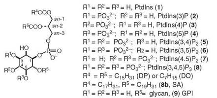
Figure 1. Structures of PIPn 1-9. For stereospecific numbering of the glycerol moiety, see Reference 6. For stereospecific numbering of myo-inositol residue, see References (5) and (7).
Regarding the lipid composition of phosphatidylinositols, the mammalian PIPn carry stearic and arachidonic acid esters at the glycerol sn-1 and sn-2-positions, respectively (11). The composition of inositol phospholipids isolated from plants varies from those present in mammalian tissues because they contain linoleoyl residue at the sn-2-position and palmitoyl residue at the sn-1-position.
In principle, only three members of PIPn family including phosphatidylinositol (1), phosphatidylinositol 4-phosphate (3), and 4,5-bisphosphate (7), are available in sufficient quantities by isolation from natural sources. Other phosphoinositides, although they play important roles in cell physiology, are formed transiently or are present in biological sources at only very low concentrations, which makes preparative isolation of these compounds unfeasible. To date, all known naturally occurring phosphoinositides, with the exception of putative prostaglandyl inositol phosphate (12) but including glycosylated phosphatidylinositols (13), have been synthesized (4), with some methodology published from the Department of Medicinal Chemistry and Pharmacognosy laboratory at the University of Illinois (14). Because of the synthetic expediency, most available synthetic inositol phospholipids are supplied as unnatural analogs that contain the saturated fatty acid chains, with either a long (dipalmitoyl) or shorter chains (dioctanoyl or dihexanoyl). These saturated analogs are not only easier to synthesize, but also are more stable chemically because they do not undergo peroxidation reactions common to unsaturated fatty acids. In addition, synthetic methods have been developed toward phosphatidyl inositide analogs that carry fluorescent, biotin, and photoaffinity residues to identify the various phosphoinositide binding proteins and to study spatiotemporal aspects of inositol-signaling pathways (15, 16).
Principal Biosynthetic and Metabolic Pathways
In plants, inositol is biosynthesized from glucose 1-phosphate via inosose 1-phosphate, which is reduced into 1D-inositol 3-phosphate (INO1 pathway). After dephosphorylation by inositol phosphate monophosphatase, myo-inositol is fed into biosynthesis of inositol phospholipids via a Kennedy-like sequence. In mammalian organisms, inositol is acquired from the diet and is also biosynthesized via INO1. The latter pathway, rather than the inositol phosphate phosphatase, seems to be a more promising target of antipsychotic drugs (17, 18) (see Fig. 2).

Figure 2. Principal metabolic pathways of inositol phospholipids.
Phosphatidylinositol
Phosphatidylinositol (PI, 1) is the most abundant member of the PIPn family and is a precursor to all known PIPns. This compound is biosynthesized from myo-inositol and CDP-diacylglycerol by phosphatidylinositol synthetase localized in the endoplasmic reticulum (19). The PI metabolism involves sequential phosphorylations at the 3 (20), 4- (21), and 5-positions (22) to provide compounds (2-8). In addition, PI is subject to degradation by phospholipases C and phospholipases A2. Although most mammalian phospholipases C prefer PI-4,5-P2 and PI-4-P (23), isolated reports of PI-specific phospholipases in mammalian tissues exist (24). In contrast to mammalian PI-PLC, the bacterial enzymes display strict specificity for un- phosphorylated PI (25). Generally, PIPn that bear a phosphate group at the inositol 3-position are regarded as resistant to cleavage by all PLC isozymes (26). PI seems also refractive to hydrolysis by two major isozymes of mammalian PLD (27). PI is also subject to the deacylation reaction by sequential action of phospholipase A and lysophospholipase that generate glycerophosphoinositols (28), which was recently shown to inhibit invasion of cancer cells (29).
Phosphatidylinositol 3-phosphate
Phosphatidylinositol 3-phosphate (PI-3-P, 2) is a rare phospholipid generated from PI by the action of Class II and Class III PI-3 kinase (PI-3K) (30, 31) and via hydrolysis of the 5-phosphate from PI-3,5-P2 by phosphatidylinositol phosphate 5-phosphatases SHIP (32).
Phosphatidylinositol 4-phosphate
Phosphatidylinositol 4-phosphate (PI-4-P, 3) is the second most abundant inositol phospholipid in biological membranes. It is a product of phosphorylation by four different species of PI 4-kinases (21), and it is a precursor to PI-4,5-P2 and PI-3,4,5-P3. These kinases are the two inositol lipids whose biological roles are understood best. The biological function of PI-4-P is not well known; however, the studies in yeast indicate its role extends beyond the substrate for PI 5-kinase (21), such as regulation of vesicular trafficking and protein secretion from Golgi. PI-4-P is removed by the subsequent phosphorylation to PI-4,5-P2 or dephosphorylation to PI by ER-localized phosphatase (21).
Phosphatidylinositol 5-phosphate
Phosphatidylinositol 5-phosphate (PI-5-P, 4) is formed by the hydrolysis of 3-phosphate from PI-3,5-P2 by myotubularin, MTM1 (33). This molecule can be removed either by phosphorylation into PI-4,5-P2 by phosphorylation with the P-form of PI-4K (34) or by dephosphorylation of PI-5-P by the novel form of PTEN-like proteins with selectivity toward phosphatidylinositol phosphorylated at the 5-position (35). Recent studies suggest a role for PI-5-P in a variety of cellular events, such as tumor suppression, response to bacterial invasion (35), and control of osmotic pressure (36). This phospholipid has been shown recently to function as a second messenger that binds to an Arabidopsis homolog of trithorax, which suggests that it may have a regulatory function that connects lipid signaling with nuclear functions (37).
Phosphatidylinositol 3,5-bisphosphate
Phosphatidylinositol 3,5-bisphosphate (PI-3,5-P2, 6) is the low abundance, newest member of PIPn family (33). It is involved in mediation of several cellular processes such as vacuolar homeostasis, membrane trafficking, and vesicular protein sorting (36, 38). The recently discovered PI-3,5-P2 effectors include a family of P-propeller, epsin, and CHMP protein families (39). The importance of PI-3,5-P2 in human physiology is demonstrated by its role in insulin signaling, myotubular myopathy, and corneal dystrophy (38).
Phosphatidylinositol 3,4-bisphosphate
Phosphatidylinositol 3,4-bisphosphate (PI-3,4-P2, 5) is biosynthesized by phosphorylation of PI-4-P at the 3-position (2a), by dephosphorylation of PI-3,4,5-P3 at the 5-position by the SHIP phosphatase (39, 40), and by phosphorylation of PI-3-P by the Type II 4-kinase (41). The significance of this lipid is underscored by the association between SHIP2 gene polymorphism and type 2 diabetes mellitus (39). Therefore, SHIP2 constitutes an important target for treatment of both type 2 diabetes and obesity (40).
Phosphatidylinositol 4,5-bisphosphate
Phosphatidylinositol 4,5-bisphosphate (PI-4,5-P2, 7) is the third most abundant inositol phospholipid in biological membranes. The predominant biosynthetic pathway is via phosphorylation of PI-4-P by a 5-kinase (1h). This inositol lipid is best known for participating in receptor-mediated cleavage by mammalian phospholipases C-β, -γ, -δ, and -ε to produce inositol 1,4,5-trisphosphate second messenger (41, 42), and for initiating extremely complex metabolic pathways of inositol phosphates, which include formation of several new second messengers, such as inositol 1,3,4,5-tetrakisphosphate, 1,3,4-trisphosphate, and 3,4,5,6-tetrakisphosphate (1e, 43). The biological role of PI-4,5-P2 is much larger because it is a substrate for Type I 3-kinases that ultimately provide PI-3,4,5-P3 as another important signaling molecule (44). In addition, PI-4,5-P2 is recognized specifically by pleckstrin homology domains of many important proteins, which include several enzymes [e.g., phospholipase D (45)], which provides an interesting crosstalk between the PLC and PLD lipolytic activities. Another example of the interrelationship between phospholipases is the regulation of cPLA2 by PI-4,5-P2 (46). Furthermore, association of certain transmembrane receptors with PI-4,5-P2 affects their functional activity [e.g., interaction of vanilloid receptor with PIP2 is necessary for receptor desensitization (47)]. Therefore, PI-4,5-P2 plays and important role in both human health and disease (48).
Phosphatidylinositol 3,4,5-trisphosphate
Phosphatidylinositol 3,4,5-trisphosphate (PI-3,4,5-P2, 8) is the most phosphorylated inositol phospholipid, and it is one whose function is related to cell growth, survival, and differentiation (49, 50). The proper physiological levels of PI-3,4,5-P3 are maintained in a major part by phosphorylation of PI-4,5-P2 by the Type I phosphatidylinositol 3-kinases and by hydrolysis of two 3-phosphoinositide phosphatases: PTEN and SHIP (20). The latter activities lead to the regeneration of PI-4,5-P2 and to formation of another second messenger, PI-3,4-P2, respectively (39, 40). Strong evidence suggests that PI-3,4,5-P3 is an important player of signaling pathways in the nucleus. Recent results also indicate that nuclear translocation of cell surface receptors could activate nuclear PI-3K, which suggests a new pathway of signal transduction (51). The most important cellular function of PIP3 at the moment seems to be its strong interaction with the PH domain of protein kinase Akt. Binding of PIP3 causes translocation of Akt to the plasma membrane where it becomes phosphorylated and activated (50). The activated Akt then phosphorylates downstream cellular proteins that promote cell proliferation and survival, which results ultimately in tumorigenesis. Therefore, the proper concentration of PI-3,4,5-P3 is pivotal to cell homeostasis, and its elevated levels promote tumor formation (50).
Phosphoinositide-Binding Protein Domains
A major advance in understanding signaling roles of phosphatidylinositols has been the discovery of several highly conserved protein domains whose function is to bind head groups of specific phosphatidylinositol phosphates. Such “cut and paste” modules are attached into a diverse array of multidomain proteins, and they enable recruitment of such proteins to specific regions in cells via binding to plasma or to intracellular membranes enriched with such phosphoinositides (52, 53). As a result, phosphoinositides can act as signal mediators in a spatially- and temporally-defined manner, and they can control various intracellular events such as cytoskeletal rearrangement and membrane trafficking. The pleckstrin homology (PH) domain was the first identified phosphoinositide-binding domain. It contains the largest number of members and is associated with the formation of signaling complexes on the plasma membrane. Recent studies identified other novel phosphoinositide-binding domains such as FYVE, Phox homology (PX), and epsin N-terminal homology (ENTH), which attest even more to the functional versatility of phosphoinositides (54). The number of known domains or modules that bind phosphoinositides has increased dramatically over the past few years. Structural analysis of interactions of inositol phospholipids with phosphoinositol-binding domains has provided significant insight into the mechanism of membrane recruitment by the different cellular phosphoinositides. Thus, the domains that target only the rare (3-phosphorylated) phosphoinositides must bind with high affinity and specificity. In the case of certain PH domains (which bind PI-3,4,5-P3 and/or PI-3,4-P2), this binding is achieved exclusively by headgroup interactions. In contrast, the PI-3-P-targeting PX and FYVE domains require an additional membrane-insertion and/or oligomerization component. Domains that target PI-4,5-P2, which is 25-fold more abundant than other phosphatidylinositol tris- and bis-phosphates, do not require the same stringent affinity and specificity and tend to be more diverse in structure. The mode of phosphoinositide binding by different domains also seems to reflect their distinct functions. For example, PH domains that serve as simple targeting domains recognize only the phosphoinositide head- groups. By contrast, certain other domains, notably the epsin ENTH domain, seem to promote bilayer curvature by inserting into the membrane on binding (55, 56). Structural specificity of the phosphoinositide binding domains is listed in Table 1.
In summary, transmembrane and intracellular signaling with phosphatidylinositol phosphates is a dynamically expanding field. The progress in this area is critical to understanding the intricate spatiotemporal effects of generation of these small molecular mediators in response to extracellular signals on the functional roles of intracellular proteins.
Table 1. Recognition of inositol phospholipids by specific phosphoinositide-binding domains
|
Phosphoinositide |
PI-3P |
PI-4P |
PI-5P |
PI-3,4-P2 |
PI-3,5-P2 |
PI-4,5-P2 |
PI-3,4,5-P3 |
|
Phosphoinositide- |
PX |
PH |
PX |
PH |
PH |
PH PX |
PH PX |
|
binding domain |
FYVE |
E/ANTH |
|
|
|
E/ANTH |
|
1. Seminal reviews on phosphoinositide metabolism: (a) Berridge, M. J.; Irvine, R. F. Nature 1984; 312:315-321.
(b) Hokin, R. H. Ann. Rev. Biochem. 1985; 54:205-235.
(c) Michell, R. H. Nature 1986; 319:176-177;
(d) Majerus, P. W.; Conolly, T. M.; Bansal, V. S.; Inhorn, R. C.; Ross, T. S.; Lips, D. L. J. Biol. Chem. 1988; 263:3051-3054;
(e) Berridge, M. J. Ann. Rev. Biochem. 1987; 56:159-193;
(f) Berridge, M. J.; Irvine, R. F. Nature 1989; 341:197-205;
(g) Clark, E. A.; Brugge, J. S.; Science 1995; 268:233-239.
(g) Duckworth, B. C.; Cantley, L. C. In Handbook of Lipid Research, Bell, R. M.; Exton, J. H.; Prescott, S. M. Eds., Plenum Press, New York, 1996, Vol. 8, p. 125.
(h) Tolias, K. F.; Cantley, L. C. Chem. Phys. Lipids 1999; 98:69-77.
2. Specific roles of phosphoinositides: (a) Rameh, L. E.; Cantley, L. C.; J. Biol. Chem. 1999; 274:8347-50.
(b) Hinchliffe, K. A.; Ciruela, A.; Irvine, R. F. Biochim. Biophys. Acta 1998; 1436:87-104.
(c) Brown, F. D.; Rozelle, A. L.; Yin, H. L.; Balla, T.; Donaldson, J. G. J. Cell. Biol. 2001; 154:1007-17.
(d) Takenawa. T.; Itoh, T. Biochim. Biophys. Acta. 2001; 1533:190-206.
(e) Exton, J. H.; Annu. Rev. Pharmacol. Toxicol. 1996; 36:481-509.
(f) Cremona, O.; De Camilli, P. J. Cell. Sci. 2001; 114:1041-52.
(g) Jones, D. R.; Varela-Nieto, I. Mol. Med. 1999,., 505-14.
3. As of 05/2007 Chemical Abstract Service database contained ca. 140,000 entries containing the ’’inositol” keyword.
4. Reviews on synthesis of phosphoinositides: (a) Potter, B. L. V. Nat. Prod. Rep. 1990, 1-24.
(b) Billington, D. C. Chem. Soc. Rev. 1989; 18:83-122.
(c) Potter, B. V. L.; Lampe, D. Angew. Chem. Int. Ed. Engl. 1995; 34:1933-1972.
(d) Billington, D. C. (1993) “The Inositol Phosphates: Chemical Synthesis and Biological Significance”, VCH, Weinheim;
(e) Prestwich, G. D. Acc. Chem. Res. 1996; 29:503-513.
5. (a) Inositol Phosphates and Derivatives. Synthesis, Biochemistry, and Therapeutic Potential, Reitz, A. B. Ed., ACS Symp. Ser. 1991, 463.
(b) Phosphoinositides: Chemistry, Biochemistry and Biomedical Applications ”, Bruzik, K. S. Ed., ACS Symp. Ser. 1998, 718.
6. IUPAC-IUB Commission on Biochemical Nomenclature (CBN). Chem. Phys. Lipids 1968; 2:156-67.
7. Parthasarathy, R., Eisenberg, F. Jr. Biochem. J. 1986; 235:313-2.
8. Ferguson, M. A. J. Cell. Sci. 1999; 112:2799-809.
9. Zacks, M. A., Garg, N. Mol. Membr. Biol. 2006; 23:209-25.
10. Larner, J. Int. J. Exp. Diabetes Res. 2002,., 47-60.
11. Allan, D., Cockroft, S. Biochem. J., 1983; 213:555-7.
12. Shashkin, P. N.; Wasner, H. K.; Ortmeyer, H. K.; Hansen, B. C. Diabetes Metab. Res. Rev. 2001; 17:273-84.
13. Baeshlin, D. K., Chaperon, A. R., Green, L. G., Hahn, M. G., Ince, S. J., Ley, S. V. Chemistry 2000; 6:172-86.
14. Kubiak, R. J., Bruzik, K. S. J. Org. Chem. 2003;68:960-968; Bruzik, K. S. NATO Symposium Series: Chemical Probes in Biology, Schneider, M. P. Ed., 2003, 51-62. The previously published synthetic procedures developed by other groups have been comprehensively referenced inside this paper.
15. Prestwich, G. D. Chem. Biol. 2004; 11:619-37.
16. Prestwich, G. D. Prostaglandins Other Lipid Mediat. 2005; 77:168-78.
17. Seelan, R. S., Parthasarathy, L. K., Parthasarathy, R. N. Arch. Biochem. Biophys. 2004; 431:95-106.
18. Agam, G., Shamir, A., Shaltiel, G., Greenberg, M. L. Bipolar Disord. 2002,., Suppl. 1, 15-20.
19. Gardocki, M. E., Jani, N., Lopes, J. M. Biochim. Biophys. Acta 2005; 1735:89-100.
20. Cantley, L. C. Science 2002; 296:1655-1657.
21. Balla, A., Balla, T. Trends Cell Biol. 2006; 16:351-361.
22. Giudici, M. L., Hinchliffe, K. A., Irvine, R. F., J. Endocrinol. Inv. 2004; 27:137-42.
23. Bruzik, K. S., Tsai, M.-D. Bioorg. Med. Chem. 1994; 2:49-72.
24. Perrella, F. W., Jankiewicz, R., Dandrow, E. A. Biochim. Biophys. Acta 1991; 1076:209-14.
25. Griffith, O. H., Ryan, M. Biochim. Biophys. Acta 1999; 1441:237-54.
26. Serunian, L. A., Haber, M. T., Fukui, T., Kim, J. W., Rhee, S. G., Lowenstein, J. M., Cantley, L. C. J. Biol. Chem. 1989; 264:17809-15.
27. Pettitt, T. R., McDermott, M., Saqib, K. M., Shimwell, N., Wakelam, M. J. Biochem. J. 2001; 360:707-15.
28. Falasca, M., Marino, M., Carvelli, A., Iurisci, C., Leoni, S., Corda, D. Eur. J. Biochem. 1996; 241:386-92.
29. Buccione, R., Baldasarre, M., Trapani, V., Catalano, C., Pompeo, A., Brancaccio, A., Giavazzi, R., Luini, A., Corda, D. Eur. J. Cancer 2005; 41:470-6.
30. Traer, C. J., Foster, F. M., Abraham, F. M., Fry, M. J., Bull Cancer 2006; 93:E53-8.
31. Wymann, M. P., Pirola, L., Biochim. Biophys. Acta 1998; 1436:127-50.
32. Cook, F. T. Arch. Bioch. Biophys. 2002; 407:143-151.
33. Tronchere, H., Laporte, J., Pendaries, C., Chaussade, C., Liaubet, L., Pirola, L., Mandel, J. L., Payrastre, B. J. Biol. Chem. 2004; 279:7304-12.
34. Doughman, R. R., Firestone, A. J., Anderson, R. A. J. Membr. Biol. 2003; 194:77-89.
35. Pagliarini, D. J., Worby, C.A., Dixon, J. E. J. Biol. Chem. 2004; 279:38950-6.
36. Dove, S. K., Cooke, F. T., Douglass, M. R., Sayers, L. G., Parker, P.J., Michell, R. H. Nature 1997; 390:187-92.
37. Alvarez-Venegas, R., Sadder, M., Hlavacka, A., Baluska, F., Xia, Y., Lu, G., Firsov, A., Sarath, G., Moriyama, H., Dubrovsky, J. G., Avramova, Z. Proc. Natl. Acad. Sci. USA 2006; 103:6049-54.
38. Michell, R. H., Heath, V. L., Lemmon, M. A., Dove, S. K. Trends Biochem. Sci. 2006; 31:52-63.
39. Dyson, J. M., Kong, A. M., Wiradjaja, F., Astle, M. V., Gurung, R., Mitchell, C. A. Int. J. Biochem. Cell Biol. 2005; 37:2260-5.
40. Banfic, H., Tang, X., Batty, I. H., Downes, C. P. J. Biol. Chem. 1998; 273:13-16.
41. Rebecchi, M. J., Pentyala, S. N. Physiol. Rev. 2000; 80:1291-335.
42. Rhee, S. G. Annu. Rev. Biochem. 2001; 70:281-312.
43. Shears, S. B. Biochim. Biophys. Acta 1998; 1436:49-67.
44. Deleris, P., Gayral, S., Breton-Douillon, M., J. Cell. Biochem. 2006; 98:469-85.
45. Henage, L. G., Exton, J. H., Brown, H. A. J. Biol. Chem. 2006; 281:3408-17.
46. Balsinde, J., Balboa, M. A., Li, W. H., Liopis, J., Dennis, E. A. J. Immunol. 2000; 164:9398-402.
47. Liu, B., Zhang, C., Qin, F. J. Neurosci. 2005; 25:4835-43.
48. Halstead, J. R., Jalink, K., Divecha, N. Trends Pharmacol. Sci. 2005; 26:654-660.
49. Wymann, M. P., Marone, R. Curr. Opin. Cell Biol. 2005; 17:141-9.
50. Robertson, G. P. Cancer Metastasis Rev. 2005; 24:273-85.
51. Neri, L. M., Borgatti, P., Capitani, S., Martelli, A. M. Biochim. Biophys. Acta 2002; 1584:73-80.
52. Cullen, P. J., Cozier, G. E., Banting, G., Mellor, H. Curr. Biol. 2001; 11:R882-93.
53. Itoh, T., Takenawa, T. Cell Signal. 2002; 14:733-43.
54. Lemmon, M. A. Traffic 2003; 4:201-13.
55. Cho, W., Stahelin, R. V. Annu. Rev. Biophys. Biochem. 2005; 34:119-51.
56. Cho, W., Stahelin, R. V. Biochim. Biophys. Acta 2006; 1761:838-49.