CHEMICAL BIOLOGY
Thrombin
EnriCO Di Cera, Department of Biochemistry and Molecular Biophysics, Washington University Medical School, St. Louis, Missouri
doi: 10.1002/9780470048672.wecb678
Thrombin is a Na+-activated, allosteric serine protease that plays opposing functional roles in blood coagulation. Binding of Na+ is the major driving force behind the procoagulant, prothrombotic, and signaling functions of the enzyme, but is dispensable for cleavage of the anticoagulant protein C. The anticoagulant function of thrombin is under the allosteric control of the cofactor thrombomodulin. Thrombin exists in three forms, E*, E, and E:Na+, which interconvert under the influence of ligand binding to distinct domains. Transitions among these forms control key functional aspects of the enzyme.
Thrombin is a serine protease of the chymotrypsin family, which includes enzymes involved in digestion and degradative processes, blood coagulation, cell-mediated immunity and cell death, complement, fibrinolysis, fertilization, and embryonic development (1). Once generated in the blood from its inactive precursor prothrombin, thrombin plays two important and paradoxically opposing functions. It acts as a procoagulant factor when it converts fibrinogen into an insoluble fibrin clot that anchors platelets to the site of lesion and initiates processes of wound repair. This action is reinforced and amplified by activation of the transglutaminase factor XIII that covalently stabilizes the fibrin clot, the inhibition of fibrinolysis via activation of TAFI, and the proteolytic activation of factors V, VIII, and XI (2). In contrast, thrombin acts as an anticoagulant through activation of protein C (3). This function unfolds in vivo on binding to thrombomodulin, which is a receptor on the membrane of endothelial cells. Binding of thrombomodulin suppresses the ability of thrombin to cleave fibrinogen and PAR1, but it enhances > 1,000-fold the specificity of the enzyme toward the zymogen protein C. Activated protein C cleaves and inactivates factors Va and VIIIa, two essential cofactors of coagulation factors Xa and IXa that are required for thrombin generation, thereby downregulating both the amplification and progression of the coagulation cascade. In addition, thrombin is inhibited irreversibly at the active site by the serine protease inhibitor antithrombin with the assistance of heparin and by the thrombin-specific heparin cofactor II. Important cellular effects are triggered by thrombin cleavage of protease-activated receptors (PARs) (4, 5), which are members of the G-protein-coupled receptor superfamily. Four PARs have been identified and share the same basic mechanism of activation: Thrombin and other proteases cleave at a specific site within the extracellular N-terminus, which exposes a new N-terminal tethered ligand domain that binds and activates the cleaved receptor. PAR1 is responsible for platelet activation in humans at low thrombin concentrations, and its action is reinforced by PAR4 at high enzyme concentrations. Activation of PAR1 and PAR4 triggers platelet activation and aggregation, and it mediates the prothrombotic role of thrombin in the blood. PAR3 is not present on human platelets, but it is expressed widely and abundantly in other cell types. PAR2 is the target of other proteases and clotting factors. In the mouse, signaling in platelets is mediated entirely by PAR4, with PAR3 facilitating PAR4 cleavage at low thrombin concentrations. The efficiency of the coagulation cascade depends on the balance between the procoagulant and anticoagulant pathways. Thrombin is the key arbiter of this balance by virtue of its dual role and has therefore received utmost attention in structure-function studies and as a target of anticoagulant therapy (6). Recent advances demonstrate that thrombin uses the transition among different conformations to perform its numerous physiological roles.
Thrombin, Na+ and Thrombomodulin
Thrombin requires Na+ for optimal activity (2) and is a member of the large family of monovalent cation activated enzymes (7, 8). Na+ binding converts thrombin from a low activity E (Na+-free) to a high activity E:Na+ (Na+-bound) form. Na+ binding is required for optimal cleavage of fibrinogen and activation of factors V, VIII, and XI necessary for the explosive generation of thrombin in the coagulation cascade, but it is dispensable for cleavage of protein C (9, 10). This finding proves that Na+ is the major driving force behind the procoagulant role of thrombin in the blood. Na+ binding also promotes the prothrombotic and signaling functions of the enzyme by enhancing cleavage of PAR1, PAR3, and PAR4. Because of the allosteric nature of thrombin, any effect that destabilizes Na+ binding stabilizes the E form and produces an anticoagulant effect by prolonging the clotting time (reduced fibrinogen cleavage) and reducing platelet activation (reduced PAR1 cleavage). Several naturally occurring mutations of the prothrombin gene affect residues linked to Na+ binding (11) and are often associated with bleeding (2). The differential effect of Na+ on fibrinogen and protein C cleavage has been the major driving force behind the rational design of anticoagulant thrombin mutants.
The anticoagulant role of thrombin is under the control of the endothelial receptor thrombomodulin (3). In the microcirculation, the density of this receptor is enhanced greatly, and thrombin is sequestered by engagement of its exosite 1, which is a structural domain separate from the active site. Binding of thrombomodulin to exosite 1 precludes interaction of fibrinogen or PAR1 and therefore shuts down both the procoagulant and prothrombotic functions of the enzyme. At the same time, the thrombin-thrombomodulin complex becomes an efficient activator of the anticoagulant protein C by markedly increasing the kcat and the rate of diffusion of substrate into the active site. Activation of protein C produces a protease that rapidly inactivates factors Va and VIIIa necessary for additional thrombin generation.
Thrombin is composed of two polypeptide chains of 36 (A chain) and 259 (B chain) residues that are covalently linked through a disulfide bond (Fig. 1). The standard orientation (12) puts the A chain in the back of the molecule, opposite to the front hemisphere of the B chain that hosts the entrance to the active site and all known functional epitopes of the enzyme. Trypsin-like specificity for Arg residues at P1 is conferred to thrombin by the presence of D189 in the S1 site that occupies the bottom of the catalytic pocket. The autolysis loop shapes the lower rim of access to the active site and contributes to recognition of fibrinogen. A loop homologous to the Ca2+ binding loop of digestive serine proteases resides 25 A away from the active site and defines exosite 1. Structural data illustrate the mode of interaction of fibrinogen (13), thrombomodulin (14), PAR1 (15), and PAR3 (16) with this thrombin exosite. Exosite 2 resides on the pole of the enzyme opposite to exosite 1 and offers a binding epitope for the platelet receptor Gplb, as well as for polyanionic ligands required for interaction of antithrombin and heparin cofactor II. Na+ binds 20 A away from residues of the catalytic triad and within 5 A from D189 in the S1 site, nestled between the 220- and 186-loops and coordinated octahedrally by two carbonyl O atoms from the protein and four buried water molecules. A Na+ binding site analogous to that of thrombin has been identified in factor Xa, factor Vila, and activated protein C.
Kinetic Mechanism of Na+ Binding: The E*, E and E:Na+ Forms
The activation effect of Na+ on thrombin has very clear kinetic signatures and specifically involves an increase in kcat and a decrease in Km. Such a “modifier” effect on kcat has long been known to be of diagnostic value (17) and unequivocally proves the existence of two active forms in equilibrium, one Na+-free with low kcat and one Na+-bound with high kcat. Rapid kinetic studies of the interaction of Na+ with thrombin have revealed important aspects on how the enzyme transitions between these forms. Na+ binding causes a significant increase in intrinsic fluorescence of the enzyme with an initial rapid phase with a kobs in the qs time scale that increases linearly with (Na+), which is followed by a slow phase with a kobs in the ms time scale that decreases hyperbolically as (Na+) increases (2, 18). These examples are signatures of a two-step mechanism for Na+ binding as follows in Scheme 1:
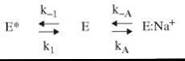
Scheme 1.
Thrombin exists in equilibrium between two forms, E* and E, that interconvert with kinetic rate constants k 1 and kobs in the μs time scale. Of these forms, only E can interact with Na+ with a rate constant kA to populate E:Na+, which may dissociate into the parent components with a rate constant k-A. The fast phase is caused by the E-E:Na+ interconversion that involves Na+ binding/dissociation, and the slow phase is caused by the E-E* interconversion that precedes Na+ binding. E and E:Na+ in Scheme 1 are the two active forms of thrombin that account for the dependence of kcat on (Na+). However, a third form E* is present in solution regardless of Na+ or any other ligand. The E*-E equilibrium of thrombin is an intrinsic property of the enzyme uncovered by its effect on the kinetics of Na+ binding. This example demonstrates how studies of Na+ binding to thrombin and related clotting proteases can advance significantly our understanding of the function and regulatory interactions of an entire class of enzymes.
Thermodynamic signatures of Na+ binding are of mechanistic and physiological importance and were also resolved from temperature studies of the kinetic mechanism. A large enthalpy change of -22 ckcal/mol is compensated by a large entropy loss of -64 cal/mol/K. The enthalpy change is caused by formation of the six ligating interactions in the coordination shell, and the entropy change reflects the uptake and ordering of water molecules within the channel embedding the primary specificity pocket and the active site linked to the occupancy of the Na+ site (11). An important consequence of the large enthalpy change is that the equilibrium association constant for Na+ binding becomes 10 M-1 at 37°C, which implies that under physiological conditions of temperature and (NaCl) = 140 mM, thrombin is only 60% bound to Na+. The fraction of thrombin in the procoagulant E:Na+ form is about 60%, and the anticoagulant form E accounts for about 40%. E* makes only a little contribution and represents <1% of the population of thrombin molecules at 37 °C.
Single-site Phe mutants of each of the nine Trp residues of thrombin were used to identify fluorophores responsible for the spectral changes associated with Na+ binding (18). The W141F and W215F mutants lose >70% of the total fluorescence change and retain only the slow phase. The environments of W141 and W215 change in the E* to E conversion and more drastically in the conversion of E to E:Na+. W215 is the closest Trp residue to the bound Na+ (13 A) and defines most of the aryl binding site involved in substrate recognition (Fig. 1). W141 is buried in a strategic location between the autolysis loop and exosite 1 and its linkage with the bound Na+, which is situated 23 A away, vouches for a pivotal role in communicating changes from the Na+ site to exosite 1 (Fig. 1). Importantly, the fast phase of fluorescence increase directly linked to the transition from E to E:Na+ is affected in all nine Trp mutants, albeit to different extent, which vouches for a global effect of Na+ binding on thrombin structure. This scenario is paradigm-shifting because it proves that the E*-E and E-E:Na+ interconversions affect the structure of the enzyme as a whole. Ala scanning mutagenesis of the epitopes of thrombin energetically linked to Na+ binding and ligand recognition confirm this scenario and vouch for long-range allosteric communications involving the active site, the Na+ site and exosite 1 (11, 19).

Figure 1. Structure of thrombin in the E:Na+ form (PDB ID code 1SG8) rendered as a ribbon in spectrum color. Relevant domains are noted. Catalytic residues (H57, D102, S195) and the S1 site (D189) are labeled, along with the two major spectral reporters W215 and W141. The bound Na+ (purple ball) is within 5 A from the side chain of D189. Numbering refers to chymotrypsin(ogen).
Molecular Basis of Thrombin Allostery
Structural information has begun to develop on how the Na+ site and exosite 1 communicate allosterically with the active site and each other. Structures of E and E:Na+ (11) are highly similar (r.m.s.d. 0.38 A), but significant differences are worth mentioning. D189 in the E:Na+ form is optimally oriented for electrostatic coupling with the P1 Arg of substrate, which may account for the lower Km observed in the presence of Na+. Subtle changes affect the catalytic triad. In the E form, the nucleophilic S195 side chain is rotated about 35° relative to the E:Na+ form, and the critical H-bond with the catalytic H57 is broken. Integrity of the H-bond is important for catalysis, and the unfavorable position of S195 in the E form may explain the lower kcat observed in the absence of Na+. Importantly, the structures of E and E:Na+ offer a plausible explanation for the long-range communication between the Na+ site and the active site. In the E:Na+ form, a network of 11 water molecules connect the bound Na+ to the Oγ atom of S195, which is located < 15 A away, through H-bonds. In the E form, only seven water molecules occupy positions in the network equivalent to those observed in the E:Na+form, and the connectivity is radically altered. Binding of Na+ organizes a network of water molecules that spans the interior of the enzyme for over 15 A up to the catalytic S195. Ordering of this network shapes the kinetic and thermodynamic signatures of Na+ binding detected experimentally.
A major advance in our understanding of thrombin allostery has come from the recent structure of the thrombin mutant D102 N depicting the elusive E* form of the enzyme (20). The mutant was engineered to inactivate thrombin with the most conservative replacement of the catalytic triad H57/D102/S195, and to produce a useful reagent for crystallization in the absence of inhibitors or in complex with physiological substrates. The structure shows changes in the active site, the S1 site, and the Na+ site that are unprecedented in thrombin and the entire realm of serine proteases and offers a compelling model of the E* form. The changes impact the conformation of the enzyme in a significant way and are consistent with a global perturbation of the structure predicted from mutagenesis and kinetic studies. The 215-219 Pβstrand collapses into the primary specificity pocket (Fig. 2) and W215 packs against the hydrophobic pocket in the active site formed by W60 d, Y60a, H57, and L99. R221a in the Na+ loop brings its guanidinium group in contact with D189 in S1 site to mimic the P1 Arg of an incoming substrate. R187 penetrates the Na+ site to occupy the space available to the cation. The structure of D102 N portrays a conformation of thrombin that cannot bind Na+ and ligands to the active site, as expected for the E* form. The drastic movement of W215 confirms the major role of this residue as a reporter of the E*-E and E-E:Na+ interconversions.

Figure 2. Structure of thrombin in the E* form (PDB ID code 3BEI) rendered as a ribbon in spectrum color in the same orientation as the structure of E:Na+ shown in Fig. (1). Note the collapse of the 215-219 P-strand against the 60-loop that completely occludes access to the active site. R221a occludes the S1 site, and R187 obliterates the Na+ site.
Binding of thrombomodulin to exosite 1 enhances the kcat/Km of thrombin for protein C < 1,000-fold, because of a 3,000-fold increase of the rate of substrate diffusion into the active site. Notwithstanding this massive functional effect, the structure of thrombin bound to a fragment of thrombomodulin at exosite 1 fails to reveal significant conformational changes in the active site or other regions (14). Such changes might have been obliterated by the presence of the active site inhibitor used in the crystallization. However, the structure of thrombin solved with hirugen bound to exosite 1 and the active site free also fails to reveal significant conformational changes in the active site (21). However, a recent structure of thrombin bound to a fragment of PAR3 bound to exosite 1 (16) reveals a rearrangement of the 60-loop that lines the upper rim of the active site entrance (Fig. 1). The indole ring of W60 d partially occludes access to the active site and restricts specificity toward physiologic substrates and inhibitors. When PAR3 binds to exosite 1, the 60-loop shifts 3.8 JA upward and causes a 180° flip of W60 d that projects the indole ring into the solvent and opens up the active site fully. This structure reveals the basis of the allosteric communication between exosite 1 and the active site cleft as well as the role of W60 d to move like a flap and regulate substrate diffusion into the active site. The allosteric effect is relevant for the cofactor function of cleaved PAR3 on PAR4 activation (4, 5), and the effect of thrombomodulin on the cleavage of protein C by thrombin (3).
More details on how exosite 1 communicates long range with the active site and the Na+ site have developed from the structure of thrombin D102 N bound to a fragment of PAR1 at exosite 1 (15). The E* conformation of D102 N is converted into the active E form by a massive conformational change that restores access to the active site and patency of the Na+ site. The structure reveals the details of this allosteric transition in terms of a network of polar interactions that can be traced from exosite 1 to the opposite side of the molecule almost 30 A away.
Conclusions and Future Directions
Thrombin continues to provide a biologically important platform for the study of the most basic aspects of protease structure, function, and regulation. Much has been learned about thrombin function and regulatory interactions, and structural details have begun to develop on the molecular basis of thrombin allostery mediated by Na+ and thrombomodulin. Kinetics studies on Na+ binding have revealed the true complexity of thrombin in terms of its ability to assume multiple conformations in equilibrium. The structure of E* confirms the prediction from kinetic studies and future studies will certainly broaden our understanding of the molecular basis of long-range communications among the active site, the Na+ site and exosite 1. Advances in our basic knowledge of mechanisms of substrate recognition and cofactor interactions will eventually foster new strategies to control or exploit the multiple functions of thrombin for pharmacological intervention.
Several questions remain on thrombin structure, function, and regulation. Future studies should focus on validation of the nature of E* and establish if this is indeed an inactive form of thrombin that cannot bind substrate at the active site. Structural studies should address more directly the role of thrombomodulin using thrombin in the E* or E forms. The physiological role of E* should be elucidated. More direct assessment of the thermodynamic signatures of thrombin interactions and regulatory transitions should rely on calorimetry. Finally, future studies should address the molecular basis of Na+and substrate specificity of thrombin and how they can be reengineered. Of particular importance will be the design of more potent anticoagulant thrombin mutants or mutants with selective activation of PARs.
References
1. Page MJ, Di Cera E. Serine peptidases: classification, structure and function. Cell Mol. Life Sci. 2008; 65:1220-1236.
2. Di Cera E. Thrombin. Mol Aspects Med. 2008; 29:203-254.
3. Esmon CT. The protein C pathway. Chest 2003; 124:26S-32S.
4. Coughlin SR. Thrombin signalling and protease-activated receptors. Nature 2000; 407:258-264.
5. Coughlin SR. Protease-activated receptors in hemostasis, thrombosis and vascular biology. J. Thromb. Haemost. 2005; 3:1800-1814.
6. Bates SM, Weitz JI. The status of new anticoagulants. Br. J. Haematol. 2006; 134:3-19.
7. Di Cera E. A structural perspective on enzymes activated by monovalent cations. J. Biol. Chem. 2006; 281:1305-1308.
8. Page MJ, Di Cera E. Role of Na+ and K+ in enzyme function. Physiol. Rev. 2006; 86:1049-1092.
9. Dang OD, Vindigni A, Di Cera E. An allosteric switch controls the procoagulant and anticoagulant activities of thrombin. Proc. Natl. Acad. Sci. U.S.A. 1995; 92:5977-5981.
10. Dang QD, Guinto ER, Di Cera E. Rational engineering of activity and specificity in a serine protease. Nat. Biotechnol. 1997; 15:146-149.
11. Pineda AO, Carrell CJ, Bush LA, Prasad S, Caccia S, Chen ZW, Mathews FS, Di Cera E. Molecular dissection of Na+ binding to thrombin. J. Biol. Chem. 2004; 279:31842-31853.
12. Bode W, Turk D, Karshikov A. The refined 1.9-A X-ray crystal structure of D-Phe-Pro-Arg chloromethylketone-inhibited human alpha-thrombin: structure analysis, overall structure, electrostatic properties, detailed active-site geometry, and structure-function relationships. Protein Sci. 1992; 1:426-471.
13. Pechik I, Madrazo J, Mosesson MW, Hernandez I, Gilliland GL, Medved L. Crystal structure of the complex between thrombin and the central “E” region of fibrin. Proc. Natl. Acad. Sci. U.S.A. 2004; 101:2718-2723.
14. Fuentes-Prior P, et al. Structural basis for the anticoagulant activity of the thrombin-thrombomodulin complex. Nature 2000; 404:518-525.
15. Gandhi PS, Chen Z, Mathews FS, Di Cera E. Structural identification of the pathway of long-range communication in an allosteric enzyme. Proc. Natl. Acad. Sci. U.S.A. 2008; 105:1832-1837.
16. Bah A, Chen Z, Bush-Pelc LA, Mathews FS, Di Cera E. Crystal structures of murine thrombin in complex with the extracellular fragments of murine protease-activated receptors PAR3 and PAR4. Proc. Natl. Acad. Sci. U.S.A. 2007; 104:11603-11608.
17. Botts J, Morales M. Analytical description of the effects of modifiers and of multivalency upon the steady state catalyzed reaction rate. Trans. Faraday Soc. 1953; 49:696-707.
18. Bah A, Garvey LC, Ge J, Di Cera E. Rapid kinetics of Na+ binding to thrombin. J. Biol. Chem. 2006; 281:40049-40056.
19. Mengwasser KE, Bush LA, Shih P, Cantwell AM, Di Cera E. Hirudin binding reveals key determinants of thrombin allostery. J. Biol. Chem. 2005; 280:23997-27003.
20. Pineda AO, Chen ZW, Bah A, Garvey LC, Mathews FS, Di Cera E. Crystal structure of thrombin in a self-inhibited conformation. J. Biol. Chem. 2006; 281:32922-32928.
21. Vijayalakshmi J, Padmanabhan KP, Mann KG, Tulinsky A. The isomorphous structures of prethrombin2, hirugen-, and PPACK- thrombin: changes accompanying activation and exosite binding to thrombin. Protein Sci 1994; 3:2254-2271.
Further Reading
Di Cera E, Page MJ, Bah A, Bush-Pel, LA, Garvey LC. Thrombin allostery. Phys. Chem. Chem. Phys. 2007; 9:1292-1306.
Davie EW, Kulman JD. An overview of the structure and function of thrombin. Semin. Thromb. Hemost. 2006; 32(suppl 1):3-15.
Cantwell AM, Di Cera E. Rational design of a potent anticoagulant thrombin. J. Biol. Chem. 2000; 275:39827-29830.
Gibbs CS, et al. Conversion of thrombin into an anticoagulant by protein engineering. Nature 1995;378:413-416.
Gruber A, Cantwell AM, Di Cera E, Hanson SR. The thrombin mutant W215A/E217A shows safe and potent anticoagulant and antithrombotic effects in vivo. J. Biol. Chem. 2002; 277:27581-27584.
See Also
Serine Proteases and Serine Protease Inhibitors