5 Steps to a 5 AP Psychology, 2014-2015 Edition (2013)
STEP 4. Review the Knowledge You Need to Score High
Chapter 7. Biological Bases of Behavior
IN THIS CHAPTER
Summary: As you read this page, lots of things are going through your mind. Your mind is what your brain does, according to many psychologists. The relationships of behavior, the mind, and the nervous system, especially the brain, have become increasingly clear as improvements in technology have enabled scientists to make better observations. In all areas of anatomy and physiology, structure is related to function. Specialized structures throughout your body enable regulatory function at all levels of organization from your neurotransmitter molecules to your nervous and endocrine systems.
Neuropsychologists explore the relationships between brain/nervous systems and behavior. Neuropsychologists are also called biological psychologists or biopsychologists, behavioral geneticists, physiological psychologists, and behavioral neuroscientists.
This chapter focuses on what we know about our nervous system and all of its parts at different levels of organization, and the tools that have enabled us to learn about them.

Key Ideas
![]() Techniques to learn about structure and function
Techniques to learn about structure and function
![]() Nervous system organization
Nervous system organization
![]() Brain structure and function
Brain structure and function
![]() Neuron structure and functions
Neuron structure and functions
![]() Endocrine system structure and function
Endocrine system structure and function
![]() Evolution and behavior
Evolution and behavior
![]() Genetics and behavior
Genetics and behavior
Techniques to Learn About Structure and Function
As technology has improved, scientists have used a wide range of techniques to learn about brain and neural function. Over 150 years ago, studying patients with brain damage linked loss of structure with loss of function. Phineas Gage was the level-headed, calm foreman of a railroad crew (1848) until an explosion hurled an iron rod through his head. After the injury severed the connections between his limbic system and frontal cortex, Gage became hostile, impulsive, and unable to control his emotions or his obscene language. Observed at autopsy, his loss of tissue (where the limbic system is connected to the frontal lobes) revealed the relationship between frontal lobes and control of emotional behavior. In another case, Paul Broca (1861) performed an autopsy on the brain of a patient, nicknamed Tan, who had lost the capacity to speak although his mouth and vocal cords weren’t damaged and he could still understand language. Tan’s brain showed deterioration of part of the frontal lobe of the left cerebral hemisphere, as did the brains of several similar cases. This connected destruction of the part of the left frontal lobe known as Broca’s area to loss of the ability to speak, known as expressive aphasia. Carl Wernicke similarly found another brain area involved in understanding language in the left temporal lobe. Destruction of Wernicke’s area results in loss of the ability to comprehend written and spoken language, known as receptive aphasia.
“Structure is always related to function in living things.”
—Adrianne, AP teacher
Gunshot wounds, tumors, strokes, and other diseases that destroy brain tissue enabled further mapping of the brain. Because the study of the brain through injury was a slow process, quicker methods were pursued. Lesions, precise destruction of brain tissue, enabled more systematic study of the loss of function resulting from surgical removal (also called ablation), cutting of neural connections, or destruction by chemical applications. Surgery to relieve epilepsy cuts neural connections at the corpus callosum, between the cerebral hemispheres. Studies by Roger Sperry and Michael Gazzaniga of patients with these “split brains” have revealed that the left and right hemispheres do not perform exactly the same functions (brain lateralization), that the hemispheres specialize in. The left cerebral hemisphere is specialized for verbal, mathematical, and analytical functions. The nonverbal right hemisphere is specialized for spatial, musical, and holistic functions such as identifying faces and recognizing emotional facial expressions.
Direct electrical stimulation of different cortical areas of the brain during surgery enabled scientists to observe the results. Stimulating the back of the frontal cortex at particular sites caused body movement for different body parts enabling mapping of the motor cortex.
In recent years, neuroscientists have been able to look inside the brain without surgery. Computerized axial tomography (CAT or CT) creates a computerized image using x-rays passed through various angles of the brain showing two-dimensional “slices” that can be arranged to show the extent of a lesion. In magnetic resonance imaging (MRI), a magnetic field and pulses of radio waves cause emission of faint radio frequency signals that depend upon the density of the tissue. The computer constructs images based on varying signals that are more detailed than CT scans. Both CT scans and MRIs show the structure of the brain, but don’t show the brain functioning.
Measuring Brain Function
Scientists have developed a number of tools to measure the brain functions of people. An EEG (electroencephalogram) is an amplified tracing of brain activity produced when electrodes positioned over the scalp transmit signals about the brain’s electrical activity (“brain waves”) to an electroencephalograph machine. The amplified tracings are called evoked potentials when the recorded change in voltage results from a response to a specific stimulus presented to the subject. EEGs have been used to study the brain during states of arousal such as sleeping and dreaming, to detect abnormalities (such as deafness and visual disorders in infants), and to study cognition. Another technology, positron emission tomography (PET) produces color computer graphics that depend on the amount of metabolic activity in the imaged brain region. When neurons are active, an automatic increase in blood flow to the active region of the brain brings more oxygen and glucose necessary for respiration. Blood flow changes are used to create brain images when tracers (such as radioactively tagged glucose) injected into the blood of the subject emit particles called positrons, which are converted into signals detected by the PET scanner. Functional MRI (fMRI) shows the brain at work at higher resolution than the PET scanner. Changes in oxygen in the blood of an active brain area alters its magnetic qualities, which is recorded by the fMRI scanner. After further computer processing, a detailed picture of that local brain activity emerges. With new brain imaging technology, psychologists can explore far more about our abilities than ever before, from well-known systems like perception to less understood systems like motivation and emotion.
Organization of Your Nervous System
Your patterns of behavior generally involve masses of neural tissue rather than a few neurons. All of the neurons in your body are organized into your nervous system. Your nervous system has subdivisions based on location and function. The two major subdivisions are your central nervous system and your peripheral nervous system. Your central nervous system consists of your brain and your spinal cord. Your peripheral nervous system includes two major subdivisions: your somatic nervous system and your autonomic nervous system. Your peripheral nervous system lies outside the midline portion of your nervous system carrying sensory information to and motor information away from your central nervous system via spinal and cranial nerves. Your somatic nervous system has motor neurons that stimulate skeletal (voluntary) muscle. Your autonomic nervous system has motor neurons that stimulate smooth (involuntary) and heart muscle. Your autonomic nervous system is subdivided into the antagonistic sympathetic nervous system and parasympathetic nervous system. Sympathetic stimulation results in responses that help your body deal with stressful events including dilation of your pupils, release of glucose from your liver, dilation of bronchi, inhibition of digestive functions, acceleration of heart rate, secretion of adrenalin from your adrenal glands, acceleration of breathing rate, and inhibition of secretion of your tear glands. Parasympathetic stimulation calms your body following sympathetic stimulation by restoring digestive processes (salivation, peristalsis, enzyme secretion), returning pupils to normal pupil size, stimulating tear glands, and restoring normal bladder contractions. Your spinal cord, protected by membranes called meninges and your spinal column of bony vertebrae, starts at the base of your back and extends upward to the base of your skull where it joins your brain. The cord is composed mainly of interneurons and glial cells, which are all bathed by cerebrospinal fluid produced by your glial cells.
The Brain
Your brain, which has the consistency of soft-serve yogurt, is covered by protective membranes called meninges and is housed in your skull. The evolutionary approach describes the brain’s evolution from more primitive organisms, reasoning that new types of behavior developed as each new layer of the brain evolved. According to one evolutionary model (triune brain), the human brain has three major divisions, overlapping layers with the most recent neural systems nearest the front and top. The reptilian brain, which maintains homeostasis and instinctive behaviors, roughly corresponds to the brainstem, which includes the medulla, pons, and cerebellum. Developmental psychologists call the brainstem the hindbrain. The old mammalian brain roughly corresponds to the limbic system that includes the septum, hippocampus, amygdala, cingulate cortex, hypothalamus, and the thalamus, which are all important in controlling emotional behavior, some aspects of memory, and vision. The new mammalian brain or neocortex, synonymous with the cerebral cortex, accounts for about 80% of brain volume and is associated with the higher functions of judgment, decision making, abstract thought, foresight, hindsight and insight, language and computing, as well as sensation and perception. Developmental psychologists call the structures of the “mammalian brains” the forebrain. The surface of your cortex has peaks called gyri and valleys called sulci, which form convolutions that increase the surface area of your cortex. Deeper valleys are called fissures. The last evolutionary development of the brain is the localization of functions on different sides of your brain.
Localization and Lateralization of the Brain’s Function
Although multiple representations of information can be located within different areas of your brain, specific regions of your brain seem most critical in handling particular functions. This localization of structure and function has been identified for numerous regions (see Figure 7.1). Association areas are regions of the cerebral cortex that do not have specific sensory or motor functions, but are involved in higher mental functions, such as thinking, planning, remembering, and communicating. In general, crossing over of nerves sending information from one side of your body to the other side of your brain results in contralaterality, control of one side of your body by the other side of your brain.
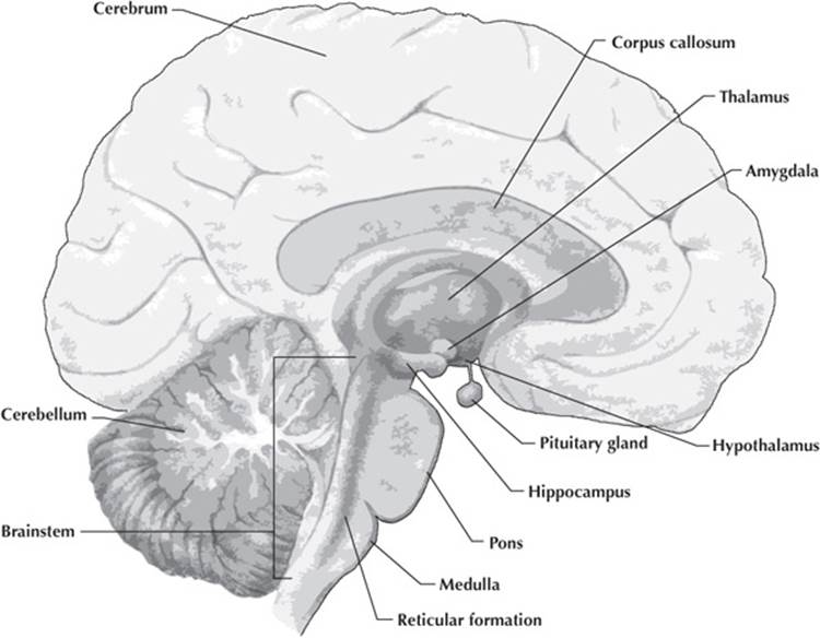
Figure 7.1 Major structures of the brain in medial view.
Just as a map or globe can be divided into hemispheres and continents, your cerebral cortex can be divided into eight lobes, four in the left cerebral hemisphere and four in the right cerebral hemisphere (see Figure 7.2). Go to www.g2conline.org and click on 3-D Brain for a more detailed view of a three-dimensional brain model. You can download it as an app.
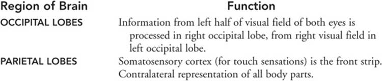
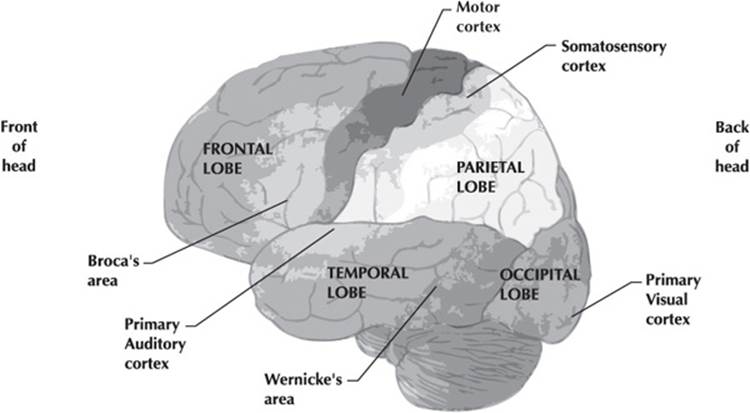
Figure 7.2 Regions of the left cerebral cortex in lateral view.
Although specific regions of the brain are associated with specific functions, if one region is damaged, the brain can reorganize to take over its function, which is called plasticity. In phantom limb syndrome, a somewhat unfortunate example of plasticity, reorganization of the somatosensory cortex leads to someone experiencing sensations where a missing limb used to be.
Structure and Function of the Neuron
Your extraordinarily complex brain is composed of trillions of neurons and glial cells. Glial cells guide the growth of developing neurons, help provide nutrition for and get rid of wastes of neurons, and form an insulating sheath around neurons that speeds conduction. The neuron is the basic unit of structure and function of your nervous system. Neurons perform three major functions: receive information, process it, and transmit it to the rest of your body. Three major regions of a neuron enable the cell to communicate with other cells (see Figure 7.3). The cell body (a.k.a. cyton or soma) contains cytoplasm and the nucleus, which directs synthesis of such substances as neurotransmitters. The dendrites are branching tubular processes capable of receiving information. The axon emerges from the cyton as a single conducting fiber (longer than a dendrite) which branches and ends in tips called terminal buttons, axon terminals, or synaptic knobs. The axon is usually covered by an insulating myelin sheath (formed by glial cells). Neurogenesis, the growth of new neurons, takes place throughout life.
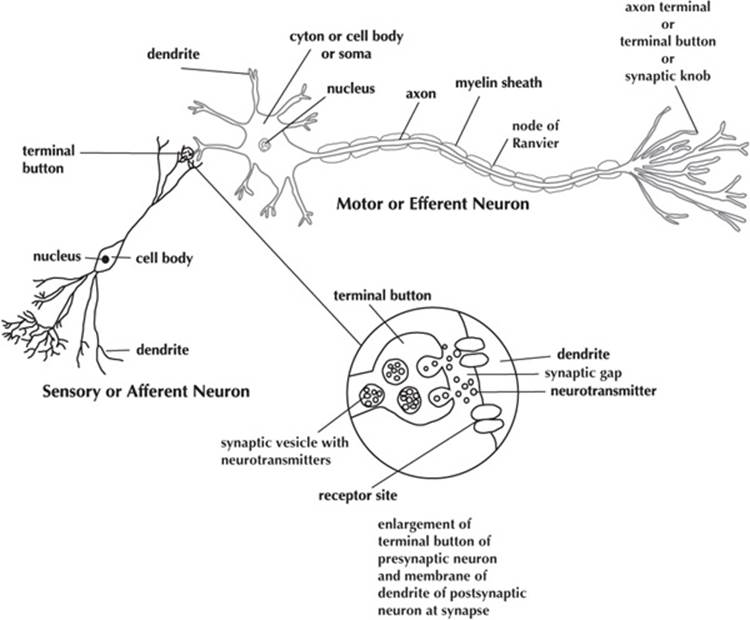
Figure 7.3 Typical neurons.
Neurotransmitters are chemicals stored in structures of the terminal buttons called synaptic vesicles. Different neurotransmitters have different chemical structures and perform different functions. For example, acetylcholine (ACh) causes contraction of skeletal muscles, helps regulate heart muscles, is involved in memory, and also transmits messages between the brain and spinal cord. Lack of ACh is associated with Alzheimer’s disease. Dopaminestimulates the hypothalamus to synthesize hormones and affects alertness and movement. Lack of dopamine is associated with Parkinson’s disease; too much dopamine is associated with schizophrenia. Glutamate is a major excitatory neurotransmitter involved in information processing throughout the cortex and especially memory formation in the hippocampus. Both schizophrenia and Alzheimer’s may involve glutamate receptors. Serotonin is associated with sexual activity, concentration and attention, moods, and emotions. Lack of serotonin is associated with depression. Opioid peptides such as endorphins are often considered the brain’s own pain killers. Gamma-aminobutyric acid (GABA) inhibits firing of neurons. Benzodiazepine (Valium) and anticonvulsant drugs increase activity of GABA. Huntington’s disease is associated with insufficient GABA-producing neurons in parts of the brain involved in coordination of movement. Seizures are associated with malfunctioning GABA systems. Other chemicals, such as drugs, can interfere with the action of neurotransmitters. Agonists may mimic a neurotransmitter and bind to its receptor site to produce the effect of the neurotransmitter. Antagonists block a receptor site inhibiting the effect of the neurotransmitter or agonist.
Neuron Functions
All your behavior begins with the actions of your neurons. A neuron gets incoming information from its receptors spread around its dendrites. That information is sent to its cell body, where it’s combined with other incoming information. Neural impulses are electrical in nature along the neuron. The neuron at rest is more negative inside the cell membrane relative to outside of the membrane. The neuron’s resting potential results from the selective permeability of its membrane and the presence of electrically charged particles called ions near the inside and outside surfaces of the membrane in different concentrations. When sufficiently stimulated (to threshold), a net flow of sodium ions into the cell causes a rapid change in potential across the membrane, known as the action potential (see Figure 7.4). If stimulation is not strong enough, your neuron doesn’t fire. The strength of the action potential is constant whenever it occurs. This is the all-or-none principle.
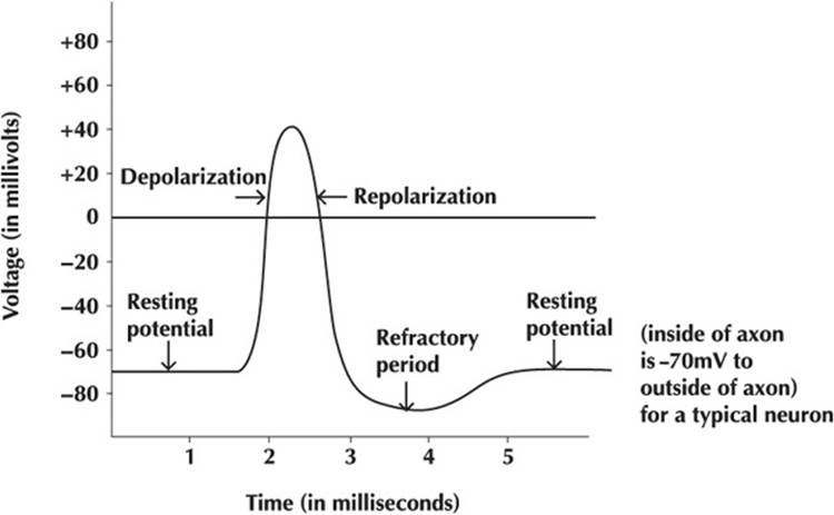
Figure 7.4 Action potential.
The wave of depolarization and repolarization is passed along the axon to the terminal buttons, which release neurotransmitters. Spaces between segments of myelin are called nodes of Ranvier. When the axon is myelinated, conduction speed is increased since depolarizations jump from node to node. This is called saltatory conduction. Chemical neurotransmitters are released into the synapse where they attach to specific receptor sites on membranes of dendrites of your postsynaptic neurons, like a key fitting into the tumbler of a lock (the lock and key concept). Some of your synapses are excitatory, the neurotransmitters cause the neuron on the other side of the synapse to generate an action potential (to fire); other synapses are inhibitory, reducing or preventing neural impulses. The sum of all excitatory and inhibitory inputs determines whether your next neuron will fire and at what rate. The constant flow of these neurochemical impulses gives your behavior its amazing complexity. It regulates your metabolism, temperature, and respiration. It also enables you to learn, remember, and decide.
Reflex Action
The simplest form of your behavior, called a reflex, involves impulse conduction over a few (perhaps three) neurons. The path is called a reflex arc. Sensory or afferent neurons transmit impulses from your sensory receptors to the spinal cord or brain. Interneurons, located entirely within your brain and spinal cord, intervene between sensory and motor neurons. Motor or efferent neurons transmit impulses from your sensory or interneurons to muscle cells that contract or gland cells that secrete. Muscle and gland cells are called effectors. Examples of your reflexes include your pupillary reflex, knee jerk, sneezing, and blinking. Neural impulses travel one way along the neuron from dendrites to axons to terminal buttons, and among neurons from the receptor to the effector.
The Endocrine System
Your endocrine system interacts with your nervous system to regulate your behavior and body functions. Your endocrine system consists of glands that secrete chemical messengers called hormones into your blood. The hormones travel to target organs where they bind to specific receptors. Endocrine glands include the pineal gland, hypothalamus, and pituitary gland in your brain; the thyroid and parathyroids in your neck; the adrenal glands atop your kidneys; pancreas near your stomach; and either testes or ovaries.
Genetics and Evolutionary Psychology
Why do you behave the way you do? To what extent is your behavior determined by your heredity? To what extent is it determined by your life history or environment? The nature-nurture controversy deals with the extent to which heredity and the environment each influence behavior. Evolutionary psychologists study how natural selection favored behaviors that contributed to survival and spread of our ancestors’ genes, and may currently contribute to our survival into the next generations. Evolutionary psychologists look at universal behaviors shared by all people. They look at behaviors conserved across related species to understand how we are adapted to maximize our success in our environments. Charles Darwin pointed out the similarities of the expressions of emotions in people and other animals, suggesting that expressions shared across cultures and species are biologically determined.
Genetics and Behavior
Behavioral geneticists study the role played by our genes and our environment in mental ability, emotional stability, temperament, personality, interests, etc.; they look at the causes of our individual differences. Your genes predispose your behavior. Studies of twins have been helping to separate the contributions of heredity and environment. Identical twins are two individuals who share all of the same genes/heredity because they develop from the same fertilized egg or zygote; they are monozygotic twins. Fraternal twins are siblings that share about half of the same genes because they develop from two different fertilized eggs or zygotes; they are dizygotic twins. Heritability is the proportion of variation among individuals in a population that is due to genetic causes. For the special case of identical twins, you might want to say that the heritability for traits of identical twins is zero, but that is not exactly correct. Like evolution, heritability is a concept applied to the population rather than the individual. When twins grow up in the same environment, the extent to which behaviors of monozygotic twins are behaviorally more similar than dizygotic twins reveals the contribution of heredity to behavior. Schizophrenia and general intelligence are more similar in monozygotic twins than dizygotic twins. If monozygotic twins are separated at birth and raised in different environments (adoption studies), behavioral differences may reveal the contribution of environment to behavior; similarities may reveal the contribution of heredity.
Adoption studies assess genetic influence by comparing resemblance of adopted children to both their adoptive and biological parents. The children must have been adopted as infants without contact with their biological parents. If the children resemble their biological parents, but not their adoptive families, with respect to a given trait, researchers infer a genetic component for that trait. Such constellations of behaviors as alcoholism, schizophrenia, and general intelligence have shown both genetic and environmental components.
Transmission of Hereditary Characteristics
Transmission of hereditary characteristics is achieved by biological processes, including formation of sex cells, fertilization, embryonic development, and protein synthesis. Each DNA segment of a chromosome that determines a trait is a gene. Chromosomes carry information stored in genes to new cells during reproduction. Normal human body cells have 46 chromosomes, except for eggs and sperms that have 23 chromosomes. Males have 44 chromosomes, plus X and Y. Females have 44 chromosomes, plus X and X. At fertilization, 23 chromosomes from the sperm unite with 23 chromosomes from the egg to form a zygote with 46 chromosomes. If the male contributes an X chromosome, the baby is female; if the male contributes a Y chromosome, the baby is male. The presence of a Y chromosome makes the baby a male. All of the cells of the embryo/baby have the same 23 pairs of chromosomes, which carry genes for the same traits. Fertilization that includes a sperm or egg with the wrong number of chromosomes results in a zygote, and subsequently an individual, with chromosomal abnormalities. Turner syndrome females have only one X sex chromosome (XO). Girls with Turner syndrome are typically short with a webbed neck, lack ovaries, and fail to develop secondary sex characteristics at puberty. Although usually of normal intelligence, they typically evidence specific cognitive deficits in arithmetic, spatial organization, and visual form perception. Klinefelter’s syndrome males arise from an XXY zygote. The syndrome becomes evident at puberty when male secondary sex characteristics fail to develop, but breast tissue does. Klinefelter’s males tend to be passive. The presence of three copies of chromosome-21 results in the expression of Down syndrome. Down syndrome individuals are typically mentally retarded and have a round head, a flat nasal bridge, a protruding tongue, small round ears, a fold in the eyelid, and poor muscle tone and coordination.
The genetic makeup for a trait of an individual is called its genotype. The expression of the genes is called its phenotype. For traits determined by one pair of genes, if they are the same (homozygous), the individual expresses that phenotypic characteristic. If the genes are different, the expressed gene is called the dominant gene; the hidden gene is the recessive gene. Numerous recessive genes are responsible for syndromes in the homozygous condition. Tay-Sachs syndrome produces progressive loss of nervous function and death in a baby. Albinism arises from a failure to synthesize or store pigment and also involves abnormal nerve pathways to the brain, resulting in quivering eyes and the inability to perceive depth or three-dimensionality with both eyes. Phenylketonuria (PKU) results in severe, irreversible brain damage unless the baby is fed a special diet low in phenylalanine within 30 days of birth; the infant lacks an enzyme to process this amino acid which can build up and poison cells of the nervous system. Thus, heredity and environment interact to determine a trait. Huntington’s disease is an example of a dominant gene defect that involves degeneration of the nervous system. Progressive symptoms involve forgetfulness, tremors, jerky motions, loss of the ability to talk, personality changes such as temper tantrums or inappropriate accusations, blindness, and death. Recessive genes for color blindness are located on the X chromosome with no corresponding gene on the Y chromosome. As a result, males show sex-linked traits like color blindness much more frequently than females. Behaviors and diseases may have variations only some of which are genetically based. A form of familial Alzheimer’s disease has been attributed to a gene on chromosome 21, but not all cases of Alzheimer’s disease are associated with that gene.
![]() Review Questions
Review Questions
1. A neuron without terminal buttons would be unable to
(A) receive information from neighboring neurons
(B) generate an action potential
(C) direct the synthesis of neurotransmitters
(D) secrete neurotransmitters to postsynaptic neurons
(E) transport ions across the cell membrane
2. Paul Broca found that the loss of the ability to speak intelligibly is associated with damage to a region of the brain in the
(A) thalamus
(B) right parietal lobe
(C) right occipital lobe
(D) left temporal lobe
(E) left frontal lobe
3. Scientists are able to see changes in the brain as it processes information by means of
(A) lesioning
(B) autopsy
(C) CT
(D) MRI
(E) PET
4. The simplest behaviors exhibit
(A) are learned when we are infants
(B) do not involve the central nervous system
(C) are called instincts
(D) include sneezing and blinking
(E) must be processed by the medulla
5. Of the following, the effect of adrenalin on the body is most similar to the effect of the
(A) cerebellum
(B) parathyroids
(C) somatic nervous system
(D) parasympathetic nervous system
(E) sympathetic nervous system
6. Mr. Jenkins suffered a “stroke” as a result of a brain injury. Although he can still move the fingers on his right hand, he has lost sensation in these parts. Of the following, the site of damage to his brain is most likely in the
(A) right frontal lobe
(B) right temporal lobe
(C) left frontal lobe
(D) left parietal lobe
(E) hypothalamus
7. Of the following, which are located exclusively in the central nervous system?
(A) afferent neurons
(B) interneurons
(C) efferent neurons
(D) glial cells
(E) effectors
8. Which of the following glands interact(s) most directly with all of the others to help regulate body processes?
(A) pituitary
(B) adrenals
(C) parathyroids
(D) thyroid
(E) ovaries
9. Gunshot wounds, tumors, and strokes all result in
(A) infections
(B) significant loss of function
(C) lesions
(D) pain
(E) necessity for surgery
10. Which of the following must be males?
(A) dizygotic twins
(B) monozygotic twins
(C) Down syndrome children
(D) Klinefelter’s syndrome children
(E) Turner’s syndrome children
11. Which includes all of the others?
(A) autonomic nervous system
(B) peripheral nervous system
(C) somatic nervous system
(D) parasympathetic nervous system
(E) sympathetic nervous system
12. Which stimulate a muscle to contract?
(A) adrenal hormones
(B) receptors
(C) sensory neurons
(D) motor neurons
(E) interneurons
13. The part of the brain most closely associated with maintaining balance and the coordination of complex sequences of movements is the
(A) hypothalamus
(B) thalamus
(C) pons
(D) medulla
(E) cerebellum
14. Loss of the ability of the brain to produce adequate levels of dopamine often leads to
(A) aphasia
(B) Alzheimer’s disease
(C) Parkinson’s disease
(D) bipolar disorder
(E) amnesia
15. Which task is primarily a right cerebral hemisphere function in most people?
(A) understanding written language
(B) understanding spoken language
(C) processing visual information from the left eye
(D) recognizing faces
(E) processing sensory information from the right leg
![]() Answers and Explanations
Answers and Explanations
1. D—Terminal buttons secrete neurotransmitters into the synapse.
2. E—Broca’s area is a region in the left frontal lobe anterior to the motor cortex.
3. E—PET scans visualize changes in the brain as it functions. While fMRI also shows changes in the brain as it functions, MRI and CT scans show structure only.
4. D—Our simplest behaviors are reflexes. Sneezing and blinking are reflexes.
5. E—Adrenalin is a hormone that speeds up breathing and heart rate, sends a message to change stored food back to glucose, etc. The sympathetic nervous system stimulates the same changes in the body.
6. D—The center for sensation in the brain is the somatosensory region of the cerebral cortex located in the front of the parietal lobes. Nerves carrying sensations from the right side of the body cross over to the left side of the brain, so the most probable site of damage is the left parietal lobe.
7. B—Interneurons are found in the brain and spinal cord only. The others can be found in the peripheral nervous system.
8. A—The pituitary gland, which is sometimes called “the master gland,” produces many hormones that stimulate other glands, including the adrenals, parathyroids, thyroid, and ovaries.
9. C—Lesions are interruptions in tissue. While the other choices may accompany wounds, tumors, and strokes, they also may not.
10. D—Presence of the Y chromosome determines the sex of a human baby. Of the choices, only a Klinefelter’s child (XXY) must have a Y chromosome.
11. B—The peripheral nervous system comprises the autonomic nervous and somatic nervous system. The autonomic nervous system is subdivided into the parasympathetic and sympathetic nervous systems.
12. D—Motor neurons or efferent neurons cause muscles to contract or glands to secrete.
13. E—The cerebellum functions in balance and coordination.
14. C—Parkinson’s disease is associated with depletion of cells that produce dopamine.
15. D—Pattern matching and picture and facial recognition are all right hemispheric functions.
![]() Rapid Review
Rapid Review
Neuropsychologists—those who explore the relationships between brain/nervous systems and behavior. Neuropsychologists are also called biological psychologists or bio-psychologists, behavioral geneticists, physiological psychologists, and behavioral neuroscientists.
Studying patients with brain damage linked loss of structure with loss of function.
Lesions—precise destruction of brain tissue, enables more systematic study of the loss of function resulting from surgical removal (also called ablation), cutting of neural connections, or destruction by chemical applications.
CT scans and MRIs show structure.
• Computerized axial tomography (CAT or CT)—creates a computerized image using x-rays passed through the brain to show structure and/or the extent of a lesion.
• Magnetic resonance imaging (MRI)—creates more detailed computerized images using a magnetic field and pulses of radio waves that cause emission of signals that depend upon the density of tissue.
EEGs, PET scans, and fMRIs show function.
• EEG (electroencephalogram)—an amplified tracing of brain activity produced when electrodes positioned over the scalp transmit signals about the brain’s electrical activity (“brain waves”) to an electroencephalograph machine.
• Evoked potentials—EEGs resulting from a response to a specific stimulus presented to the subject.
• Positron emission tomography (PET)—shows brain activity when radioactively tagged glucose rushes to active neurons and emits positrons.
• Functional MRI (fMRI)—shows brain activity at higher resolution than the PET scan when changes in oxygen concentration near active neurons alter magnetic qualities.
Central nervous system (CNS)—brain and spinal cord.
Peripheral nervous system (PNS)—portion of the nervous system outside the brain and spinal cord; includes all of the sensory and motor neurons, and subdivisions called the autonomic and somatic nervous systems.
Autonomic nervous system (ANS)—subdivision of PNS that includes motor nerves that innervate smooth (involuntary) and heart muscle. Its sympathetic nervous system prepares the body for “fight or flight”; the parasympathetic nervous system causes bodily changes for maintenance or rest.
• Sympathetic nervous system—subdivision of PNS and ANS whose stimulation results in responses that help your body deal with stressful events.
• Parasympathetic nervous system—subdivision of PNS and ANS whose stimulation calms your body following sympathetic stimulation by restoring normal body processes.
Somatic nervous system—subdivision of PNS that includes motor nerves that stimulate skeletal (voluntary) muscles.
Spinal cord—portion of the central nervous system below the level of the medulla.
Brain—portion of the central nervous system above the spinal cord.
According to the evolutionary model, the brain consists of three sections: reptilian brain (medulla, pons, cerebellum); old mammalian brain (limbic system, hypothalamus, thalamus); and the new mammalian brain (cerebral cortex).
According to the developmental model, it consists of three slightly different sections: the hindbrain (medulla, pons, cerebellum), the midbrain (small region with parts involved in eye reflexes and movements), and the forebrain (including the limbic system, hypothalamus, thalamus, cerebral cortex).
Convolutions—folding-in and out of the cerebral cortex that increases surface area of the brain.
Contralaterality—control of one side of your body by the other side of your brain.
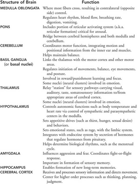
The parts of the brain with the functions associated with each are:
• Medulla oblongata—regulates heart rhythm, blood flow, breathing rate, digestion, vomiting.
• Pons—includes portion of reticular activating system or reticular formation critical for arousal and wakefulness; sends information to and from medulla, cerebellum, and cerebral cortex.
• Cerebellum—controls posture, equilibrium, and movement.
• Basal ganglia—regulates initiation of movements, balance, eye movements, and posture.
• Thalamus—relays visual, auditory, taste, and somatosensory information to/from appropriate areas of cerebral cortex.
• Hypothalamus—controls feeding behavior, drinking behavior, body temperature, sexual behavior, threshold for rage behavior, activation of the sympathetic and parasympathetic systems, and secretion of hormones of the pituitary.
• Amygdala—influences emotions such as aggression, fear, and self-protective behaviors.
• Hippocampus—enables formation of new long-term memories.
• Cerebral cortex—center for higher-order processes such as thinking, planning, judgment; receives and processes sensory information and directs movement.
• Association areas—areas of the cerebral cortex that do not have specific sensory or motor functions, but are involved in higher mental functions such as thinking, planning, and communicating.
Geographically, the cerebral cortex can be divided into eight lobes, four on the left side and four on the right side:
• Occipital lobes—primary area for processing visual information.
• Parietal lobes—front strip is somatosensory cortex that processes sensory information including touch, temperature, and pain from body parts; association areas perceive objects.
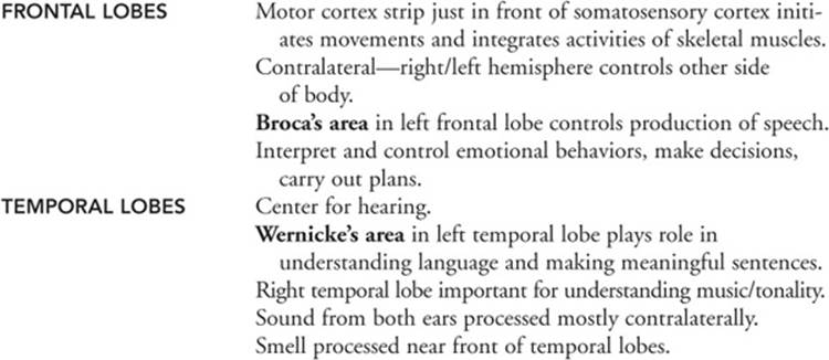
• Frontal lobes—interpret and control emotional behaviors, make decisions, carry out plans; motor cortex strip just in front of somatosensory cortex initiates movements and integrates activities of skeletal muscles; produces speech (Broca’s area).
• Temporal lobes—primary area for hearing, understanding language (Wernicke’s area), understanding music/tonality, processing smell.
Aphasia—impairment of the ability to understand or use language.
Glial cells—supportive cells of the nervous system that guide the growth of developing neurons, help provide nutrition for and get rid of wastes of neurons, and form an insulating sheath around neurons that speeds conduction.
Neuron—the basic unit of structure and function of your nervous system. Neurons perform three major functions: receive information, process it, and transmit it to the rest of your body. Terms relating to the structure and function of the neuron include:
• Cell body—also called the cyton or soma; the part of the neuron that contains cytoplasm and the nucleus, which directs synthesis of such substances as neurotransmitters.
• Dendrites—branching tubular processes of a neuron that have receptor sites for receiving information.
• Axon—a long, single conducting fiber extending from the cell body of a neuron that transmits an action potential and that branches and ends in tips called terminal buttons (a.k.a. axon terminals, or synaptic knobs), which secrete neurotransmitters.
• Myelin sheath—a fatty covering of the axon made by glial cells, which speeds up conduction of the action potential.
• Terminal buttons (a.k.a. axon terminals, end bulbs, or synaptic knobs)—tips at the end of axons that secrete neurotransmitters when stimulated by the action potential.
Neurotransmitters—chemical messengers released by the terminal buttons of the presynaptic neuron into the synapse. Different neurotransmitters have different chemical structures and perform different tasks:
• Acetylcholine (ACh)—a neurotransmitter that causes contraction of skeletal muscles, helps regulate heart muscles, is involved in memory, and also transmits messages between the brain and spinal cord. Lack of ACh is associated with Alzheimer’s disease.
• Dopamine—a neurotransmitter that stimulates the hypothalamus to synthesize hormones and affects alertness, attention, and movement. Lack of dopamine is associated with Parkinson’s disease; too much is associated with schizophrenia.
• Glutamate—a neurotransmitter that stimulates cells throughout the brain, but especially in the hypothalamus, and is associated with memory formation and information processing.
• Serotonin—a neurotransmitter associated with arousal, sleep, appetite, moods, and emotions. Lack of serotonin is associated with depression.
• Endorphin—a neurotransmitter similar to the opiate morphine that relieves pain and may induce feelings of pleasure.
• Gamma-aminobutyric acid (GABA)—a neurotransmitter that inhibits firing of postsynaptic neurons. Huntington’s disease and seizures are associated with malfunctioning GABA systems.
Action potential—also called an impulse, the “firing” of a neuron; a net flow of sodium ions into the cell that causes a rapid change in potential across the membrane when stimulation reaches threshold.
All-or-none principle—the law that the neuron either generates an action potential when the stimulation reaches threshold or doesn’t fire when stimulation is below threshold. The strength of the action potential is constant whenever it occurs.
Nodes of Ranvier—spaces between segments of myelin on the axons of neurons.
Saltatory conduction—rapid conduction of impulses when the axon is myelinated since depolarizations jump from node (of Ranvier) to node.
Synapse—region of communication between the transmitting presynaptic neuron and receiving postsynaptic neuron, muscle, or gland, consisting of the presynaptic terminal buttons, a tiny space, and receptor sites typically on the postsynaptic dendrites.
Excitatory neurotransmitter—chemical secreted at terminal button that causes the neuron on the other side of the synapse to generate an action potential (to fire).
Inhibitory neurotransmitter—chemical secreted at terminal button that reduces or prevents neural impulses in the postsynaptic dendrites.
Reflex—the simplest form of behavior.
Reflex arc—the path over which the reflex travels, which typically includes the following:
• Sensory receptor—cell typically in sense organs that initiates action potentials, which then travel along sensory/afferent neurons to the CNS.
• Afferent neuron—also called sensory neuron; nerve cell in your PNS that transmits impulses from receptors to the brain or spinal cord.
• Interneuron—nerve cell in the CNS that transmits impulses between sensory and motor neurons. Neural impulses travel one way along the neuron from dendrites to axons to terminal buttons, and among neurons from the receptor to the effector.
• Efferent neuron—also called motor neuron; nerve cell in your PNS that transmits impulses from sensory or interneurons to muscle cells that contract or gland cells that secrete.
• Effector—muscle cell that contracts or gland cell that secretes.
Endocrine system—ductless glands that typically secrete hormones directly into the blood, which help regulate body and behavioral processes. Components of the endocrine system include:
• Hormone—chemical messenger that travels through the blood to a receptor site on a target organ.
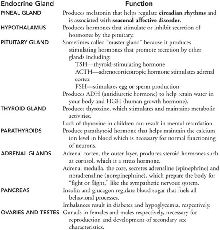
• Pineal gland—endocrine gland in brain that produces melatonin that helps regulate circadian rhythms and is associated with seasonal affective disorder.
• Hypothalamus—portion of brain part that acts as endocrine gland and produces hormones that stimulate (releasing factors) or inhibit secretion of hormones by the pituitary.
• Pituitary gland (sometimes called “master gland”)—endocrine gland in brain that produces stimulating hormones, which promote secretion by other glands including TSH–thyroid-stimulating hormone; ACTH–adrenocorticotropic hormone, which stimulates the adrenal glands; FSH, which stimulates egg or sperm production; ADH (antidiuretic hormone) to help retain water in your body; and HGH (human growth hormone).
• Thyroid gland—endocrine gland in neck that produces thyroxine, which stimulates and maintains metabolic activities.
• Parathyroids—endocrine glands in neck that produce parathyroid hormone, which helps maintain calcium ion level in blood necessary for normal functioning of neurons.
• Adrenal glands—endocrine glands atop kidneys. Adrenal cortex—the outer layer—produces steroid hormones such as cortisol, which is a stress hormone. Adrenal medulla—the core—secretes adrenaline (epinephrine) and noradrenaline (norepinephrine), which prepare the body for “fight or flight” like the sympathetic nervous system.
• Pancreas—gland near stomach that secretes the hormones insulin and glucagon, which regulate blood sugar that fuels all behavioral processes. Imbalances result in diabetes and hypoglycemia.
• Ovaries and testes—gonads in females and males, respectively, that produce hormones necessary for reproduction and development of secondary sex characteristics.
Evolutionary psychologists—study how Charles Darwin’s theory of natural selection favored behaviors that contributed to survival and spread of our ancestors’ genes; evolutionary psychologists look at universal behaviors shared by all people.
Behavioral geneticists—study the role played by our genes and our environment in mental ability, emotional stability, temperament, personality, interests, etc.; they look at the causes of our individual differences.
Zygote—fertilized egg.
Studies of twins help separate the contributions of heredity from environment.
• Identical twins—also called monozygotic twins; two individuals who share all of the same genes/heredity because they develop from the same zygote.
• Fraternal twins—also called dizygotic twins; siblings that share about half of the same genes because they develop from two different zygotes.
Heritability—the proportion of variation among individuals in a population that is due to genetic causes.
When twins grow up in the same environment, the extent to which behaviors of monozygotic twins are behaviorally more similar than dizygotic twins reveals the contribution of heredity to behavior.
If monozygotic twins are separated at birth and raised in different environments (adoption studies), behavioral differences may reveal the contribution of environment to behavior; similarities reveal the contribution of heredity.
In adoption studies, if the children resemble their biological parents, but not their adoptive families, with respect to a given trait, researchers infer a genetic component for that trait.
Gene—each DNA segment of a chromosome that determines a trait.
Chromosome—structure in the nucleus of cells that contains genes determined by DNA sequences.
Human cells contain 23 pairs of chromosomes, 23 of which come from the sperm of the father and 23 of which come from the egg of the mother at fertilization. If the father contributes a Y sex chromosome, the baby is male; otherwise the baby is female.
Errors during fertilization can result in the wrong number of chromosomes in cells of a baby. These can result in:
• Turner syndrome—females with only one X sex chromosome who are short, often sterile, and have difficulty calculating.
• Klinefelter’s syndrome—males with XXY sex chromosomes.
• Down syndrome—usually with three copies of chromosome-21 in their cells, individuals who are typically mentally retarded and have a round head, flat nasal bridge, protruding tongue, small round ears, a fold in the eyelid, and poor muscle tone and coordination.
Genotype—the genetic makeup of an individual.
Phenotype—the expression of the genes.
Homozygous—the condition when both genes for a trait are the same.
Heterozygous—also called hybrid; the condition when the genes for a trait are different.
Dominant gene—the gene expressed when the genes for a trait are different.
Recessive gene—the gene that is hidden or not expressed when the genes for a trait are different.
Tay-Sachs syndrome—recessive trait that produces progressive loss of nervous function and death in a baby.
Albinism—recessive trait that produces lack of pigment and involves quivering eyes and inability to perceive depth with both eyes.
Phenylketonuria (PKU)—recessive trait that results in severe, irreversible brain damage unless the baby is fed a special diet low in phenylalanine.
Huntington’s disease—dominant gene defect that involves degeneration of the nervous system, characterized by tremors, jerky motions, blindness, and death.
Sex-linked traits—recessive genes located on the X chromosome with no corresponding gene on the Y chromosome, which result in expression of recessive trait, more frequently in males.
Color blindness—sex-linked trait with which individual cannot see certain colors, most often red and green.