Barron's AP Psychology, 7th Edition (2016)
Chapter 3. Biological Bases of Behavior
KEY TERMS
Neuroanatomy
Neuron
Dendrites
Cell body (also called the soma)
Axon
Myelin sheath
Terminal buttons (also called end buttons, axon terminal, terminal branches of axon, and synaptic knobs)
Neurotransmitters
Synapse
Receptor sites
Threshold
Action potential
All-or-none principle
Neural firing
Excitatory neurotransmitters
Inhibitory neurotransmitters
Acetylcholine
Dopamine
Endorphins
Serotonin
GABA
Glutamate
Norepinephrine
Afferent neurons (or sensory neurons)
Efferent neurons (or motor neurons)
Central nervous system
Spinal cord
Peripheral nervous system
Somatic nervous system
Autonomic nervous system
Sympathetic nervous system
Parasympathetic nervous system
Accidents
Lesions
Electroencephalogram (EEG)
Computerized axial tomography (CAT or CT scan)
Magnetic resonance Imaging (MRI scan)
Positron emission tomography (PET scan)
Functional MRI (fMRI)
Hindbrain
Medulla
Pons
Cerebellum
Midbrain
Reticular formation
Forebrain
Thalamus
Hypothalamus
Amygdala
Hippocampus
Limbic system
Cerebral cortex
Hemispheres
Left hemisphere
Right hemisphere
Brain lateralization (or hemispheric specialization)
Corpus callosum
Lobes
Association area
Frontal lobes
Broca’s area
Wernicke’s area
Motor cortex
Parietal lobes
Sensory cortex
Occipital lobes
Temporal lobes
Brain plasticity
Endocrine system
Adrenal glands
Monozygotic twins
KEY PEOPLE
Roger Sperry
Michael Gazzaniga
Paul Broca
Carl Wernicke
Thomas Bouchard
OVERVIEW
The influence of biology (sometimes called the neuroscience or biopsychological perspective) is growing. Some researchers predict that someday psychology will be a specialty within the field of biology. An understanding of the biological principles relevant to psychology is needed not only for the AP exam but for any understanding of current psychological thinking.
NEUROANATOMY
Neuroanatomy refers to the study of the parts and function of neurons. Neurons are individual nerve cells. These cells make up our entire nervous system, from the brain to the neurons that fire when you stub your toe. Every neuron is made up of discrete parts (see Fig. 3.1).
Dendrites — rootlike parts of the cell that stretch out from the cell body. Dendrites grow to make synaptic connections with other neurons (see Synapse, below).
Cell body (also called the soma) — contains the nucleus and other parts of the cell needed to sustain its life.
Axon — wirelike structure ending in the terminal buttons that extends from the cell body.
Myelin sheath — a fatty covering around the axon of some neurons that speeds neural impulses.
Terminal buttons (also called end buttons, terminal branches of axon, and synaptic knobs) — the branched end of the axon that contains neurotransmitters.
Neurotransmitters — chemicals contained in terminal buttons that enable neurons to communicate. Neurotransmitters fit into receptor sites on the dendrites of neurons like a key fits into a lock.
Synapse — the space between the terminal buttons of one neuron and the dendrites of the next neuron.
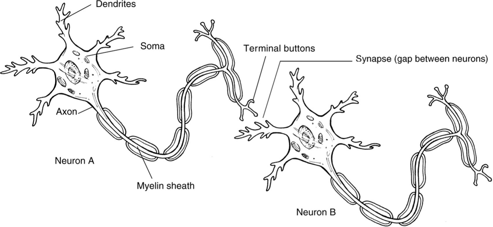
Figure 3.1. A neuron.
Neural firing is an electrochemical process. Electricity travels within the cell (from the dendrites to the terminal buttons), and chemicals (neurotransmitters) travel between cells in the synapse. Electricity does not jump between the neurons.
How a Neuron “Fires”
All of the different parts of the neuron work in sequence when a neuron transmits a message. In its resting state, a neuron has an overall slightly negative charge (–70mv) because mostly negative ions are within the cell and mostly positive ions are surrounding it. The cell membrane of the neuron is selectively permeable and prevents these ions from mixing. Visualize a two-neuron chain (see Fig. 3.1). The reaction begins when the terminal buttons of neuron A are stimulated and release neurotransmitters into the synapse. These neurotransmitters fit into receptor sites on the dendrites of neuron B. If enough neurotransmitters are received (this level is called the threshold), the cell membrane of neuron B becomes permeable and positive ions rush into the cell bringing the charge within the cell to approximately +40mv. The change in charge spreads down the length of neuron B like a bullet from a gun. This electric message firing is called an action potential. It travels quickly: 120 meters per second. When the charge reaches the terminal buttons of neuron B, the buttons release their neurotransmitters into the synapse. The process may begin again if enough neurotransmitters are received by that next cell to pass the threshold. Notice that a neuron either fires completely or it does not fire; this is called the all-or-none principle. If the dendrites of a neuron receive enough neurotransmitters to push the neuron past its threshold, the neuron will fire completely every time. A neuron cannot fire a little or a lot; the impulse is the same every time.
Neurotransmitters
You already know that neurotransmitters are chemicals held in the terminal buttons that travel in the synaptic gap between neurons. It is important to understand that different types of neurotransmitters exist. Some neurotransmitters are excitatory, meaning that they excite the next cell into firing. Other neurotransmitters are inhibitory, meaning that they inhibit the next cell from firing. Each synaptic gap at any time may contain many different kinds of inhibitory and excitatory neurotransmitters. The amount and type of neurotransmitters received on the receptor sites of the neuron determine whether it will pass the threshold and fire. Researchers are identifying different types and functions of neurotransmitters every year. This ongoing research makes generalizing about what each neurotransmitter does difficult. However, Table 3.1 indicates some of the more important types and functions of neurotransmitters to psychologists.
NERVOUS SYSTEM
We can sense the world because our nervous system brings information from our senses to our brain. Since a neuron fires in only one direction (from dendrite to terminal buttons), our body needs two sets of wires: one to take information to the brain and one to take instructions back from the brain to the muscles.
Afferent Neurons (or Sensory Neurons)
Afferent neurons take information from the senses to the brain. (You can think of afferent nerves as taking information in at the brain.)
Table 3.1. Neurotransmitters Important to Psychologists
|
Neurotransmitter |
Function |
Problems Associated with |
|
Acetylcholine |
Motor movement |
Lack of acetylcholine is associated with Alzheimer’s disease |
|
Dopamine |
Motor movement and alertness |
Lack of dopamine is associated with Parkinson’s disease, an overabundance is associated with schizophrenia |
|
Endorphins |
Pain control |
Involved in addictions |
|
Serotonin |
Mood control |
Lack of serotonin is associated with clinical depression |
|
GABA |
Important inhibitory neurotransmitter |
Seizures, sleep problems |
|
Glutamate |
Excitatory neurotransmitter, involved in memory |
Migraines, seizures |
|
Norepinephrine |
Alertness, arousal |
Depression |
Interneurons
Once information reaches the brain or spinal cord, interneurons take the messages and send them elsewhere in the brain or on to efferent neurons.
Efferent Neurons (or Motor Neurons)
Efferent neurons take information from the brain to the rest of the body. (You can think of efferent nerves as carrying information that exits the brain.)
ORGANIZATION OF THE NERVOUS SYSTEM
Our nervous system is divided into different categories based on function. The two main divisions are the central nervous system and the peripheral nervous system. These are then subdivided further (see Fig. 3.2).
The Central Nervous System
The central nervous system (CNS) consists of our brain and spinal cord—all the nerves housed within bone (the skull and vertebrae). Information about the structure and function of different parts of the brain is available in a later section. The spinal cord is a bundle of nerves that run through the center of the spine. It transmits information from the rest of the body to the brain.
The Peripheral Nervous System
The peripheral nervous system (PNS) consists of all the other nerves in your body—all the nerves not encased in bone. The peripheral nervous system is divided into two categories: the somatic and the autonomic nervous systems.
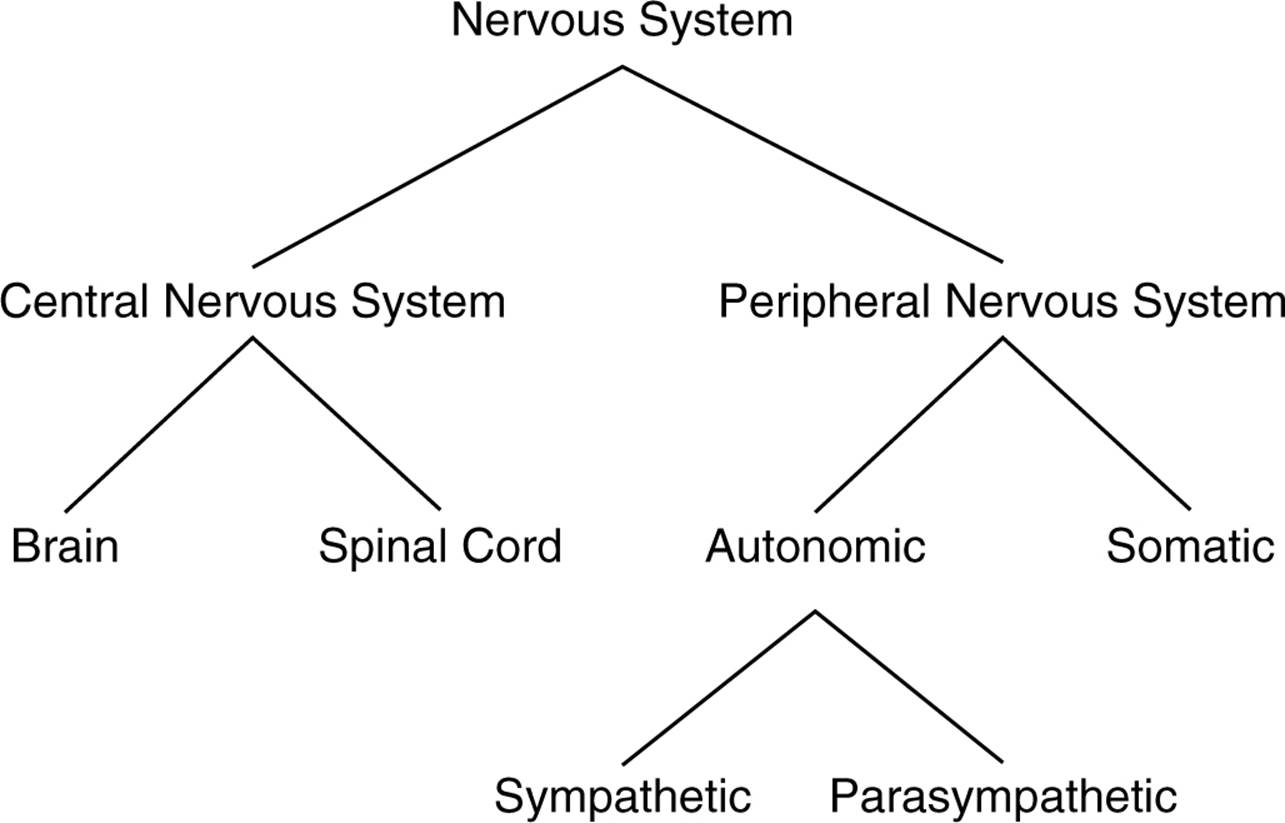
Figure 3.2. The nervous system.
SOMATIC NERVOUS SYSTEM
The somatic nervous system controls our voluntary muscle movements. The motor cortex of the brain sends impulses to the somatic nervous system, which controls the muscles that allow us to move.
AUTONOMIC NERVOUS SYSTEM
The autonomic nervous system controls the automatic functions of our body—our heart, lungs, internal organs, glands, and so on. These nerves control our responses to stress—the fight or flight response that prepares our body to respond to a perceived threat. The autonomic nervous system is divided into two categories: the sympathetic and parasympathetic nervous systems.
Sympathetic Nervous System
The sympathetic nervous system mobilizes our body to respond to stress. This part of our nervous systems carries messages to the control systems of the organs, glands, and muscles that direct our body’s response to stress. This is the alert system of our body. It accelerates some functions (such as heart rate, blood pressure, and respiration) but conserves resources needed for a quick response by slowing down other functions (such as digestion).
Parasympathetic Nervous System
The parasympathetic nervous system is responsible for slowing down our body after a stress response. It carries messages to the stress response system that causes our body to slow down. Think of the parasympathetic nervous system as the brake pedal that slows down the body’s autonomic nervous system.
Normal Peripheral Nervous System Transmission
Let us use an example to demonstrate how sensory information gets to our brain. While on a late-night quest for a snack, you stub your toe on a cast-iron coffee table. Sensory neurons in your toe are activated, and this message is transmitted up a neuron that runs from your toe to the base of your spine (afferent nerves). The message continues up your spinal cord on more afferent nerves until it enters your brain through the brain stem and is transmitted to the brain’s sensory cortex (see the next section, “Brain”) and you know you have stubbed your poor little toe. Your motor cortex now sends impulses down the spinal cord to the muscles controlling your leg and foot (efferent nerves), causing you to hop up and down holding your damaged limb, muttering under your breath.
Reflexes: An Important Exception
Most sensory information and muscle movements are controlled by the process described above. However, humans have a few reflexes that work differently. Certain reactions occur the moment sensory impulses reach the spinal cord. If you stimulate the correct area just below your kneecap, your leg will jerk without your conscious control. This sensory information is processed by the spine, and the spine tells your leg to move. The information reaches your brain and you realize your knee has been stimulated but only after this reflex has occurred. Another important reflex occurs in response to intense heat or cold. If we touch an object that is very hot or cold, our spine will send back a message jerking us away from that object. This might help keep us from harming ourselves, so it has adaptive value (it might help us survive, and therefore this trait is passed on to our children).
THE BRAIN
Possibly the most relevant part of biology to psychologists is the brain. As far as we can tell, the brain controls most of human thought and behavior. Researchers know quite a bit about brain anatomy and function, but many mysteries still remain about how the brain functions. Studying how the brain works is challenging because we cannot simply observe brain function the way we might observe a heart beating. To our eyes, a brain thinking looks exactly like a brain not thinking. Researchers are discovering many new details about how the brain works through experimentation and the use of technology. However, we still have a long way to go before we really understand how the brain controls our thoughts and behavior.
Ways of Studying the Brain
As mentioned previously, the first challenge of brain research is creating a way of detecting brain function. The following describes some of the methods researchers use.
ACCIDENTS
In 1848, a railroad worker named Phineas Gage was involved in an accident that damaged the front part of his brain. Gage’s doctor took notes documenting the brain damage and how Gage’s behavior and personality changed after the accident. Accidents like this give researchers clues about brain function. Gage became highly emotional and impulsive after the accident. Researchers concluded that the parts of the brain damaged in the accident are somehow involved in emotional control.
LESIONS
Lesioning is the removal or destruction of part of the brain. This is, of course, never done purely for experimental purposes. Sometimes doctors decide that the best treatment for a certain condition involves surgery that will destroy or incapacitate part of the brain. For example, a person may develop a brain tumor that cannot be removed without removing part of the surrounding brain. When these types of surgeries are performed, doctors closely monitor the patient’s subsequent behavior for changes. Any time brain tissue is removed (lesioning), researchers can examine behavior changes and try to infer the function of that part of the brain.
A famous historical example of lesioning is the frontal lobotomy. In the past, this surgery was used (many historians say overused) to control mentally ill patients with no other treatment options. Researchers knew that lesioning part of the frontal lobe would make the patients calm and relieve some serious symptoms. Drug treatments have now replaced frontal lobotomies.
ELECTROENCEPHALOGRAM
An electroencephalogram (EEG) detects brain waves. Researchers can examine what type of waves the brain produces during different stages of consciousness and use this information to generalize about brain function. The EEG is widely used in sleep research to identify the different stages of sleep and dreaming.
COMPUTERIZED AXIAL TOMOGRAPHY
A computerized axial tomography (CAT or CT) scan is a sophisticated X-ray. The CAT scan uses several X-ray cameras that rotate around the brain and combine all the pictures into a detailed three-dimensional picture of the brain’s structure. Note that the CAT scan can show only the structure of the brain, not the functions or the activity of different brain structures. A doctor could use a CAT scan to look for a tumor in the brain but would not get any information about how active different parts of the brain are.
MAGNETIC RESONANCE IMAGING
The magnetic resonance imaging (MRI) is similar to a CAT scan in a way: both scans give you pictures of the brain. The MRI, however, uses different technology to create more detailed images. An MRI uses magnetic fields to measure the density and location of brain material. Since the MRI does not use X-rays like the CAT scan does, the patient is not exposed to carcinogenic radiation. Like the CAT scan, the MRI gives doctors information about only the structure of the brain, not the function.
POSITRON EMISSION TOMOGRAPHY
The positron emission tomography (PET) scan lets researchers see what areas of the brain are most active during certain tasks. A PET scan measures how much of a certain chemical (glucose, for example) parts of the brain are using. The more used, the higher the activity. Different types of scans are used for different chemicals such as neurotransmitters, drugs, and oxygen flow.
FUNCTIONAL MRI
Functional MRI (fMRI) is a new technology that combines elements of the MRI and PET scans. An fMRI scan can show details of brain structure with information about blood flow in the brain, tying brain structure to brain activity during cognitive tasks.
Brain Structure and Function
All the different methods of studying the brain give researchers different types of information about brain structure and function. The brain is the most complicated organ in the body (in some ways, it is the most complex object we know of). Researchers categorize hundreds of different parts and functions of different parts of the brain. Because of this complexity, we need to divide the brain into separate categories in order to keep track of the information. When you study and think about the brain, think about three separate major categories or sections: the hindbrain, midbrain, and forebrain. Some evolutionary psychologists organize these categories into two major divisions: the “old brain” (hindbrain and midbrain) and the “new brain” (forebrain).
Some of the descriptions of brain function may seem vague or redundant when you read about the functions of other structures. Remember that some of the ways in which the brain works are still being investigated and the functions are just summarized here for our purposes. Keep the areas and general functions in mind instead of spending your time trying to figure out exact specific functions and locations.
HINDBRAIN
The hindbrain consists of structures in the top part of the spinal cord. The hindbrain is our life support system; it controls the basic biological functions that keep us alive. Some of the important specific structures within the hindbrain are the medulla, pons, and cerebellum (refer to Fig. 3.3 for locations of these structures).
Medulla
The medulla is involved in the control of our blood pressure, heart rate, and breathing. It is also known as the medulla oblongata and is located above the spinal cord.
Pons
The pons (located just above the medulla and toward the front) connects the hindbrain with the midbrain and forebrain. It is also involved in the control of facial expressions.
Cerebellum
The cerebellum (located on the bottom rear of the brain) looks like a smaller version of our brain stuck onto the underside of our brain. Cerebellum means little brain. The cerebellum coordinates some habitual muscle movements, such as tracking a target with our eyes or playing the saxophone.
MIDBRAIN
The midbrain (located just above the spinal cord but still below areas categorized as the forebrain) is very small in humans, but this area of the brain controls some very important functions. In general, your midbrain coordinates simple movements with sensory information. For example, if you turn your head right now, your midbrain coordinates with muscles in your eyes to keep them focused on this text. Different parts of the midbrain are important in various muscle coordinations. For purposes of the AP test, though, you should remember that this area is between the hindbrain and the forebrain and integrates some types of sensory information and muscle movements. One specific structure in the midbrain you should be familiar with is the reticular formation. It is a netlike collection of cells throughout the midbrain that controls general body arousal and the ability to focus our attention. If the reticular formation does not function, we fall into a deep coma.
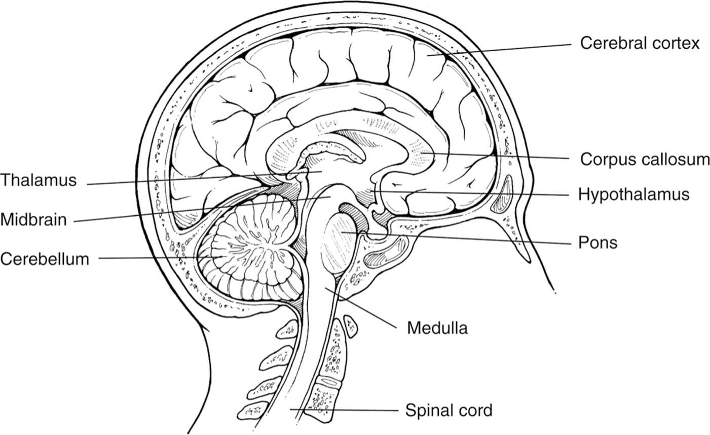
Figure 3.3. The brain.
FOREBRAIN
The various areas of the forebrain are very important to psychologists (and to students taking the AP psychology test). Areas of the forebrain control what we think of as thought and reason. Notice in Figure 3.3 how large the forebrain is in comparison with the other areas. The size of our forebrain makes humans human, and most psychological researchers concentrate their efforts in this area of the brain. Specific areas of interest to us in the forebrain are the thalamus, hypothalamus, amygdala, and hippocampus (the amygdala and hippocampus are not illustrated in Fig. 3.3).
Thalamus
The thalamus is located on top of the brain stem. It is responsible for receiving the sensory signals coming up the spinal cord and sending them to the appropriate areas in the rest of the forebrain (see the specific areas listed in the section about the cerebral cortex for specific examples of where some of these messages end up).
Hypothalamus
The hypothalamus is a small structure right next to the thalamus. The small size of the hypothalamus belies the importance of its functions. The hypothalamus controls several metabolic functions, including body temperature, sexual arousal (libido), hunger, thirst, and the endocrine system (see “Endocrine System” section later in this chapter). If you consider yourself a morning person or a night person, the hypothalamus might be involved since it controls our biological rhythms.
Amygdala and Hippocampus
There are two arms surrounding the thalamus. These are called the hippocampus. Structures near the end of each hippocampal arm are called the amygdala. The amygdala is vital to our experiences of emotion, and the hippocampus is vital to our memory system. Memories are not permanently stored in this area of the brain, however. Memories are processed through this area and then sent to other locations in the cerebral cortex for permanent storage. Researchers now know that memories must pass through this area first in order to be encoded because individuals with brain damage in this area are unable to retain new information.
Cerebral Cortex
When most people think of the human brain, they think of and picture the cerebral cortex. The gray wrinkled surface of the brain is actually a thin (0.039-inch [1-mm]) layer of densely packed neurons. This layer covers the rest of the brain, including most of the structures we have described. When we are born, our cerebral cortex is full of neurons (more than we have now, actually) but the neurons are not yet well connected. As we develop and learn, the dendrites of the neurons in the cerebral cortex grow and connect with other neurons. This process forms the complex neural web you now have in your brain. The surface of the cerebral cortex is wrinkled (the wrinkles are called fissures) to increase the available surface area of the brain. The more wrinkles, the more surface area contained within our skull. If our cerebral cortex were not wrinkled, our skull would have to be 3 square feet (0.3 m2) to hold all those neural connections!
These parts of the brain (thalamus, hypothalamus, amygdala, and hippocampus) are grouped together and called the limbic system because they all deal with aspects of emotion and memory. When you study the parts of the brain, grouping structurestogether according to function should help you remember them.
Hemispheres
The cerebral cortex is divided into two hemispheres: left and right. The hemispheres look like mirror images of one another, but they exert some differences in function. The left hemisphere gets sensory messages and controls the motor function of the right half of the body. The right hemisphere gets sensory messages and controls the motor function of the left half of the body (this is called contralateral control). Researchers are currently investigating other differences between the hemispheres, such as the possibility that the left hemisphere may be more active during logic and sequential tasks and the right during spatial and creative tasks. However, these generalizations need to be researched further before conclusions are drawn. This specialization of function in each hemisphere is called brain lateralization or hemispheric specialization. Most of this research in differences between the hemispheres is done by examining split-brain patients—patients whose corpus callosum (the nerve bundle that connects the two hemispheres, see Fig. 3.3) has been cut to treat severe epilepsy. The operation was pioneered by neuropsychologists Roger Sperry (1913–1994) and Michael Gazzaniga (1939–present). Split-brain patients also cannot orally report information only presented to the right hemisphere, since the spoken language centers of the brain are usually located in the left hemisphere.
Areas of the Cerebral Cortex
When you study the cerebral cortex, think of it as a collection of different areas and specific cortices. Think of the cerebral cortex as eight different lobes, four on each hemisphere: frontal, parietal, temporal, and occipital. Some of the major functions of these parts of the brain that are relevant to the AP test are mentioned here. Any area of the cerebral cortex that it is not associated with receiving sensory information or controlling muscle movements is labeled as an association area. Although specific functions are not known for each association area, these areas are very active in various human thoughts and behaviors. For example, association areas are thought to be responsible for complex, sophisticated thoughts like judgment and humor.
FRONTAL LOBES
The frontal lobes are large areas of the cerebral cortex located at the top front part of the brain behind the eyes (see Fig. 3.4). The anterior or front of the frontal lobe is called the prefrontal cortex and is thought to play a critical role in directing thought processes. The prefrontal cortex is said to act as the brain’s central executive and is believed to be important in foreseeing consequences, pursuing goals, and maintaining emotional control. Researchers believe this part of the brain is responsible for abstract thought and emotional control. The story of Phineas Gage mentioned previously exemplifies some of the functions of the prefrontal cortex. Phineas Gage’s limbic system was separated from his frontal lobes in an accident. Doctors reported that he lost control of his emotions and became impulsive and animalistic.
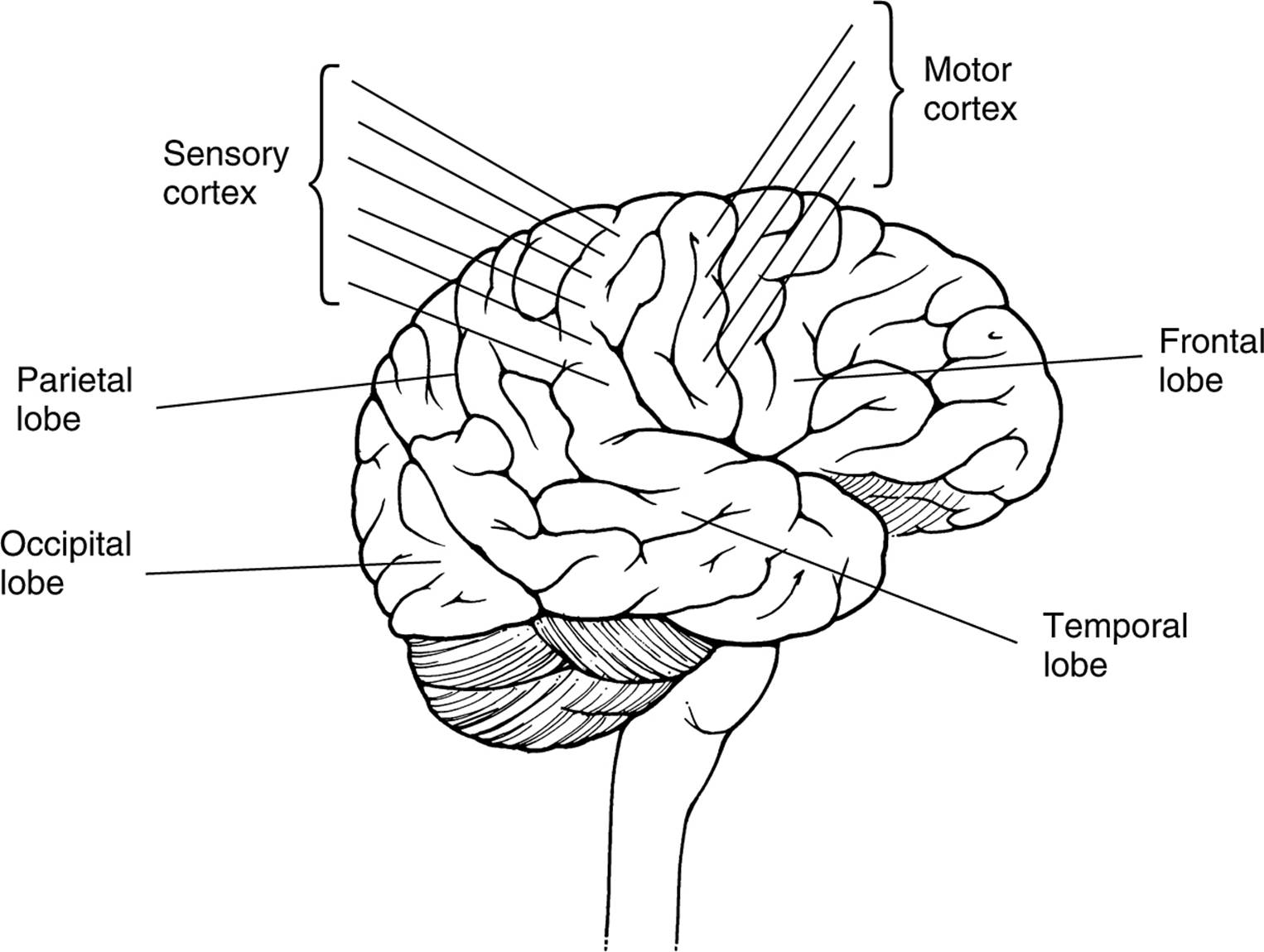
Figure 3.4. The lobes of the cerebral cortex.
In most people, the frontal lobe in the left hemisphere contains one of the two special areas responsible for language processing (some left-handed people’s language centers are in the right hemisphere). Broca’s area (Paul Broca,1824–1880) is in the frontal lobe and is responsible for controlling the muscles involved in producing speech. Damage to Broca’s area might leave us unable to make the muscle movements needed for speech. (The other area is Wernicke’s area (Carl Wernicke, 1848–1905) and is located in the temporal lobe—see that section for more information.)
A thin vertical strip at the back of the frontal lobe (farthest from the eyes, see Fig. 3.4) is called the motor cortex. This part of the cerebral cortex sends signals to our muscles, controlling our voluntary movements. The top of the body is controlled by the neurons at the bottom of this cortex (by the ears), progressing down the body as you go up the cortex. So the top of the motor cortex controls the feet and toes of the body.
PARIETAL LOBES
The parietal lobes are located behind the frontal lobe but still on the top of the brain (see Fig. 3.4). The parietal lobes contain the sensory cortex (also known as the somato-sensory cortex), which is located right behind the motor cortex in the frontal lobe. The sensory cortex is a thin vertical strip that receives incoming touch sensations from the rest of our body. The sensory cortex is organized similarly to the motor cortex. The top of the sensory cortex receives sensations from the bottom of the body, progressing down the cortex to the bottom, which processes signals from our face and head.
OCCIPITAL LOBES
Our occipital lobes are at the very back of our brain, farthest from our eyes. This is somewhat anti-intuitive since one of the major functions of this lobe is to interpret messages from our eyes in our visual cortex. (Study hint: the term occipital looks like the word optical to some students.) Impulses from the retinas in our eyes are sent to the visual cortex to be interpreted. Impulses from the right half of each retina are processed in the visual cortex in the right occipital lobe. Impulses from the left part of each retina are sent to the visual cortex in our left occipital lobe.
TEMPORAL LOBES
The temporal lobes process sound sensed by our ears. Sound waves are processed by the ears, turned into neural impulses, and interpreted in our auditory cortices. The auditory cortex is not lateralized like the visual cortices are. Sound received by the left ear is processed in the auditory cortices in both hemispheres. The second language area is located in the temporal lobe (the first was Broca’s area in the frontal lobe). Wernicke’s area interprets both written and spoken speech. Damage to this area would affect our ability to understand language. Our speech might sound fluent but lack the proper syntax and grammatical structure needed for meaningful communication.
Brain Plasticity
Researchers know some of the functions of different areas of the cerebral cortex, but they have also discovered that the brain is somewhat plastic or flexible. While these cortices and lobes usually perform the functions already mentioned, other parts of the brain can adapt themselves to perform other functions if needed. You already know that the cerebral cortex is made up of a complex network of neurons connected by dendrites that grow to make new connections. Since dendrites grow throughout our lives, if one part of the brain is damaged, dendrites might be able to make new connections in another part of the brain that would be able to take over the functions usually performed by the damaged part of the brain. Dendrites grow most quickly in younger children. Researchers know that younger brains are more plastic and are more likely to be able to compensate for damage.
ENDOCRINE SYSTEM
Another part of human biology relevant to psychology is the endocrine system. This is a system of glands that secrete hormones that affect many different biological processes in our bodies. As mentioned previously, the endocrine system is controlled in the brain by the hypothalamus. The endocrine system is complex, but a few elements of the entire process are especially relevant to psychologists.
Adrenal Glands
The adrenal glands produce adrenaline, which signals the rest of the body to prepare for fight or flight. This response was mentioned earlier in connection with the autonomic nervous system—the part of our nervous system that controls involuntary responses, such as heart rate and blood pressure.
Ovaries and Testes
Women’s ovaries and men’s testes produce our sex hormones, estrogen for women and testosterone for men. Research shows that levels of these hormones in men and women may partially explain gender differences demonstrated in certain experiments and situations. See the chapter “Developmental Psychology” for examples of these differences.
GENETICS
Besides the functioning of the brain and nervous system, another biological factor that affects human thought and behavior is genetics. Most human traits, like body shape, introversion, or temper, result from the combined effects of nature (our genetic code) and nurture (the environment where we grow up and live). Psychological researchers attempt to determine how much nature and nurture contribute to human traits.
Basic Genetic Concepts
Every human cell contains 46 chromosomes in 23 pairs. The genetic material that makes up chromosomes is DNA—deoxyribonucleic acid. Certain segments of DNA control the production of specific proteins that control some human traits. These discrete segments are called genes. Genes can be dominant or recessive. If we inherit two recessive genes for a particular trait, that trait will be expressed. In any other combination of genes, the dominant trait is expressed. Psychological researchers investigate how different combinations of genes create tendencies for physical and behavioral traits.
Twins
Since identical twins (called monozygotic twins since they develop from one fertilized egg called a zygote) share all the same genetic material, researchers study them in order to examine the influence of genes on human traits. In one famous study, Thomas Bouchard found more than 100 identical twins who were given up for adoption and raised in different families. The study compared hundreds of traits and concluded about the relative influences of genetics and the environment on specific traits. For example, the study found a correlation coefficient of 0.69 on the IQ test for identical twins raised apart and a 0.88 for identical twins living together. This shows that the environment has some effect on IQ score since twins raised in the same family have more similar IQs. However, the IQs of twins raised apart are still highly correlated, demonstrating that IQ is also heavily influenced by genetics. Twin studies like this one have been criticized in one important way, however. Even twins raised in separate families obviously share very similar physical appearances. This physical similarity may cause others to treat them in similar ways, creating the same effective psychological environment for both twins. This similarity in environment might explain the high correlations that Bouchard attributed to genetic influence.
Chromosomal Abnormalities
Our gender is determined by our twenty-third pair of chromosomes. Men have an X and Y chromosome, and women have two X chromosomes. Usually a man will contribute either an X chromosome to a child (resulting in a girl) or a Y (resulting in a boy). Occasionally, chromosomes will combine (or fail to) in an unusual way, resulting in a chromosomal abnormality. For example, babies with Turner’s syndrome are born with only a single X chromosome in the spot usually occupied by the twenty-third pair. Turner’s syndrome causes some physical characteristics, like shortness, webbed necks, and differences in physical sexual development. Babies born with Klinefelter’s syndromehave an extra X chromosome, resulting in an XXY pattern. The effects of this syndrome vary widely, but it usually causes minimal sexual development and personality traits like extreme introversion.
Other chromosomal abnormalities may cause mental retardation. The most common type is Down syndrome. Babies with Down syndrome are born with an extra chromosome on the twenty-first pair. Some physical characteristics are indicative of Down syndrome: rounded face, shorter fingers and toes, slanted eyes set far apart, and some degree of mental retardation.
PRACTICE QUESTIONS
Directions: Each of the questions or incomplete statements below is followed by five suggested answers or completions. Select the one that is best in each case.
1.Blindness could result from damage to which cortex and lobe of the brain?
(A)visual cortex in the frontal lobe
(B)visual cortex in the temporal lobe
(C)sensory cortex in the parietal lobe
(D)visual cortex in the occipital lobe
(E)cerebral cortex in the occipital lobe
2.Paralysis of the left arm might be explained by a problem in the
(A)motor cortex in the frontal lobe in the left hemisphere.
(B)motor cortex in the frontal lobe in the right hemisphere.
(C)sensorimotor cortex in the temporal lobe in the left hemisphere.
(D)motor cortex in the parietal lobe in the left hemisphere.
(E)motor cortex in the occipital lobe in the right hemisphere.
3.Deafness can result from damage to the inner ear or damage to what area of the brain?
(A) connections between the auditory nerve and the auditory cortex in the frontal lobe
(B)connections between the auditory nerve and the auditory cortex in the temporal lobe
(C)connections between the areas of the sensory cortex that receive messages from the ears and the auditory cortex
(D)connections between the hypothalamus and the auditory cortex in the temporal lobe
(E)connections between the left and right sensory areas of the cerebellum
4.According to the theory of evolution, why might we call some parts of the brain the old brain and some parts the new brain?
(A)Old brain parts are what exist in very young children, and the new brain develops later.
(B)The old brain developed first according to evolution.
(C)The old brain becomes more active as we grow older.
(D)The new brain deals with new information, while the old brain deals with information gathered when we were children.
(E)The old brain is most affected by age deterioration (dementias) while the new brain remains unaffected.
5.Which chemicals pass across the synaptic gap and increase the possibility the next neuron in the chain will fire?
(A)synaptic peptides
(B)inhibitory neurotransmitters
(C)adrenaline-type exciters
(D)excitatory neurotransmitters
(E)potassium and sodium
6.You eat some bad sushi and feel that you are slowly losing control over your muscles. The bacteria you ingested from the bad sushi most likely interferes with the use of
(A)serotonin.
(B)insulin.
(C)acetylcholine.
(D)thorazine.
(E)adrenaline.
7.The three major categories researchers use to organize the entire brain are the
(A)old brain, new brain, and cerebral cortex.
(B)lower, middle, and upper brain.
(C)hindbrain, midbrain, and forebrain.
(D)brain stem, limbic system, and cerebral cortex.
(E)neurons, synapses, and cerebral cortex.
8.A spinal reflex differs from a normal sensory and motor reaction in that
(A)a spinal reflex occurs only in response to extremely stressful stimuli.
(B)in a spinal reflex, the spine moves the muscles in response as soon as the sensory information reaches the spine while usually the impulse must reach the brain before a response.
(C)in a normal sensory/motor reaction, the spine transmits the information through afferent nerve fibers, while reflex reactions are transmitted along special efferent nerves.
(D)spinal reflexes are part of the central nervous system response, while normal sensory/motor reactions are part of the peripheral nervous system.
(E)spinal reflexes occur only in animals because humans are born without instinctual responses.
9.Antidepressant drugs like Prozac are often used to treat mood disorders. According to what you know about their function, which neurotransmitter system do these types of drugs try to affect?
(A)serotonin
(B)adrenaline
(C)acetylcholine
(D)endorphins
(E)morphine
10.Which sentence most closely describes neural transmission?
(A)An electric charge is created in the neuron, the charge travels down the cell, and chemicals are released that cross the synapse to the next cell.
(B)A chemical change occurs within the cell, the change causes an electric charge to be produced, and the charge jumps the gap between the nerve cells.
(C)The electric charge produced chemically inside a group of neurons causes chemical changes in surrounding cells.
(D)Neurotransmitters produced in the hindbrain are transmitted to the forebrain, causing electric changes in the cerebral cortex.
(E)Neural transmission is an electrochemical process both inside and outside the cell.
11.Dr. Dahab, a brain researcher, is investigating the connection between certain environmental stimuli and brain processes. Which types of brain scans is he most likely to use?
(A)MRI and CAT
(B)CAT and EKG
(C)PET and EEG
(D)EKG and CAT
(E)lesioning and MRI
12.Split-brain patients are unable to
(A)coordinate movements between their major and minor muscle groups.
(B)speak about information received exclusively in their right hemisphere.
(C)speak about information received exclusively in their left hemisphere.
(D)solve abstract problems involving integrating logical (left-hemisphere) and spatial (right-hemisphere) information.
(E)speak about information received exclusively through their left ear, left eye, or left side of their bodies.
13.When brain researchers refer to brain plasticity, they are talking about
(A)the brain’s ability to quickly regrow damaged neurons.
(B)the surface texture and appearance caused by the layer known as the cerebral cortex.
(C)the brain’s versatility caused by the millions of different neural connections.
(D)our adaptability to different problems ranging from survival needs to abstract reasoning.
(E)new connections forming in the brain to take over for damaged sections.
14.Mr. Spam is a 39-year-old male who has been brought into your neurology clinic by his wife. She has become increasingly alarmed by her husband’s behavior over the last four months. You recommend a CAT scan to look for tumors in the brain. Which two parts of the brain would you predict are being affected by the tumors?
List of symptoms: vastly increased appetite, body temperature fluctuations, decreased sexual desire, jerky movements, poor balance when walking and standing, inability to throw objects, and exaggerated efforts to coordinate movements in a task
(A)motor cortex and emotion cortex
(B)somato-sensory cortex and hypothalamus
(C)hypothalamus and cerebellum
(D)cerebellum and medulla
(E)thalamus and motor cortex
15.In most people, which one of following is a specific function of the left hemisphere that is typically not controlled by the right hemisphere?
(A)producing speech
(B)control of the left hand
(C)spatial reasoning
(D)hypothesis testing
(E)abstract reasoning