Classic Human Anatomy in Motion: The Artist's Guide to the Dynamics of Figure Drawing (2015)
Chapter 6
Muscles of the Arm and Hand
The muscles of the torso, examined in the previous chapter, include a few that attach directly into the upper arm and help move the humerus at the shoulder joint. In this chapter we will look at the other arm muscles as well as the muscles of the hands, seeing how they move the lower arm at the elbow joint, the hand at the wrist joint, and the fingers and thumb at the interphalangeal (IP) joints.
Some figures’ arms are fairly easy to depict, but others’ arms are more challenging. “Average” arms, which are basically cylindrical, streamlined structures, are relatively simple. In these arms, most muscles are inconspicuous because they are covered by a subcutaneous layer of fatty tissue, softening the surface form. A few muscles—the deltoid, biceps, triceps, and brachioradialis—may, however, exhibit interesting shapes even on nonmuscular arms, altering shape in various arm movements.
By contrast, the arms of a person with an athletic build have many well-defined muscular bulges and ridges. One potential difficulty in depicting a muscular arm has to do with the spiraling of muscle forms that occurs when the lower arm twists, which may cause confusion about what you are seeing on the surface form.
There are, however, ways to identify the basic muscles within specific arm regions using bony landmarks as guides. And learning the locations of the muscles of the upper and lower arm in a systematic way, as well as their roles in movement, will improve the accuracy of your depictions.
To comprehend the vast complexity of the arm region, anatomists organize arm muscles into groups. There are several ways to do this. Some texts categorize arm muscles according to which body parts they move: muscles that move the forearm, muscles that move the wrist, and so on. A variation of this is to group the muscles according to the roles they generally play in movement: flexors, extensors, pronators, supinators, abductors, and adductors.
Arm muscles can also be classified by their compartments or regions. Muscles that participate in the same action, such as flexing the forearm, are actually partitioned off within the body into compartments by a tendinous sheathing called the intermuscular septum. Each of these various compartments is related to a certain region of the arm—anterior, posterior, medial, or lateral—which helps clarify its general location.
Yet another way to categorize arm muscles is to group them according to their relationship to the surface. The muscles closest to the surface belong to the superficial muscle layer. If a region has more than one muscle layer, muscles farther from the surface may be identified as belonging to the intermediate muscle layer or the deep muscle layer.
In this chapter, I use a combination of such systems, identifying muscle groups by the regions they occupy, the actions they perform, and the layers in which they are positioned. The drawings here provide a visual overview of the muscles and muscle groups of the entire arm as seen from various views. (Note that the deltoid muscle was covered at the end of the previous chapter.)
MUSCLES OF THE UPPER AND LOWER ARM
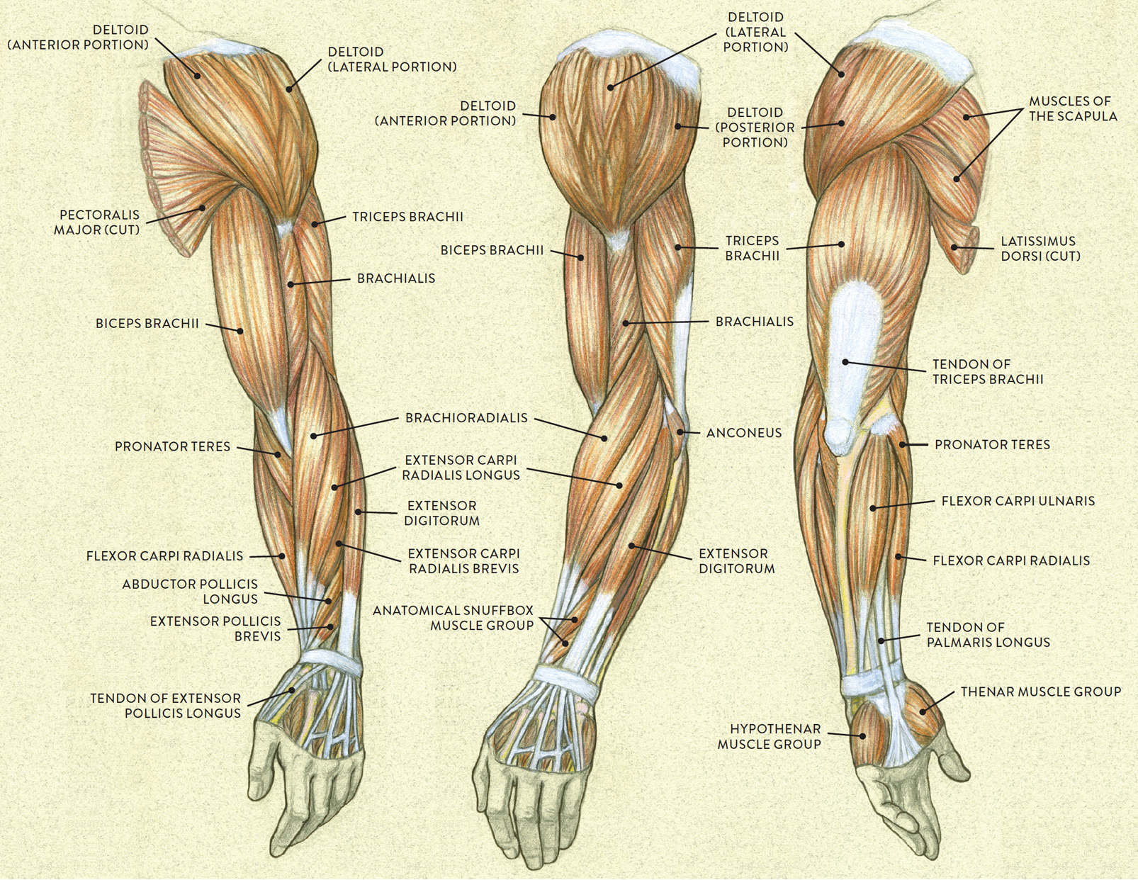
Left arm
LEFT: Anterior three-quarter view
CENTER: Lateral view
RIGHT: Posterior three quarter view
MUSCLES OF THE UPPER AND LOWER ARM
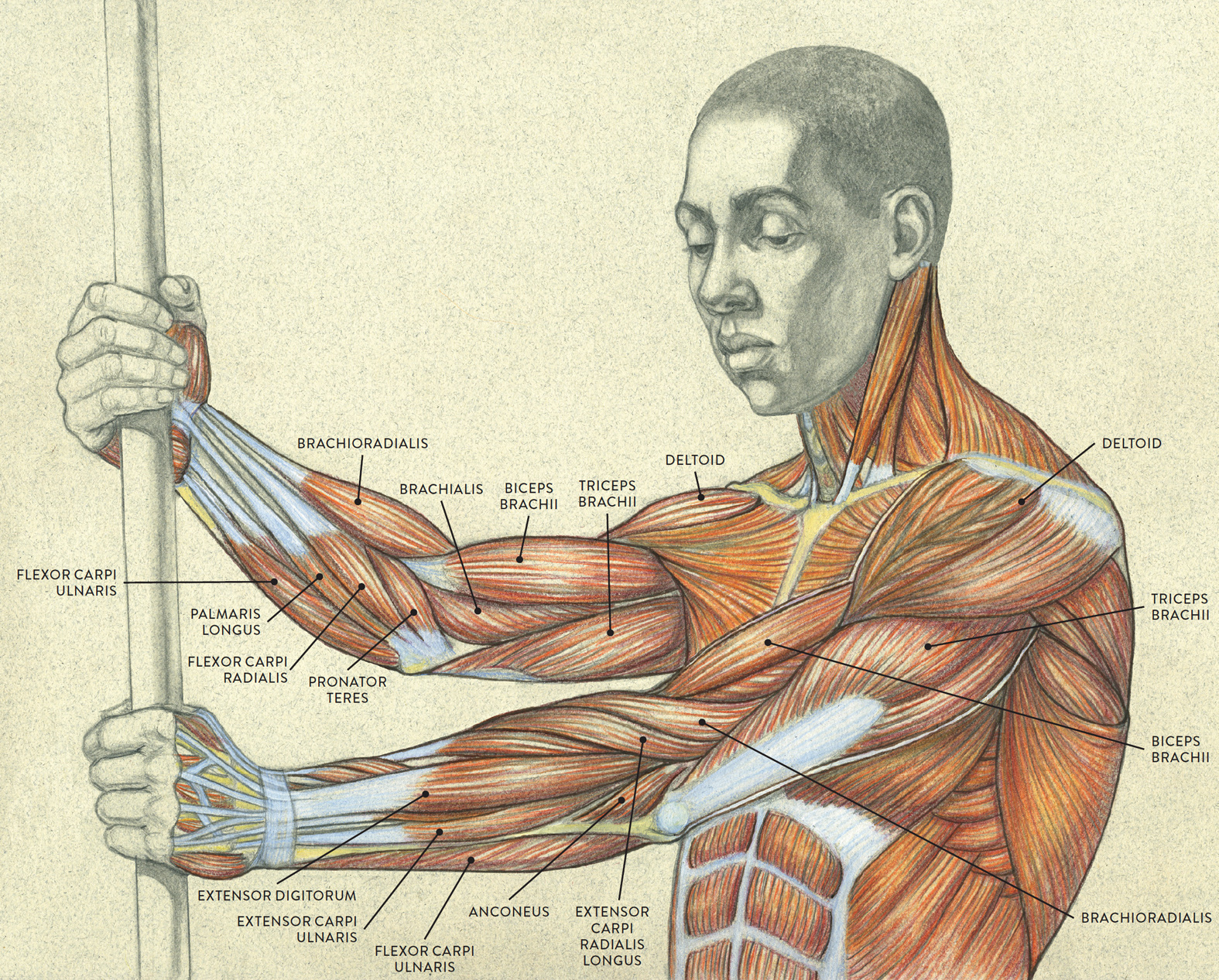
Upper torso, three-quarter view
MUSCLE GROUPS OF THE UPPER AND LOWER ARM, INCLUDING HAND
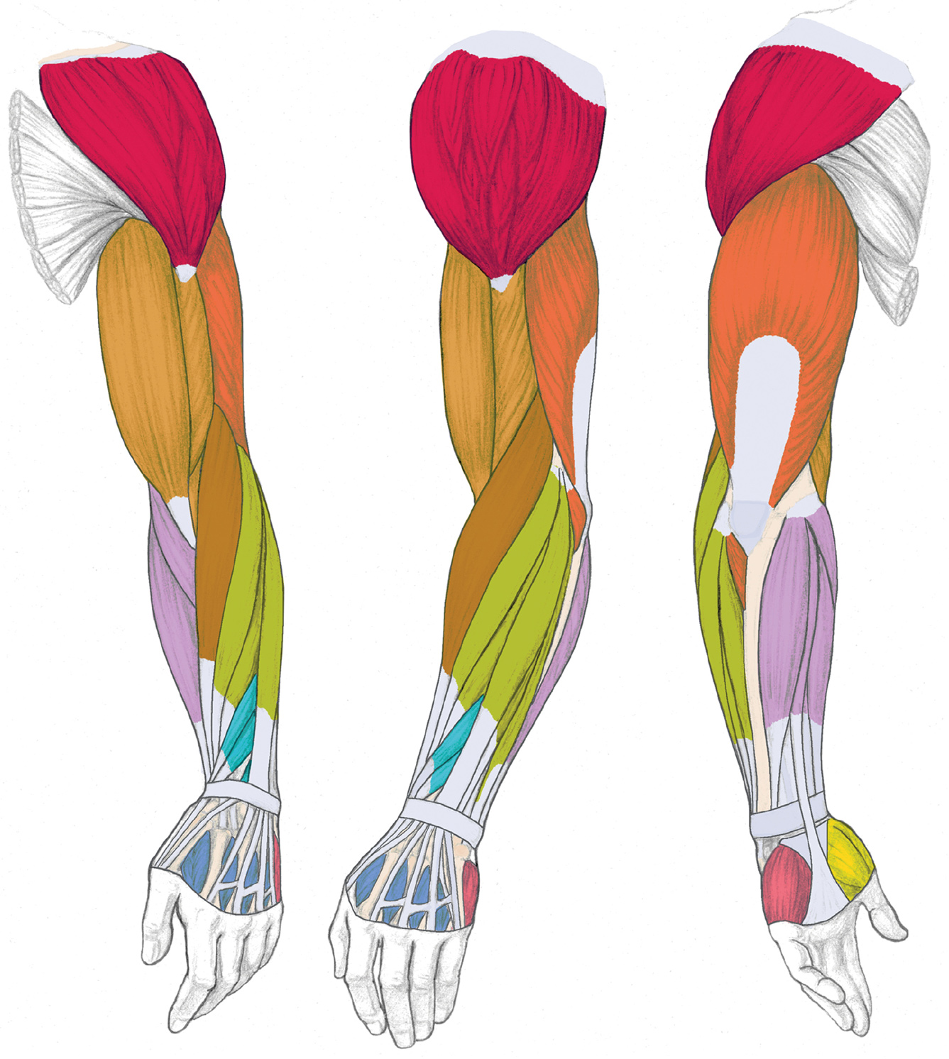
Left arm
LEFT: Anterior three-quarter view
CENTER: Lateral view
RIGHT: Posterior three quarter view
MUSCLES AND MUSCLE GROUPS OF THE UPPER AND LOWER ARM

DELTOID MUSCLE
FLEXOR MUSCLE GROUP OF UPPER ARM
EXTENSOR MUSCLE GROUP OF UPPER ARM
BRACHIORADIALIS (PART OF RADIAL MUSCLE GROUP)

EXTENSOR MUSCLE GROUP OF LOWER ARM (SUPERFICIAL LAYER)

FLEXOR MUSCLE GROUP OF LOWER ARM

EXTENSOR MUSCLE GROUP OF LOWER ARM (DEEP LAYER), ANATOMICAL SNUFFBOX MUSCLES
MUSCLE GROUPS OF THE HAND
THENAR MUSCLE GROUP

HYPOTHENAR MUSCLE GROUP
DORSAL INTEROSSEOUS MUSCLE GROUP
Muscle Groups of the Upper Arm
We begin our closer examination with the muscles of the upper arm by dividing them into two basic groups: the flexor muscle group, located on the anterior region of the upper arm, and the extensor muscle group, located on the posterior region.
Names of Arm and Hand Muscles
The names of arm and hand muscles provide clues to their location, function, or size.
· Brachialis, brachio, and brachii pertain to the upper arm.
· Carpi pertains to the carpal bones of the wrist.
· Coraco pertains to the coracoid process of the scapula.
· Digitorum and digiti pertain to the fingers (digits).
· Dorsal pertains to the back of the hand.
· Interosseous means “between bones.”
· Palmar and palmaris pertain to the palm side of the hand.
· Pollicis pertains to the thumb.
· Radialis pertains to the radius bone of the lower arm.
· Ulnaris pertains to the ulna bone of the lower arm.
· Abductor pertains to moving a body part away from the midline.
· Adductor pertains to moving a body part back toward the midline.
· Extensor pertains to stretching.
· Flexor pertains to bending.
· Brevis means “short.”
· Longus means “long.”
· Minimi means “little.”
· Profundus means “deep.”
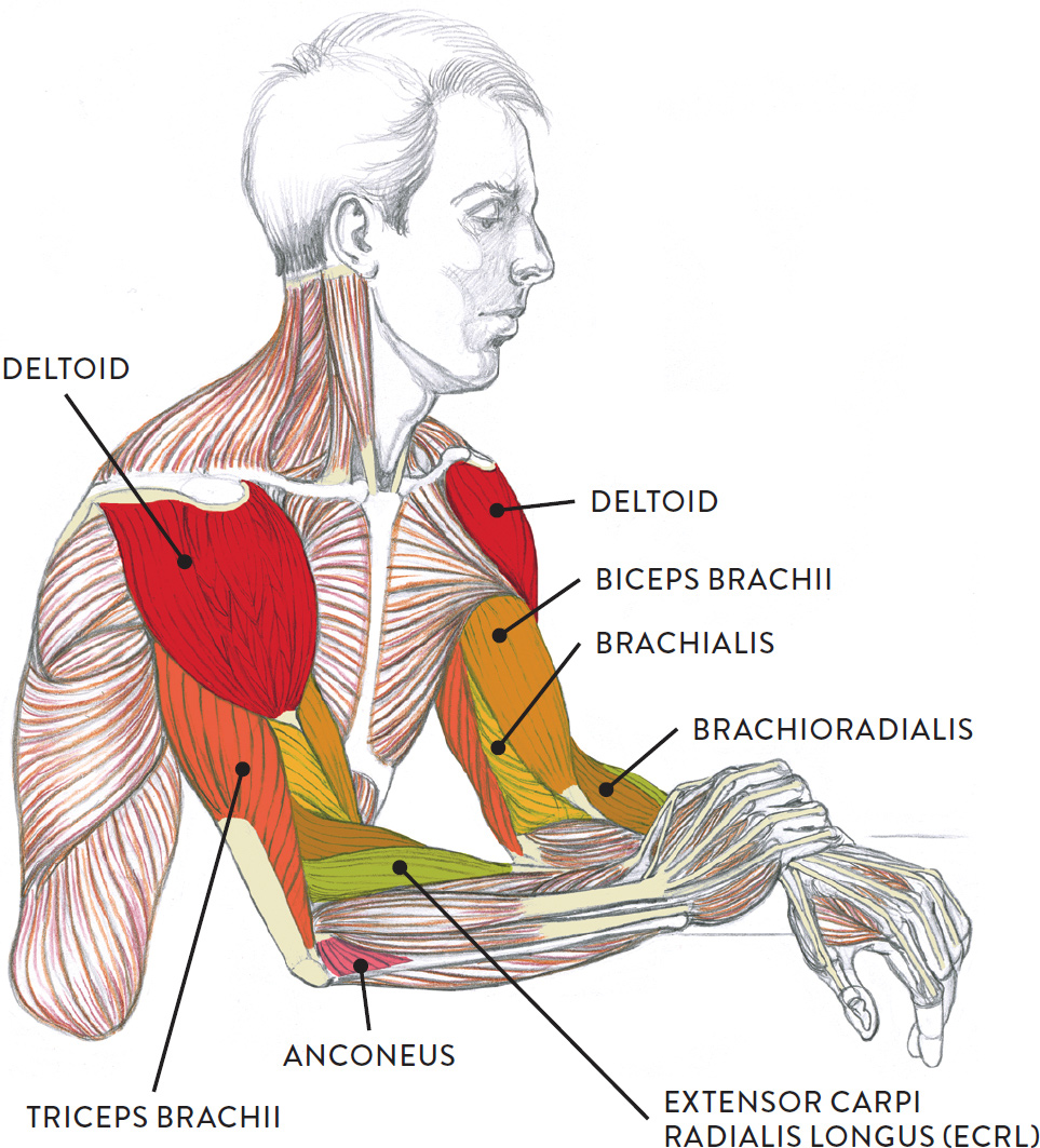
The Flexor Muscle Group of the Upper Arm
The flexor group of the upper arm is positioned on the anterior region of the humerus. Muscles belonging to this group are the biceps brachii, brachialis, and coracobrachialis. The biceps brachii is the most prominent, taking up most of the front of the upper arm region. The brachalis is mainly hidden by other muscles, with only a small area seen on the surface. The coracobrachialis, positioned behind the biceps brachii, can only be seen when the arm is lifted overhead. The biceps brachii and brachialis appear in the drawing opposite; the coracobrachialis is shown separately, in the following drawing.
CORACOBRACHIALIS
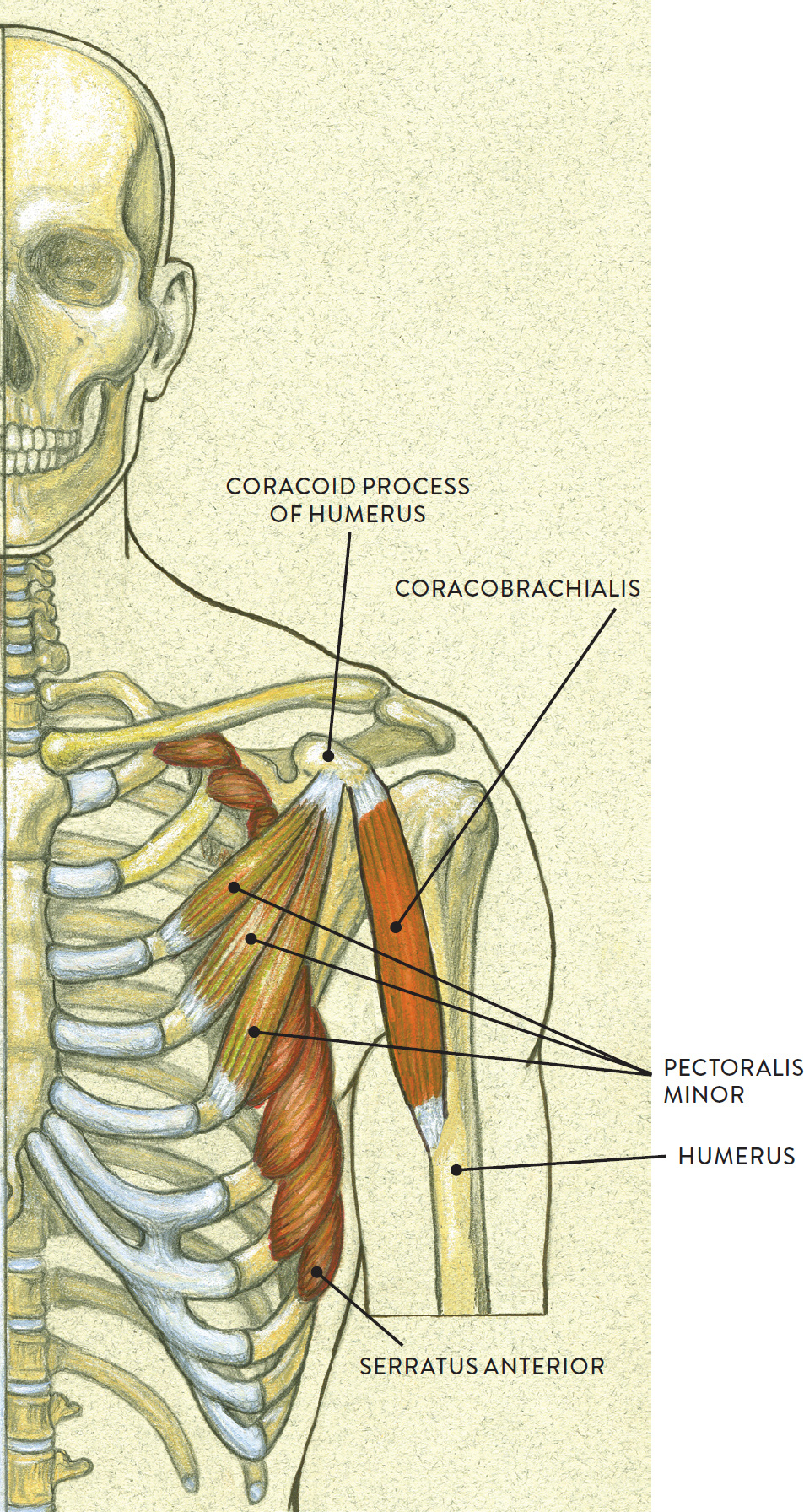
Skeletal torso, anterior view
The biceps brachii (pron., BI-seps BRAY-kee-ee or BI-seps BRAKE-ee-eye) is a two-headed fusiform muscle located on the front region of the upper arm, or humerus. Elongated when relaxed, the biceps brachii changes shape when its muscle fibers contract, becoming tightly compact. On muscularly defined arms, the biceps looks like a small melon when contracted.
The long head of the muscle begins on the scapula above the glenoid fossa at the supraglenoid tubercle, which is a small projection of bone. The short head begins on the coracoid process of the scapula. At the other end of the biceps brachii is a single tendon, which attaches on the radius bone of the lower arm on a small protrusion called the radial tuberosity. The biceps brachii moves the lower arm toward the upper arm from the elbow joint (flexion) and also rotates the lower arm from the elbow joint (supination).
The brachialis (pron., BRAY-kee-al-iss or bray-kee-AA-liss) is a slightly flattened fusiform muscle mostly covered by the biceps brachii. On an average arm the brachialis is hard to detect, but on a muscularly defined arm the muscle can been seen as a slight bulge between the triceps and the biceps. The brachialis begins on the lower half of the humerus and inserts into the coronoid process of the ulna. The muscle assists the biceps brachii in bending the forearm toward the upper arm from the elbow joint (flexion).
The coracobrachialis (pron., KOR-ah-ko-BRAY-kee-al-iss) is a small elongated fusiform muscle positioned beneath the biceps brachii of the upper arm. The muscle can only be seen on the surface when the upper arm is raised overhead, appearing as small ridgelike form next to the short head of the biceps brachii in the armpit region. The muscle begins on the coracoid process, a small bony projection, shaped like a bird’s beak, on the scapula. It then inserts into the medial side of the humerus. The coracobrachialis helps bend the humerus (flexion), rotates the humerus in an inward direction (medial rotation), and brings the abducted humerus back to its normal location (adduction).
FLEXOR MUSCLE GROUP OF THE UPPER ARM
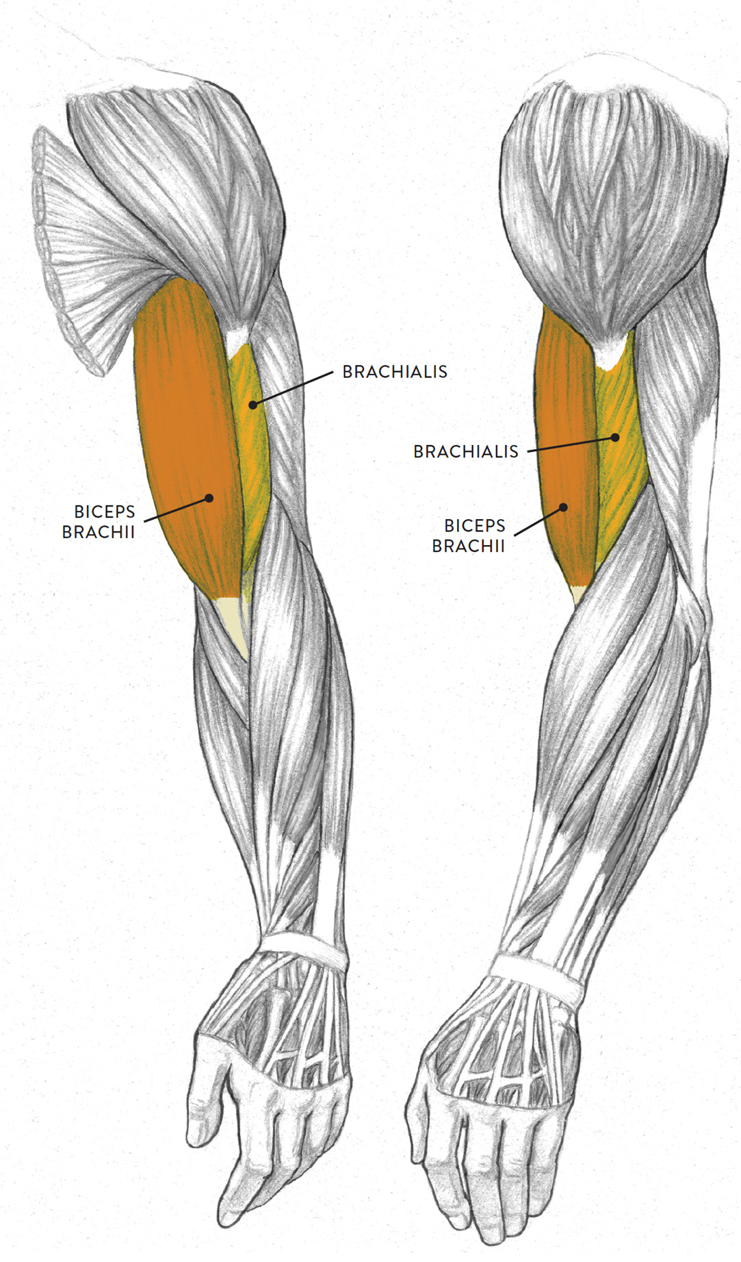
Left upper and lower arm
LEFT: Anterior three-quarter view
RIGHT: Lateral view
The Extensor Muscle Group of the Upper Arm
The two muscles of the extensor group of the upper arm are the triceps brachii and the anconeus, as shown in the following drawing. The triceps brachii is located on the posterior region of the upper arm, while the anconeus is located on the posterior region of the lower arm, near the elbow. They help straighten the lower arm (ulna) from a flexed or bent position.
EXTENSOR MUSCLE GROUP OF THE UPPER ARM
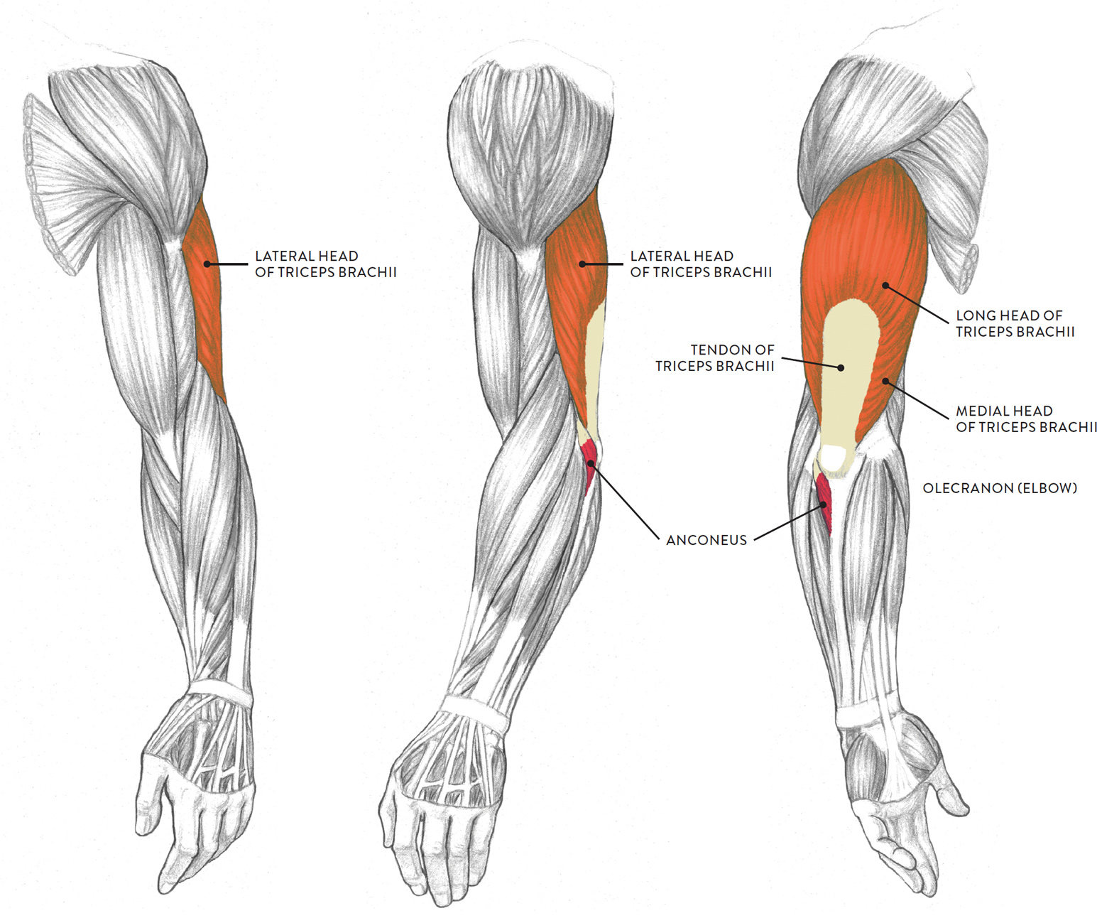
Left upper and lower arm
LEFT: Anterior three-quarter view
CENTER: Lateral view
RIGHT: Posterior three-quarter view
The triceps brachii (pron., TRI-seps BRAY-kee-ee or TRI-seps BRAKE-ee-eye) is a large three-headed muscle occupying most of the posterior aspect of the upper arm. The three heads are the lateral (outer) head, the long head, and the medial (inner) head. If the triceps of the upper arm is significantly developed, a fleshy horseshoe shape will appear on the surface when the muscle contracts—the combined shapes of the heads surrounding the neutral tendon. If the muscle is not well developed, then the back of the relaxed upper arm registers as a cylindrical shape, with very little indication of the triceps heads. On an “average” upper arm, the outer head and long head will appear as two bulges on the surface form only when the muscle is contracting.
The lateral head of the triceps brachii begins on the posterior and lateral surfaces of the humerus. The medial head begins on the posterior and medial surfaces of the humerus. The long head begins on the scapula below the glenoid fossa at a small triangular region called the infraglenoid tubercle. The muscle fibers of all three heads merge into a large flat tendon that inserts into the olecranon process (elbow) of the ulna. The triceps brachii straightens the lower arm at the elbow joint (extension). The long head assists in the action of adduction of the humerus, which is moving an abducted humerus back to the side of the body.
The anconeus (an-KOH-nee-us) is a small triangular muscle located between the olecranon (elbow) of the ulna and the radial muscle group. Its fleshy mass is at times noticeable on the surface, depending on the position of the arm. The muscle begins on the humerus, at the lateral epicondyle, and inserts into the ulna on the upper part of the bone’s posterior surface. Along with the triceps brachii, the anconeus helps straighten the lower arm at the elbow (extension).
The muscles of the upper arm can be clearly seen in the well-defined arms of the model in the life study Male Figure Resting with One Hand over the Other Arm. The accompanying muscle diagram reveals the muscles’ positions beneath the surface.
MALE FIGURE RESTING WITH ONE HAND OVER THE OTHER ARM
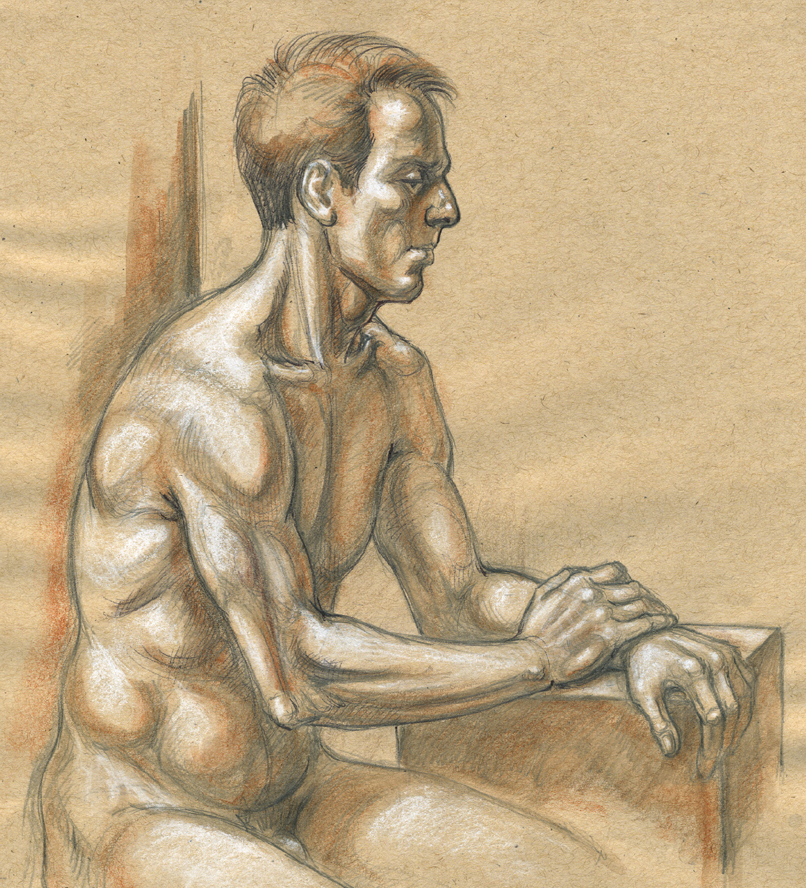
Graphite pencil, ballpoint pen, watercolor pencil, and white chalk on toned paper.
MUSCLE DIAGRAM
Muscle Groups of the Lower Arm
Of all the body’s muscles, those of the lower arm may be the most challenging to learn, for several reasons:
1. Some of these muscles have very similar names (e.g., flexor carpi ulnaris and extensor carpi ulnaris), which often causes confusion.
2. The various lower arm muscles look very similar to each other on écorché charts, appearing as slender muscles with extremely long tendons. (Écorché charts are diagrams showing the figure without skin or subcutaneous tissue, so that the muscles are clearly visible.)
3. On the surface, the muscles of the lower arm generally register as a single form rather than as distinct, obvious shapes like the biceps brachii or deltoid, making them difficult to locate.
4. When the lower arm rotates at the elbow joint, these muscles engage in a spiraling action, further contributing to the complexity of this region.
It is possible, however, to locate the muscles’ general whereabouts as the lower arm moves to different positions. One key is to keep an eye on the bony landmarks of the arms, such as the medial and lateral condyles of the humerus, which are important attachment sites for various lower arm muscles. Muscles also attach along the ulna, which can be located by tracing a line from the elbow (olecranon) down the little-finger side of the hand to the ulna head, the small protrusion of bone at the outer wrist. (The radius bone, located on the thumb side of the hand, is harder to detect on the surface.)
The lower arm has three muscle layers on the anterior region: the superficial layer, the intermediate layer, and the deep layer. On the posterior region, there are just two: the superficial layer and the deep layer. Although the discussion that follows focuses mostly on the superficial layer, the intermediate and deep layers are introduced to give you an idea of how the forms are built atop one another.
Like the muscles of the upper arm, some of the muscles of the lower arm are divided into flexor and extensor muscle groups located in the three layers (superficial, intermediate, and deep). Knowing the meanings of these terms can help you locate certain muscles and give you a better sense of their roles in movement. A muscle with the term flexor in its name is probably located on the anterior region of the lower arm, and its primary action is to flex, or bend, the hand and fingers (depending on the layer). A muscle with the term extensor in its name is probably located on the posterior region of the lower arm, and its primary action is to extend, or straighten, the hand, fingers, and thumb (again depending on the layer). In addition to the flexor and extensor muscle groups, the muscles of the lower arm include the radial muscle group, which is located in the lateral (outer) region of the lower arm. As its name implies, this group is positioned along the radius bone of the lower arm. Note that some lower arm muscles (e.g., the extensor carpi radialis longus) may be classified as belonging to more than one group.
The Flexor Muscle Group of the Lower Arm
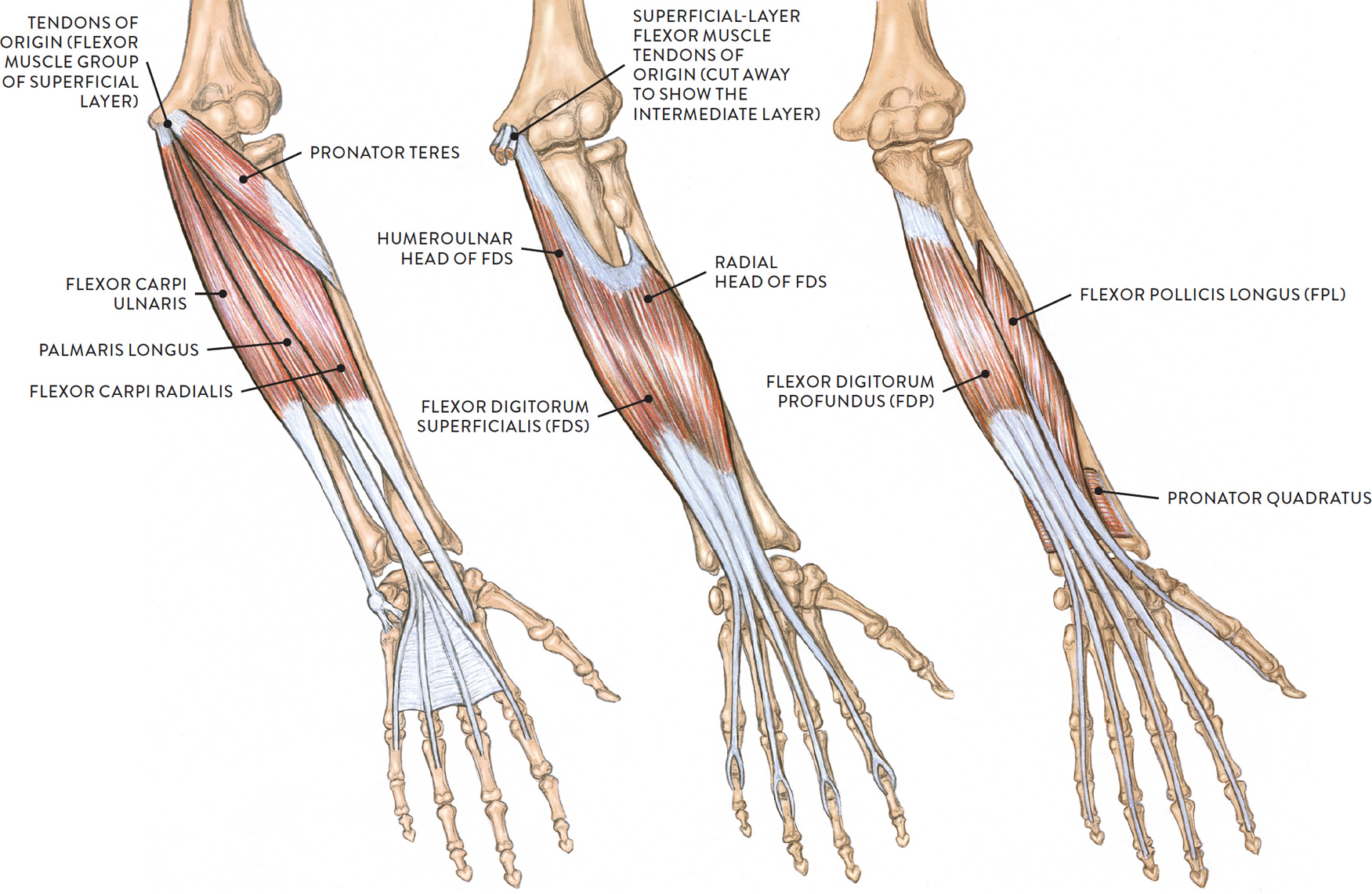
Let’s look first at the flexor muscle group of the anterior region of the lower arm. There are three muscle layers in this region: superficial, intermediate, and deep. The following drawing shows the three layers from the anterior view. In all three views, the lower left arm is positioned with the palm facing toward the viewer.
FLEXOR MUSCLE GROUP OF THE LOWER ARM
Lower left arm, anterior view, palm facing front
LEFT: Superficial layer
CENTER: Intermediate layer
RIGHT: Deep layer
Anterior Region, Superficial Muscle Layer (of the Flexor Muscle Group)
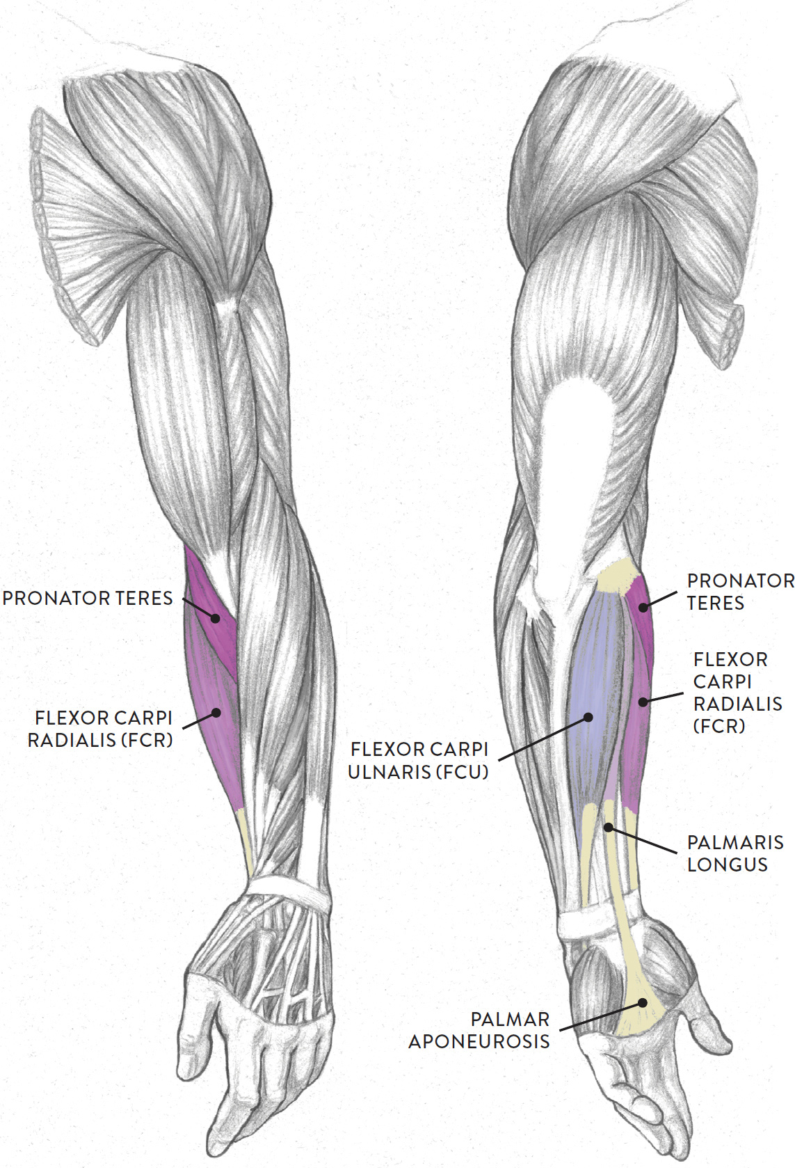
Four main muscles of the anterior region of the lower arm begin on the medial epicondyle of the humerus. These are the pronator teres, flexor carpi radialis, palmaris longus, and flexor carpi ulnaris. With the exception of the pronator teres, these muscles insert into the general anterior region of the hand and are responsible for moving the hand at the wrist joint.
The pronator teres (pron., pro-NAY-tor TEH-reez or PRO-nay-tor TEH-reez) has two heads: a humeral head, which begins on the medial epicondyle of the humerus, and an ulnar head, which begins on the corocoid process of the ulna bone. Both heads merge and insert about midway along the shaft of the radius bone. The pronator teres muscle assists in rotating the lower arm (pronation) and helps bend the lower arm at the elbow joint (flexion).
The flexor carpi radialis (pron., FLEK-sor KAR-pea ray-dee-AL-iss or FLEK-sor KARP-eye RAY-dee-ah-liss), or FCR, is a slender muscle positioned on the thumb side of the lower arm. The muscle begins on the medial epicondyle of the humerus and the antebrachial fascia, which is the deep fascia of the lower arm. It inserts by way of a long slender tendon into the bases of the second and third metacarpal bones of the hand. The flexor carpi radialis bends the hand toward the inner lower arm (flexion of the hand at the wrist joint) and assists in bending the hand sideways at the wrist (radial abduction of the hand at the wrist joint).
The palmaris longus (pron., pahl-MAR-iss LON-gus) is a thin muscle positioned in the middle of the lower arm. The muscle begins on the medial epicondyle of the humerus. Halfway down the shaft of the lower arm it becomes a slender tendon that, when it passes the wrist, flares out into a fan shape and inserts into the palmar aponeurosis, a fibrous sheathing of the palm. The palmaris longus helps bend the hand toward the inner lower arm (flexion of hand at wrist joint). It also helps tense or tighten the palmar aponeroneus and skin during certain hand movements, such as gripping an object.
The flexor carpi ulnaris (pron., FLEK-sor KAR-pea ull-NARE-iss or FLEK-sor KARP-eye ull-NARE-iss), or FCU, is positioned on the outer edge of the ulna bone for nearly its entire length. It has two heads: a humeral head and an ulnar head. The humeral head begins on the medial epicondyle of the humerus, and the ulnar portion begins near the olecranon (elbow) and the upper two-thirds of the posterior border of the ulna. The muscle inserts into the pisaform and hamate carpal bones, the fifth metacarpal bone, and the flexor retinaculum, which is a bracelet-like fibrous sheathing of the wrist region. The flexor carpi ulnaris bends the hand toward the inner lower arm (flexion of the hand at the wrist joint) and assists in bending the hand sideways (ulnar adduction at the wrist joint).
FLEXOR MUSCLE GROUP OF THE LOWER ARM, SUPERFICIAL LAYER
Left upper and lower arm
LEFT: Anterior three-quarter view
RIGHT: Posterior three-quarter view
Anterior Region, Intermediate Muscle Layer (of the Flexor Muscle Group)
The flexor digitorum superficialis (pron., FLEK-sor dij-ih-TOR-um SOO-pur-FISH-ee-AL-iss), or FDS, is positioned beneath the superficial-layer flexor muscles discussed just above. Some experts consider it part of the superficial muscle layer, hence its name. Others, however, classify it as belonging to an intermediate muscle layer between the superficial and deep layers of the lower arm.
The flexor digitorum superficialis has two heads: the humeroulnar head and the radial head. The humeroulnar head begins on the medial epicondyle of the humerus and the coronoid process of the ulna. The radial head begins on the radius. The muscle splits into four tendons, each of which inserts into the middle phalange of each finger. The flexor digitorum superficialis bends the fingers at the metacarpophalangeal (MCP) joints and proximal interphalangeal (PIP) joints (flexion of fingers). It also helps bend the hand (flexion of the hand at the wrist joint).
Anterior Region, Deep Muscle Layer (of the Flexor Muscle Group)
The three flexor muscles of the deep muscle layer are situated beneath the flexor digitorum superficialis muscle. These muscles—the flexor digitorum profundus, flexor pollicis longus, and pronator quadratus—move the fingers and thumb and help move the hand and lower arm (radius) at the wrist joint.
The flexor digitorum profundus (pron., FLEK-sor dij-ih-TOR-um pro-FUN-dus), or FDP, influences the surface form by creating a muscular mass of the inner forearm, along with the flexor carpi ulnaris. The muscle begins on the ulna and interosseous membrane (the connective tissue between the two bones of the lower arm) and then splits into four separate tendons, each inserting into a finger. The flexor digitorum profundus bends the fingers (flexion of fingers) at the distal interphalangeal (DIP) joints and helps bend the hand (flexion of hand at the wrist joint).
The flexor pollicis longus (pron., FLEK-sor pawl-lih-kiss LON-gus or FLEK-sor poe-LEE-siss LON-gus), or FPL, while being a deep-layer muscle, also contributes to the surface form on the anterior region of the lower arm, on the radial side. The muscle attaches on the radius and interosseous membrane and inserts into the base of the distal phalanx of the thumb. The flexor pollicis longus bends the thumb (flexion of the thumb) at the MCP and IP joints.
The pronator quadratus (pro-NAY-tor kwa-DRAH-tus) is a quadrangular muscle located on the lower portions of the radius and ulna bones. The muscle is so deep that some texts categorize it as belonging to a fourth muscle layer of the lower arm. The pronator quadratus begins on the ulna (medial and anterior surfaces) and inserts into the radius (anterior lateral surface). As its name implies, it helps rotate the lower arm and hand in the action of pronation.
The Extensor Muscle Group of the Lower Arm
Now we move on to the extensor muscle group of the posterior side of the lower arm, which has two muscle layers: superficial and deep. The muscles of these layers are shown in the following drawing. In both views, the lower left arm is positioned with the back (dorsal side) of the hand facing toward the viewer.
EXTENSOR MUSCLE GROUP OF THE LOWER ARM
Lower left arm, posterior view, back of hand facing front
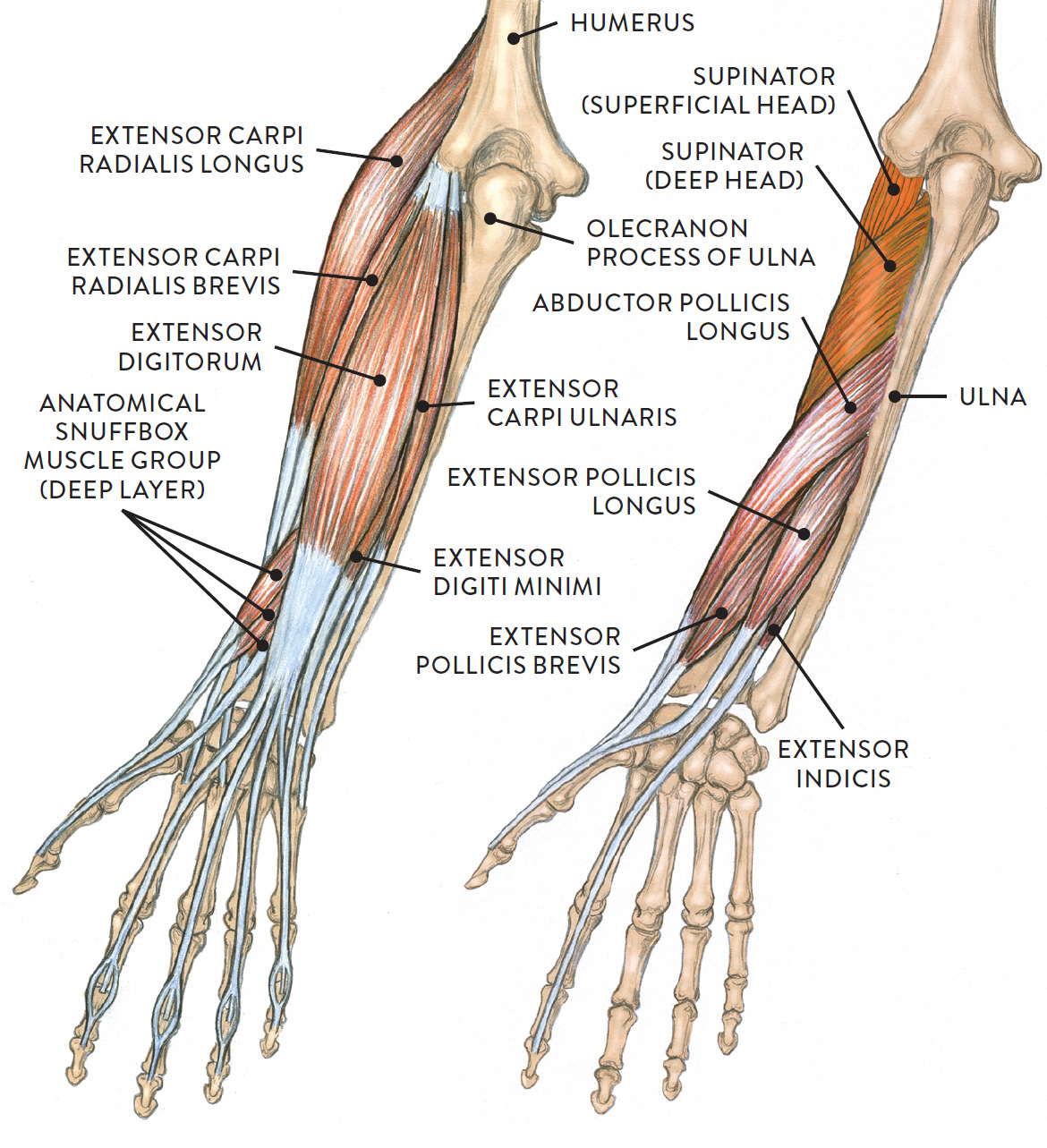
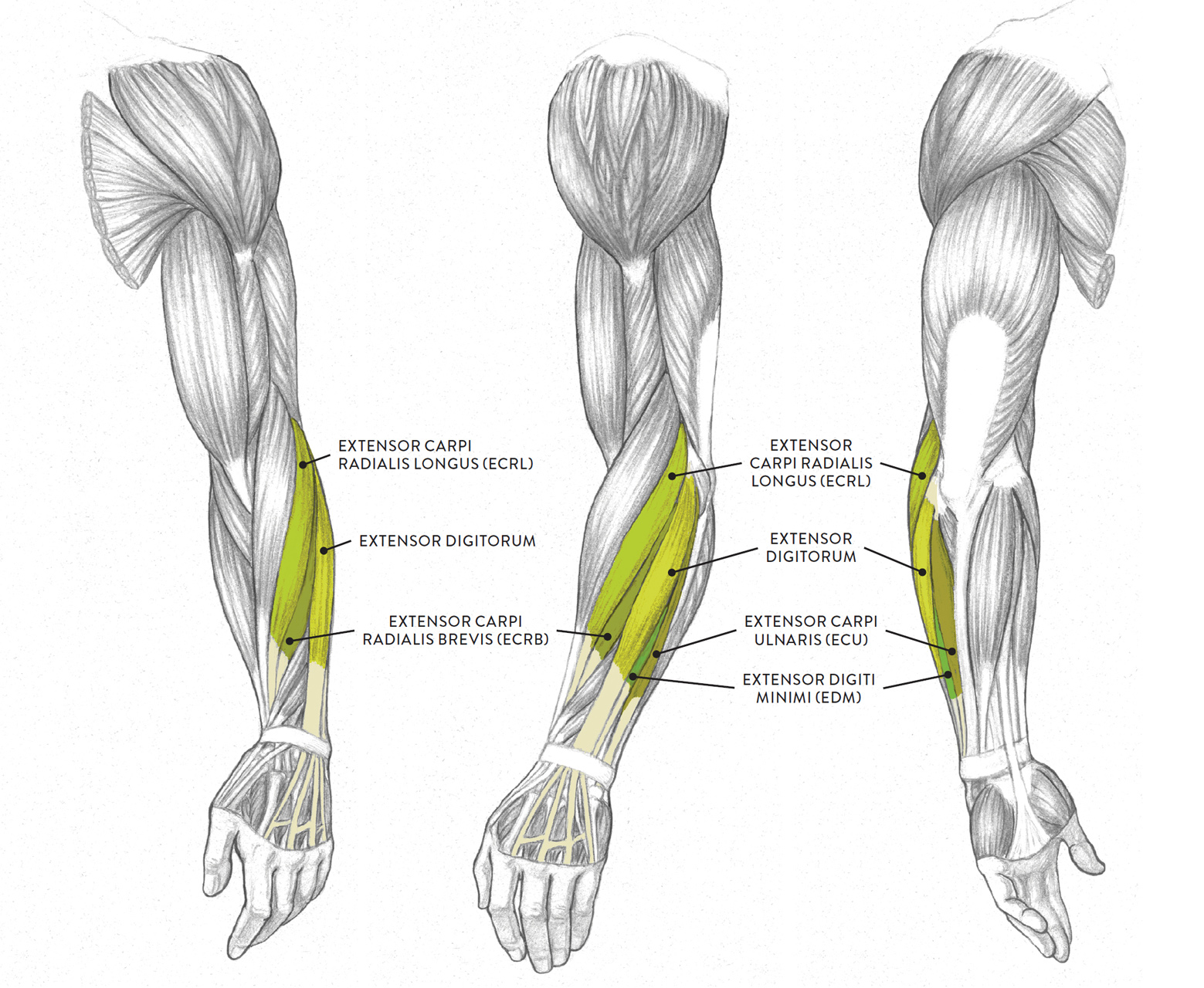
Posterior Region, Superficial Muscle Layer (of the Extensor Muscle Group)
The extensor muscle group of the superficial layer of the lower arm includes the extensor carpi radialis longus (ECRL), the extensor carpi radialis brevis (ECRB), the extensor digitorum, the extensor carpi ulnaris (ECU), and a small muscle called the extensor digiti minimi (EDM), all shown in the next drawing. All these muscles except the ECRL begin on the lateral epicondyle of the humerus, sharing a common tendon at this location. The ECRL begins above the lateral epicondyle of the humerus and does not share the common tendon of origin. The individual muscles insert by way of elongated tendons into various bones of the hand on the dorsal side. All four extensor muscles help move the hand at the wrist joint, and the extensor digitorum moves the fingers at the metacarpophalangeal (MCP) joints, proximal interphalangeal (PIP) joints, and the distal interphalangeal (DIP) joints.
EXTENSOR MUSCLE GROUP OF THE LOWER ARM, SUPERFICIAL LAYER
Left upper and lower arm
LEFT: Anterior three-quarter view
CENTER: Lateral view
RIGHT: Posterior three-quarter view
The extensor carpi radialis longus (pron., ek-STEN-sor KAR-pea ray-dee-AA-lis LON-gus or ek-STEN-sor KARP-eye RAY-dee-ah-liss LON-gus), or ECRL, is an elongated fusiform muscle that travels parallel to the brachioradialis. This muscle, which is also part of the radial muscle group of the lower arm, begins on the lower part of the humerus, above the lateral epicondyle and slightly below where the brachioradialis begins. It eventually inserts by way of a long, slender tendon on the second metacarpal bone of the hand. The ECRL helps straighten the hand at the wrist joint (extension) and also moves the hand in a side direction on the radial side of the lower arm (radial abduction).
The extensor carpi radialis brevis (pron., ek-STEN-sor KAR-pea ray-dee-AA-liss BREH-viss or ek-STEN-sor KARP-eye RAY-dee-ah-liss BREV-iss), or ECRB, is a slender muscle positioned between the ECRL and the extensor digitorum muscle on the posterior side of the lower arm. It begins on the lateral epicondyle of the humerus and inserts into the third metacarpal bone of the hand. The ECRB assists in straightening the hand at the wrist joint (extension) and in moving the hand in a sideways direction at the wrist (radial abduction).
The extensor digitorum (pron., ek-STEN-sor dij-ih-TOR-um) is positioned on the posterior side of the lower arm between the ECRB and the extensor carpi ulnaris. The muscle begins on the lateral epicondyle of the humerus. About halfway down its length, the muscle merges into a flat broad tendon, which then splits into four slender tendons at the wrist, which insert into the fingers via the four tendons. The extensor digitorum helps straightens the hand at the wrist joint and straightens the four fingers at the MCP joints (extension), PIP joints, and DIP joints.
The extensor digiti minimi (pron., ek-STEN-sor DIH-jih-tee MIN-ih-mee or ek-STEN-sor DIJ-ih-tie MIN-ih-my), or EDM, is an extremely slender fusiform muscle with an elongated tendon. It is linked to the extensor digitorum muscle. It begins on the lateral epicondyle of the humerus and inserts (along with the tendon of the extensor digitorum) into the fifth finger (little finger). The EDM straightens the fifth finger at the PIP and DIP joints.
The extensor carpi ulnaris (pron., ek-STEN-sor KAR-pea ull-NARE-riss or ek-STEN-sor KARP-eye ull-NARE-riss), or ECU, is a two-headed fusiform muscle with a long tendon that is positioned on the medial/posterior region of the lower arm. The humeral head begins on the lateral epicondyle, and the ulnar head begins on the posterior border of the ulna and inserts into the fifth metacarpal bone of the hand. The ECU straightens the hand at the wrist (extension) and moves the hand in a sideways direction at the wrist (ulnar adduction).
Posterior Region, Deep Muscle Layer (of the Extensor Muscle Group)
Located in the deep muscle layer of the posterior region of the lower arm is a group of muscles colloquially called the anatomical snuffbox muscles because their long tendons create an elongated triangular depression, called the anatomical snuffbox, in the skin near the wrist. In times past, it was fashionable for people to place a pinch of ground tobacco (snuff) in this depression and inhale it through their nostrils. Also known as the extrinsic thumb muscle group, the group has three muscles: the abductor pollicis longus, the extensor pollicis longus, and the extensor pollicis brevis. They begin on the ulna and radius bones, as well as on the interosseous membrane, and insert their tendons into the thumb bones. These tendons can be quite prominent on the surface when the thumb is extended in a “thumbs up” movement. These three muscles emerge in a diagonal direction from a location between the extensor digitorum and the extensor carpi radialis brevis (ECRB) muscles, unlike the muscles of the other layers, which descend in a vertical direction.
ANATOMICAL SNUFFBOX MUSCLES (EXTRINSIC THUMB MUSCLE GROUP)
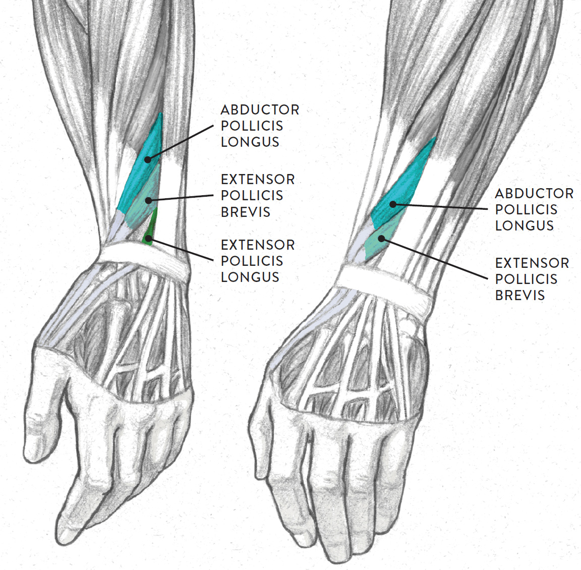
Dorsal region of left hand, two views
|
Anatomical Snuffbox Muscles—Pronunciation Guide |
|
|
MUSCLE |
PRONUNCIATION |
|
abductor pollicis longus |
ab-DUCK-tor PAWL-lih-kiss LON-gus |
|
extensor pollicis brevis |
ek-STEN-sor PAWL-lih-kiss BREV-iss |
|
extensor pollicis longus |
ek-STEN-sor PAWL-lih-kiss LON-gus |
Also located in the deep layer of the lower arm are two other muscles: the supinator and the extensor indicis. The supinator (pron., SOO-pih-NAY-tor) muscle is covered by the superficial muscles of the extensor group and is not seen on the surface form. This two-headed muscle begins on the humerus (superficial head) and ulna (deep head) and inserts into the radius. As its name implies, the supinator rotates the lower arm in the action of supination.
The extensor indicis (pron., ek-STEN-sor IN-dih-siss) is a narrow fusiform muscle that runs parallel to one of the anatomical snuffbox muscles (extensor pollicis longus). It begins on the posterior surface of the ulna and the interosseous membrane and inserts into the index finger. The extensor indicis straightens the index finger (extension) at the MCP, DIP, and PIP joints and also helps straighten the hand (extension) at the wrist joint.
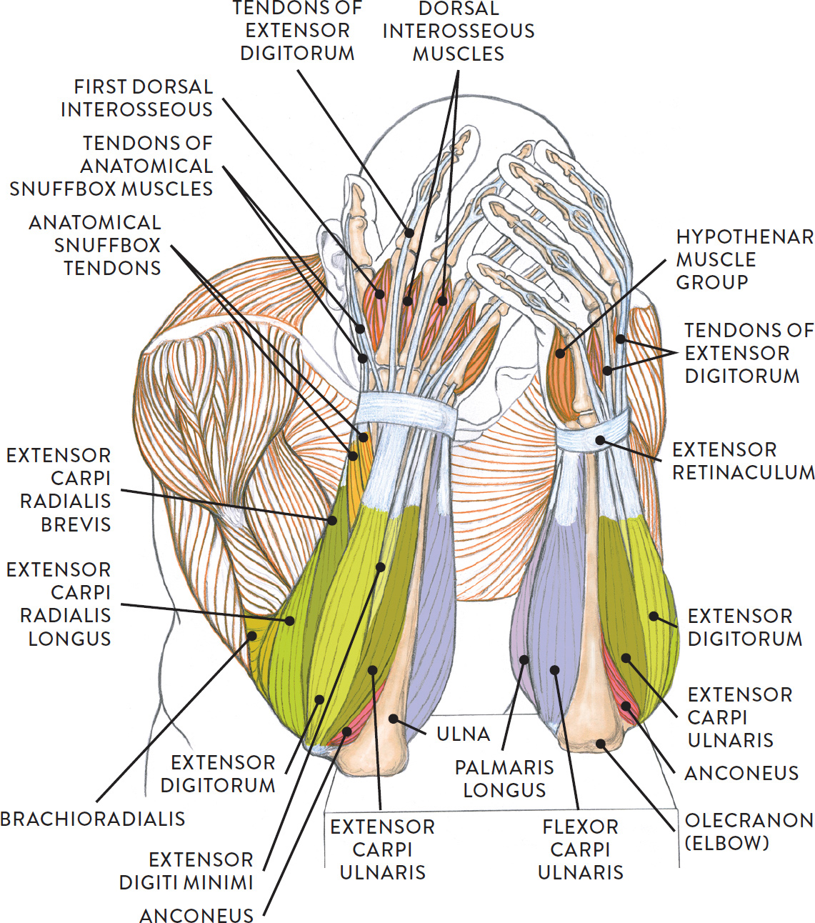
The life study Male Figure Clasping His Head in His Hands, shows a man with muscular lower arms and large hands. The accompanying muscle diagram reveals the positions of the lower arm muscles and their tendons in this pose.
MALE FIGURE CLASPING HIS HEAD IN HIS HANDS
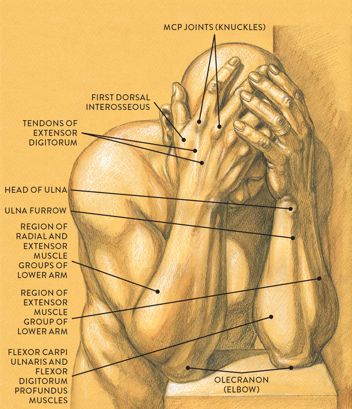
Graphite pencil, watercolor wash, and white chalk on toned paper.
MUSCLE DIAGRAM
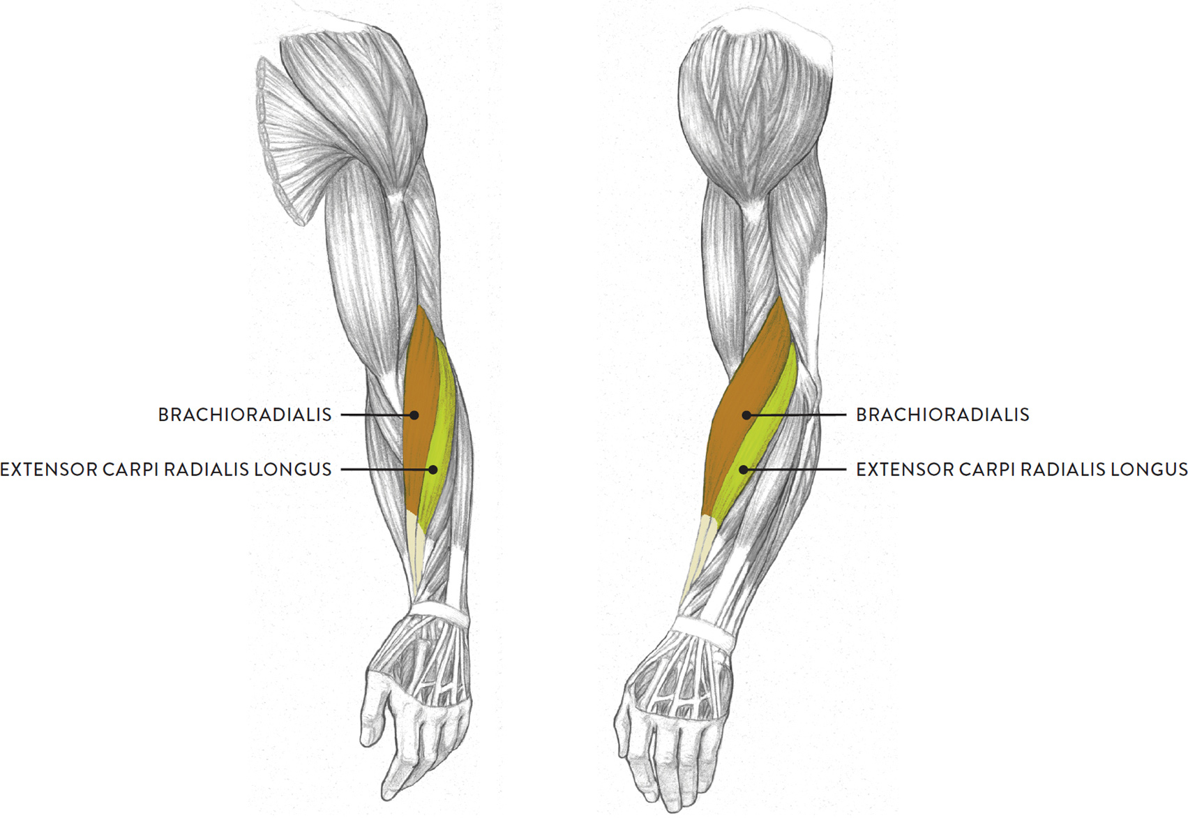
The Radial Muscle Group
Located in the superficial muscle layer of the lateral region of the lower arm, the radial muscle group provides an interesting transition between the upper arm and the lower arm. The radial group consists of two muscles: the brachioradialis and the extensor carpi radialis longus (ECRL). Some anatomists add a third muscle, called the extensor carpi radialis brevis (ECRB), to the group, but since this muscle is rather slender and hard to detect on the surface form except in muscularly defined arms, we focus here on the other two muscles, shown in the lower drawing opposite. The radial group moves the lower arm (radius and ulna) at the elbow joint and the hand at the wrist joint.
RADIAL MUSCLE GROUP
Left upper and lower arm
LEFT: Anterior three-quarter view
RIGHT: Lateral view
The radial group serves as an important surface landmark for artists. Its rich, elongated shape begins on the outer part of the humerus and spirals downward along the radius bone toward the thumb side of the wrist. The shapes of these two muscles register as a single form on the average arm but as individual muscular ridges on the athletically defined arm.
The next drawing shows the right arm in the anatomical position, slowly rotating from the elbow joint to move the hand from a position in which the palm faces forward (supination) to a position in which the back of the hand faces forward (pronation). While the extensor carpi radialis longus does not assist in this movement, its presence in the radial group is important in that it clearly shows the spiraling action of the lower arm, along with the brachioradialis. The brachioradialis (formerly known as the supinator longus) participates in both pronation and supination. Other muscles participating in the action of pronation are the pronator teres and the pronator quadratus; the biceps brachii and the supinator muscles participate in the action of supination.
ROTATION (SUPINATION AND PRONATION) OF THE LOWER ARM
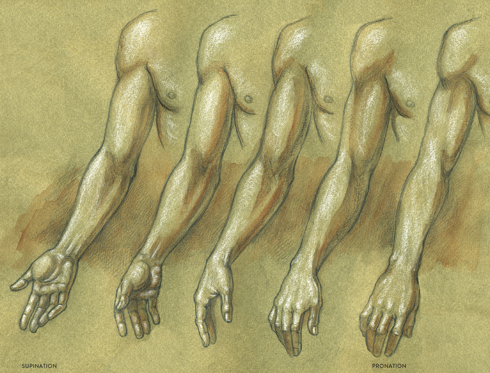
Right upper and lower arm, anterior view
The brachioradialis (pron., bray-kee-oh-ray-dee-AL-iss) is the most prominent shape of the radial group, appearing as a thick spiraling form near the elbow region. The brachioradialis begins on the outer side of the humerus and inserts by way of a long slender tendon into the lower portion of the radius. It is positioned between the extensor carpi radialis longus (of the radial muscle group) and the pronator teres and flexor carpi radialis (of the flexor muscle group). The brachioradialis helps bend the lower arm (flexion), and as stated above, it also assists in the supination and pronation of the lower arm. (The extensor carpi radialis longus has already been covered; see this page.)
Muscles of the Hand
The two sides of the hand have differing and very distinctive characteristics. The palmar (palm) region contains rich muscle forms and fibrous padding, while on the dorsal (back) side the skin pulls tightly over bones and tendons, creating a streamlined appearance.
MUSCLES AND TENDONS OF THE HAND
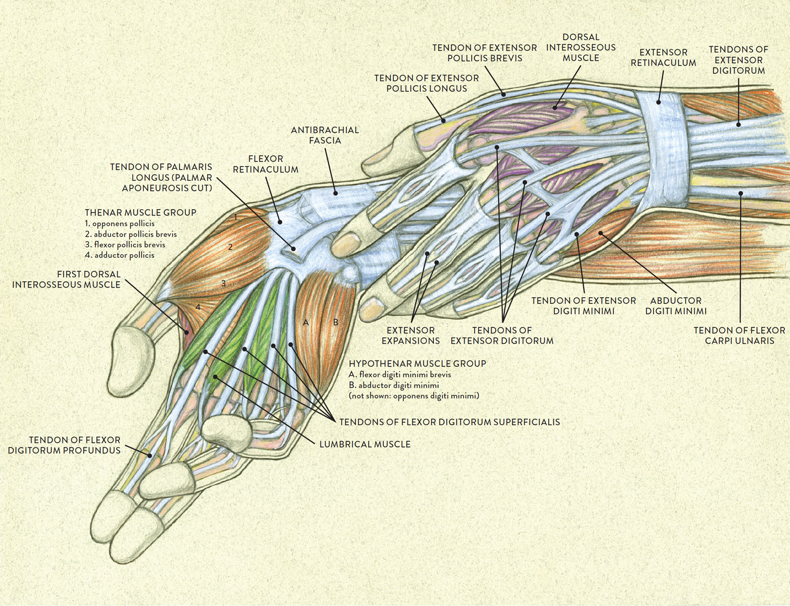
LEFT: Palmar side of right hand, anterior view
RIGHT: Dorsal side of left hand, posterior view
The dorsal region of the hand contains only one group of muscles, the dorsal interosseous muscle group. Except for the first dorsal interosseous muscle, these muscles are hidden from view on the surface. The palmar region of the hand has two muscle groups: the thenar muscle group and the hypothenar muscle group.
There are no muscles below the metacarpophalangeal joints (MCP joints, or knuckles) of the hand; instead, the fingers (including the thumb) contain sheaths for the many tendons of the muscles that move them, which originate on the lower arm. The palm and the palmar side of the fingers, especially at their tips, also contain fibrous padding (see this page).
In the following drawing Study of Theresa’s Hands, you can see the tendons of the extensor digitorum muscle pulling toward the knuckles (MCP joints). On the outer sides of the hands the hypothenar muscle group is partly visible.
STUDY OF THERESA’S HANDS
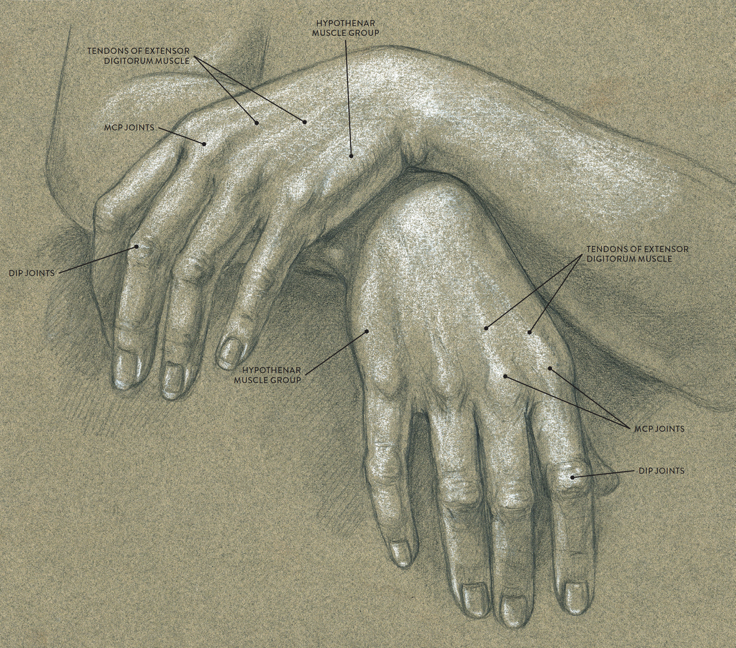
Graphite pencil and white chalk on toned paper.
The Interosseous Muscle Group
The term interosseous (pron., in-tur-OSS-ee-us) means “between bones,” and that is exactly where the muscles of the interosseous group attach—between the metacarpal bones of the hand. There are two different groups of interosseous muscles, each containing four muscles. The dorsal interosseous muscle group shown in the following drawing, is located on the dorsal region of the hand; when these muscles contract they mainly spread the fingers apart (abduction). The palmar interosseous muscle group (not shown) is located on the palm side of the hand; when these muscles contract they return the spread fingers to their normal position (adduction).
DORSAL INTEROSSEOUS MUSCLE GROUP
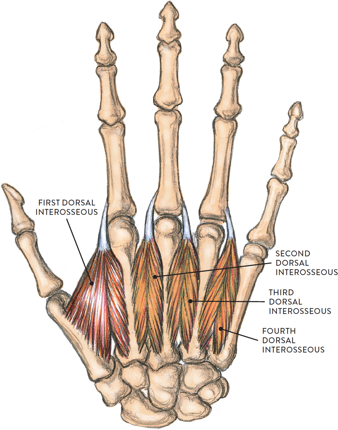
Dorsal side of right hand, posterior view
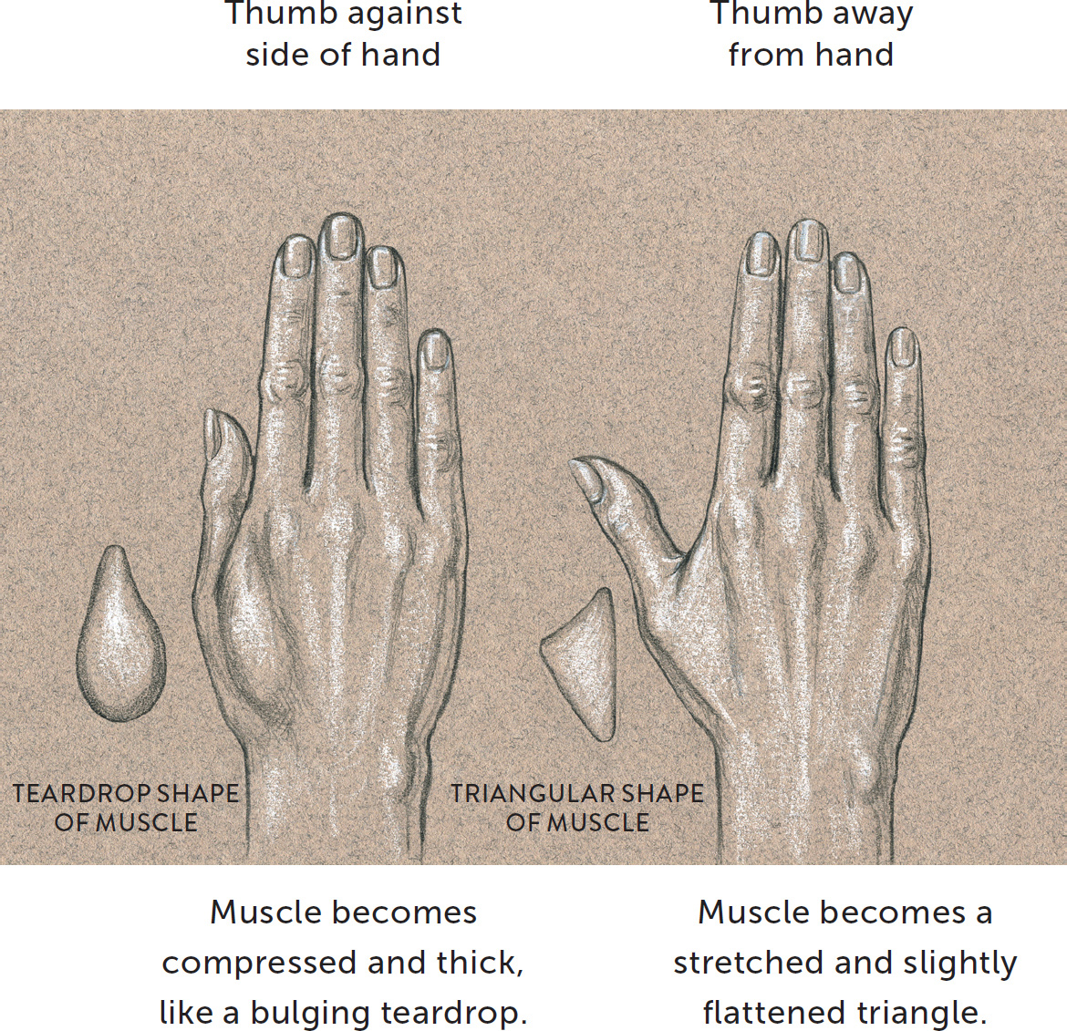
Only one interosseous muscle—the first dorsal interosseous—can be detected on the surface. The muscle fills the space between the metacarpal bone of the thumb and the metacarpal of the index finger. This triangular muscular space is sometimes referred to as the triangle of the thumb. The muscle changes shape depending on the placement of the thumb, as can be seen in the next drawing. It is easily seen when the thumb is pulled away from the hand in a sideways or downward direction. When the thumb is pressed against the side of the hand, the triangular shape becomes a thicker muscular mound—a teardrop shape on the dorsal side of the hand. Depending on how a person uses his or her hands, this shape may be large and quite thick or softer and subtler.
FIRST DORSAL INTEROSSEOUS MUSCLE—CHANGES IN SHAPE
The Thenar Muscle Group
The thenar group consists of four muscles that form the rich muscular mound found on the palm side of the hand below the thumb. It is commonly referred to as the thumb prominence or thumb eminence because of its large, distinctive shape on the palm. The muscles of the thenar group are the adductor pollicis (adductor of the thumb), abductor pollicis brevis (short abductor of the thumb), flexor pollicis brevis (short flexor of the thumb), and the opponens pollicis (which enables the thumb to oppose the other fingers). All except the opponens pollicis are shown in the following drawing. The names of these muscles are actually easier to remember than you might think. The word pollicis, which appears in each, means “thumb,” and each name tells you how the muscle makes the thumb move: adduction, abduction, flexion, and opposition. The muscle group attaches on various carpal bones, metacarpal bones, and the bracelet-like fibrous sheathing of the wrist region called the flexor retinaculum.
THENAR MUSCLE GROUP
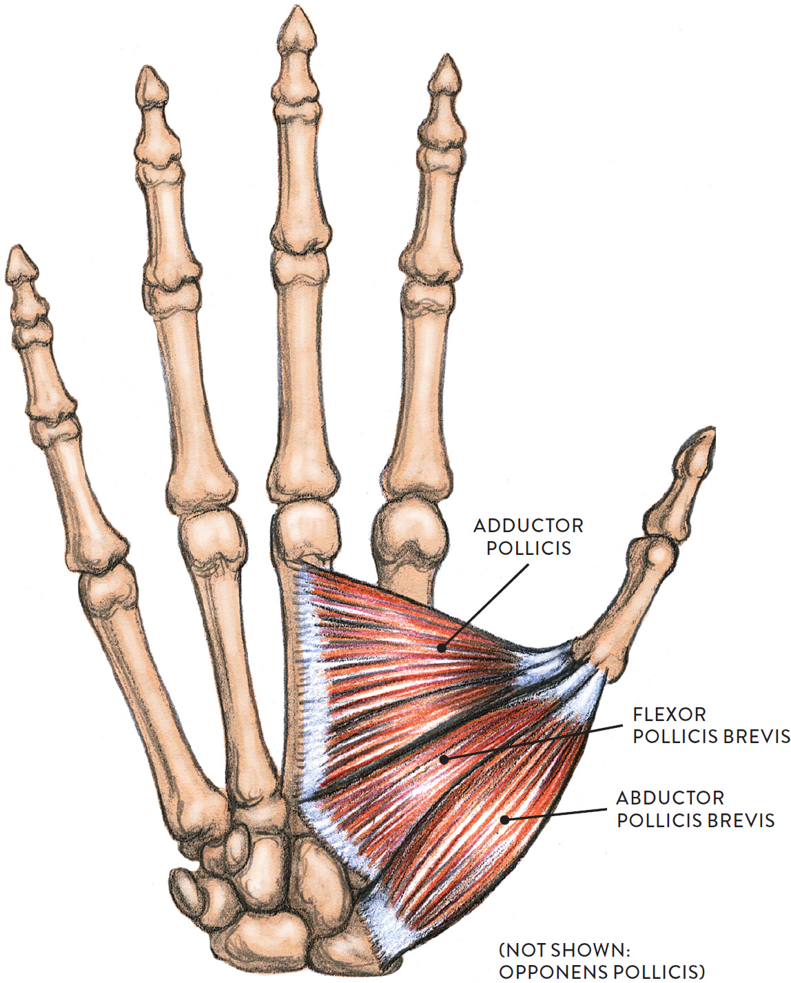
Palmar side of right hand
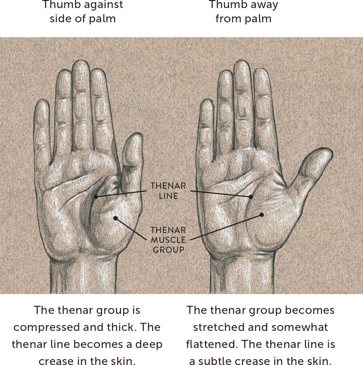
When drawing the palm side of the hand, you will usually detect the thenar group easily because of its large round shape positioned below the thumb. Since the muscles are covered with fibrous padding they usually cannot be distinguished individually, so it is best to approach the thenar group as a single form. The overall shape of the thumb and the thenar group is similar to that of a poultry drumstick, with the thumb representing the bony portion of the leg and the thenar muscles representing the thicker, meatier portion.
The shape and surface characteristics of the thenar group change depending on the placement of the thumb, as shown in the next drawing. When the thumb is positioned against the side of the palm, the thenar portion becomes compressed and thick, and the thenar line that travels around the outer perimeter of the thenar mass becomes a deep crease or fold within the skin. When the thumb is pulled away from the palm in a sideways direction, the thenar group becomes stretched and slightly flattened. The thenar line is still noticeable, but now as a subtle crease on the surface of the skin.
|
Thenar Group Muscles—Pronunciation Guide |
|
|
MUSCLE |
PRONUNCIATION |
|
adductor pollicis |
ah-DUCK-tor PAWL-lih-kiss |
|
abductor pollicis brevis |
ab-DUCK-tor PAWL-lih-kiss BREH-viss |
|
flexor pollicis brevis |
FLEK-sor PAWL-lih-kiss BREH-viss |
|
opponens pollicis |
oh-POE-nenz PAWL-lih-kiss |
THENAR MUSCLE GROUP—CHANGES IN SHAPE
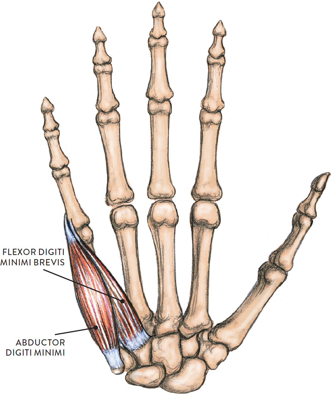
The Hypothenar Muscle Group
The hypothenar muscle group is an elongated teardrop form occupying the entire length of the side of the hand from the little finger to the heel of the hand. It consists of three muscles: the abductor digiti minimi, the flexor digiti minimi brevis, and the opponens digiti minimi. The abductor digiti minimi and flexor digiti minimi brevis are shown on this page (the opponens digiti minimi is not shown). As with the thenar group, it is not so hard to remember these muscles’ names, since the term digiti minimi means “little finger” and each muscle’s name tells you how it makes the little finger move: abduction, flexion, and opposition (in this case, enabling the little finger to oppose the thumb).
The opponens digiti minimi is positioned beneath the other two muscles and is hard to detect in most écorché views. Its shape, however, contributes to the fleshiness of the hypothenar group. The muscles are covered with fibrous padding, and so it is easier to see and depict them as a single form rather than individual muscles. The group is seen in palmar and side views of the hand on the little finger side.
HYPOTHENAR MUSCLE GROUP
(NOT SHOWN: OPPONENS DIGITI MINIMI)
Palmar side of right hand
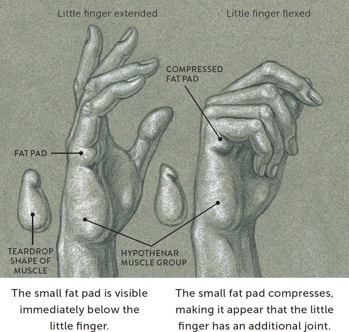
Immediately below the little finger is a small fat pad that is part of an elongated fat pad that runs horizontally across the palm at the base of the fingers. At the lower border of this elongated pad is a flexor crease called the distal palmar crease, or heart line. When the little finger bends, this outer portion of the fat pad form becomes compressed and the flexor crease is etched more deeply in the skin, creating the illusion that the little finger has an additional joint.
|
Hypothenar Group Muscles—Pronunciation Guide |
|
|
MUSCLE |
PRONUNCIATION |
|
abductor digiti minimi |
ab-DUCK-tor DIH-jih-tee MIN-ih-mee |
|
flexor digiti minimi brevis |
FLEK-sor DIH-jih-tee MIN-ih-mee BREV-iss |
|
opponens digiti minimi |
oh-POE-nenz DIH-jih-tee MIN-ih-mee |
The following drawing consists of two life studies of the right hand, showing the overall teardrop appearance of the hypothenar group. On one hand the fingers are extended but relaxed; on the other they are flexed, enabling you to see how the fat pad at the base of the little finger becomes compressed when the little finger bends.
HYPOTHENAR MUSCLE GROUP—TEARDROP SHAPE
Right hand, lateral view
I recommend that you practice drawing hands in many positions—relaxed, dynamically positioned, or holding objects. After sketching the initial shape of the hand and fingers, add any anatomical forms (thenar and hypothenar muscle groups, triangle of the thumb, tendons, knuckles) that can be seen on the surface. Such studies can be done from your own non-drawing hand, other people’s hands, or photo sources. The drawings in Three Sketchbook Studies of Hands, were done from a model holding his hands in different positions.
STUDY OF TWO HANDS—ONE HOLDING AN ACE CARD
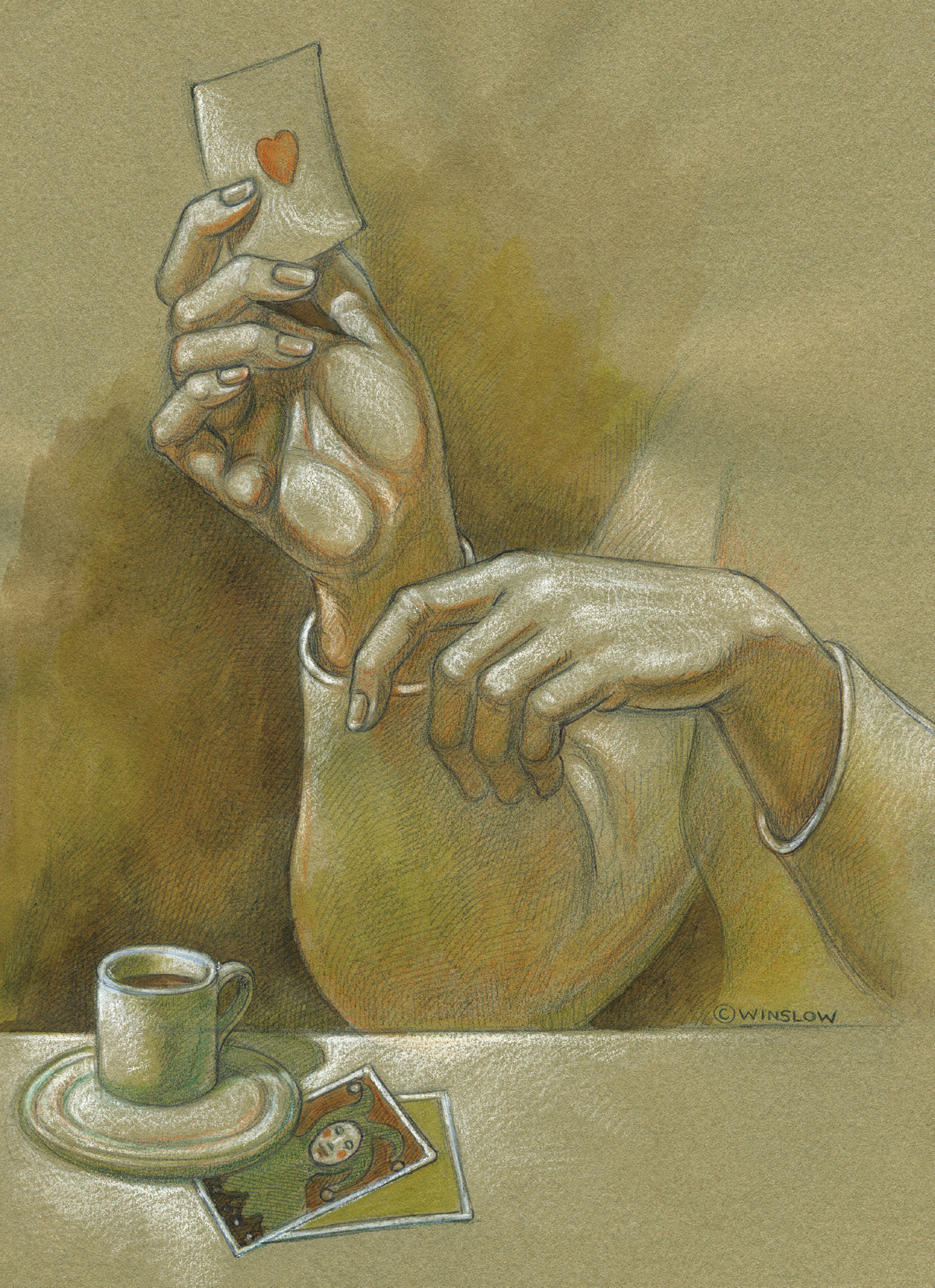
Graphite pencil, watercolor pencils, white chalk on toned paper.
THREE SKETCHBOOK STUDIES OF HANDS
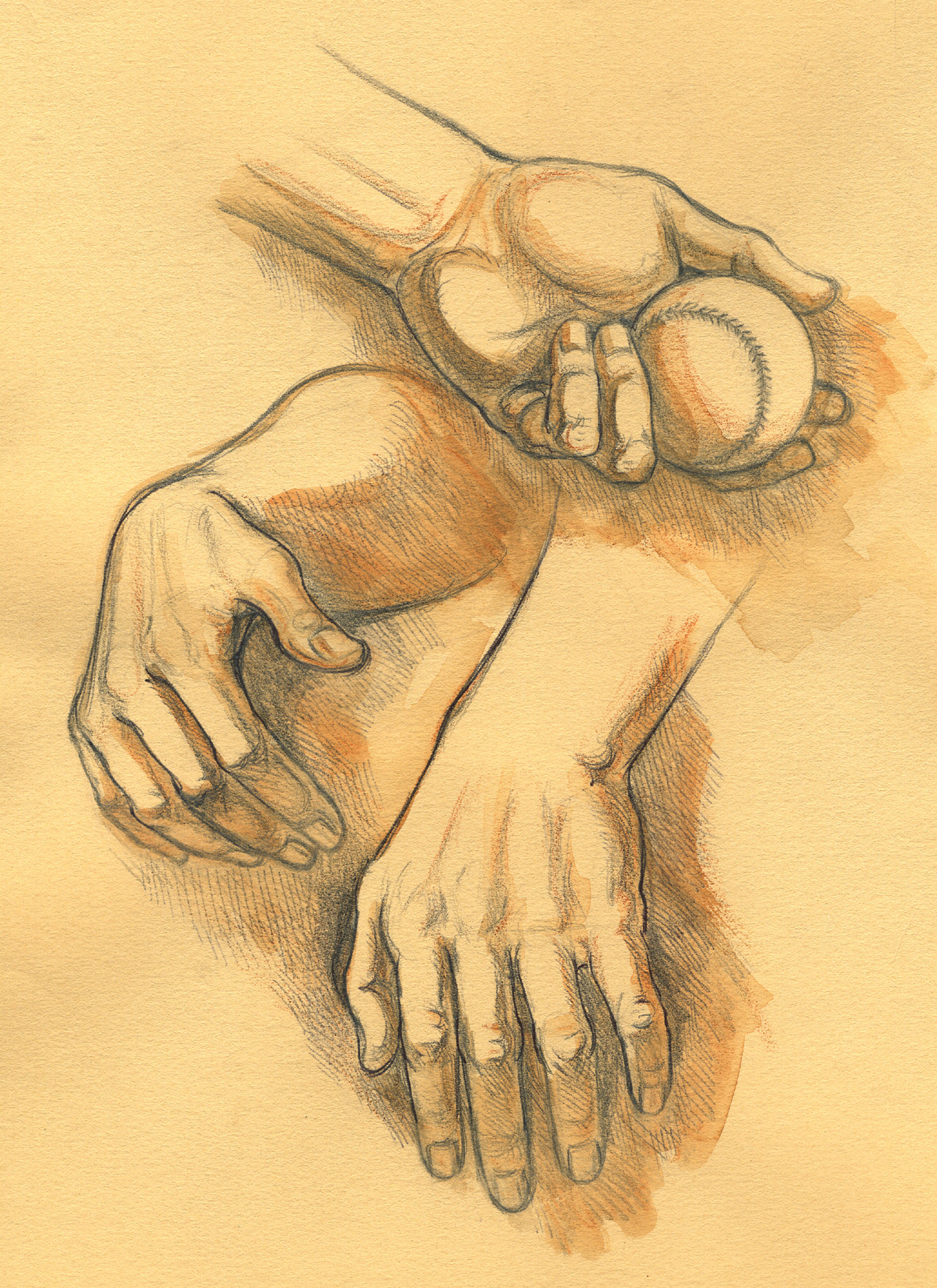
Graphite pencil, ballpoint pen, and watercolor wash on toned paper.
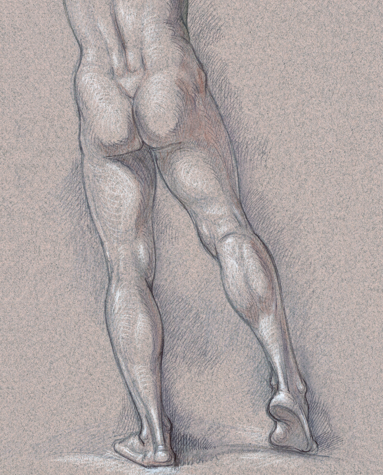
MALE FIGURE WITH RIGHT LEG POSITIONED ON THE BALL OF THE FOOT
Graphite pencil, ballpoint pen, watercolor wash, and white chalk on toned paper.