Biology Premium, 2024: 5 Practice Tests + Comprehensive Review + Online Practice - Wuerth M. 2023
UNIT 4 Cell Communication and Cell Cycle
11 The Cell Cycle
Learning Objectives
In this chapter, you will learn:
➜Phases of the Cell Cycle
➜Regulation of the Cell Cycle, Cancer, and Apoptosis
Overview
The cell cycle is important in the growth, repair, and reproduction of cells in living organisms. Controlling the rate of the cell cycle ensures that these processes occur in a timely manner while also preventing the development of uncontrolled cell growth or tumors. This chapter will review the phases of the cell cycle and the factors that control the rate of the cell cycle.
Phases of the Cell Cycle
Figure 11.1 shows the phases of the cell cycle: interphase (G1, S, and G2), mitosis (also known as the M phase), and cytokinesis. Nondividing cells will leave the cell cycle and enter a stage called G0.
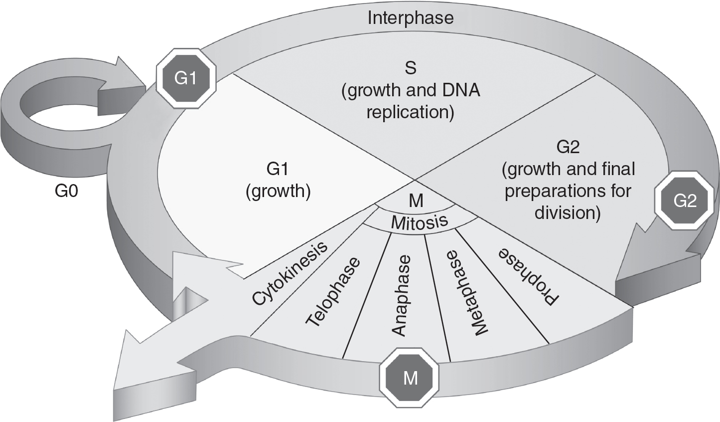
Figure 11.1 The Cell Cycle
Interphase
Interphase is the longest phase of the cell cycle. During interphase, the cell grows so that it has enough material to divide between two daughter cells. The cell also replicates its genetic material (DNA) during interphase. Interphase consists of the following three sequential stages:
1.G1—During this stage, the cell grows and prepares for the replication of DNA, and some cellular organelles (such as centrioles) are replicated.
2.S—During the S (synthesis) stage, DNA is replicated. When the S stage begins, each chromosome consists of one chromatid. After DNA replication is completed, each chromosome has two identical chromatids held together by one centromere. At the end of the S stage, the cell contains twice the amount of DNA it had at the end of G1.
3.G2—During the G2 stage, the cell continues to grow and prepares the materials needed for mitosis, such as the proteins that will make up the spindle fibers.
Mitosis (M Phase)
The goal of mitosis is to make sure there is an accurate transfer of a parent cell’s complete genome to each of the two resulting daughter cells. Mitosis consists of four stages: prophase, metaphase, anaphase, and telophase.
§ In prophase, the nuclear membrane dissolves and the chromosomes condense and become visible. Spindle fibers also begin to form.
§ In metaphase, the spindle fibers have fully attached to the centromeres of each chromosome. Chromosomes are then aligned along the “equator” of the cell in a single column. The center of the mitotic spindle is called the metaphase plate.
§ During anaphase, each chromosome splits at its centromere as opposing spindle fibers begin to shorten. The identical chromatids are pulled toward opposite ends of the cell. At this point, each chromatid now has its own centromere and is considered a separate chromosome. At the end of anaphase, the cell has twice the number of chromosomes that it had at the start of the cell cycle.
§ In telophase, two new nuclear membranes form. Each of the two nuclei now contain the same number of chromosomes and the same genetic information as the parent cell.
Cytokinesis
Cytokinesis is the division of the cytoplasm, along with all of its cellular contents, between the two daughter cells. Cytokinesis occurs after mitosis. During cytokinesis in animal cells, a cleavage furrow is formed, which partitions the cytosol and its contents between the two new cells. Plant cells have a cell wall and accomplish cytokinesis differently. In plant cells, a cell plate is built within the dividing cell, providing new cell wall material for each daughter cell.
Nondividing Cells
Some cells may stop dividing either temporarily or permanently. Cells may stop dividing when they reach their mature, fully differentiated state or when environmental conditions are not favorable for continued growth. These nondividing cells have exited the cell cycle and are in G0. Cells may enter G0 at any point in the cell cycle and may reenter the cell cycle if stimulated to do so by appropriate molecular signals.
Regulation of the Cell Cycle, Cancer, and Apoptosis
Proper regulation of the cell cycle is critical to appropriate growth, repair, and reproduction of cells in living organisms. This regulation is achieved through the use of checkpoints during the cell cycle. Some of these checkpoints are controlled by the interactions between cyclins and cyclin-dependent kinases. Cyclin-dependent kinases are present at constant levels throughout the cell cycle. These kinases add phosphate groups to other molecules, activating those molecules. However, cyclin-dependent kinases themselves are inactive until they are bound to cyclin proteins. The levels of cyclin proteins vary during the cell cycle, reaching their maximum just before mitosis starts. When cyclins are bound to cyclin-dependent kinases, a complex called mitosis-promoting factor (MPF) is formed. MPF triggers the start of mitosis. See Figure 11.2.
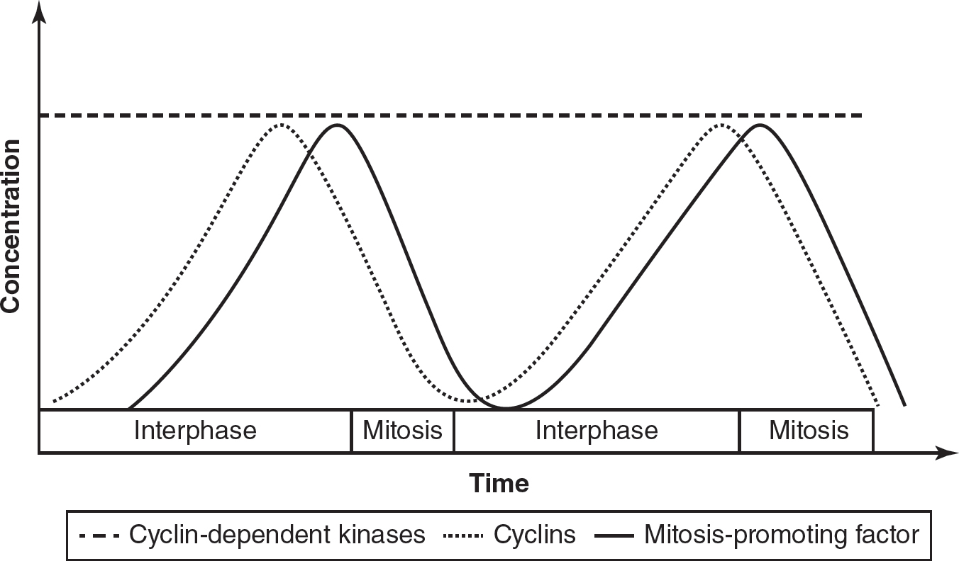
Figure 11.2 Levels of Cyclins, Cyclin-Dependent Kinases, and Mitosis-Promoting Factor in the Cell Cycle
All of the cells in an organism that are not involved with sexual reproduction are referred to as somatic body cells. The division of somatic cells, like those in tissues, can also be regulated by density-dependent inhibition. When cells in tissues, for example, become too crowded, they will stop dividing. Many types of somatic cells also exhibit anchorage dependence, which is when cells need to be attached to a surface in order to divide. Cancer cells are not regulated by density-dependent inhibition nor anchorage dependence and can continue to grow and divide under conditions that would cause normally functioning somatic cells to stop dividing.
TIP
While it IS important to know that cyclins and cyclin-dependent kinases work together to regulate the cell cycle, you do NOT need to know the names of specific cyclin/cyclin-dependent kinase pairs.
Many genes are also involved in the regulation of the cell cycle. Proto-oncogenes propel cell division at a specific rate, much as an accelerator propels a car. Proto-oncogenes are necessary for regulated and controlled cell growth. If mutated, proto-oncogenes may become oncogenes, which promote abnormally high rates of cell division. An oncogene acts in a similar way to how an accelerator stuck in the down position would cause a car to go too fast. These oncogenes can cause tumors to form when cell division occurs too quickly and too often without regard for the neighboring cells. A mutation in a single allele of a proto-oncogene can cause a cell to grow out of control and can cause a tumor to form. Since a mutation in a single allele can cause a cell to grow out of control, proto-oncogenes are said to function in a dominant way.
Tumor suppressor genes code for proteins that detect mutations in cells that may cause tumors to develop. Tumor suppressor genes function much like the brakes on a car, preventing cell division from occurring at an abnormally fast rate. If a single mutation in a tumor suppressor gene allele occurs, the cell will still possess one remaining unmutated tumor suppressor allele that is functional. The tumor suppressor allele that is not mutated will help the organism identify cells that are dividing at a rate that is too fast. However, if both alleles of a tumor suppressor gene are mutated, the growth of a tumor may occur. Tumor suppressor genes are said to function in a recessive way because both alleles of a tumor suppressor gene must be nonfunctional for a cell to grow out of control.
BRCA (sometimes called the “breast cancer gene”) is a mutation in a tumor suppressor gene. If a person has a BRCA mutation, he or she has a greatly increased risk of getting certain types of cancer compared to someone without the mutation. A person with the mutation in one allele would still have one functional tumor suppressor allele, so the occurrence of cancer would not be a certainty, but it would be a more likely occurrence than in a person without the mutation who has two functional copies of the tumor suppressor allele.
Sometimes a living organism’s survival depends on some cells dying and not reproducing. This programmed cell death is called apoptosis. Apoptosis may be triggered when a cell acquires a mutation that could cause cancer. During embryonic development, apoptosis may also occur to ensure proper development of various organs or structures. For example, during early embryonic development, the digits of the hand are initially attached with a weblike structure. During the sixth to eighth weeks of embryonic development, apoptosis eliminates the webbing between the digits of the hand, resulting in the formation of separated fingers.
Practice Questions
Multiple-Choice
1.A chemotherapy drug stops the replication of DNA. During which stage of the cell cycle would this drug have the greatest effect?
(A)G0
(B)G1
(C)G2
(D)S
2.A cell has reached full maturity, is fully differentiated, and no longer divides. Which of the following describes this stage of the cell cycle?
(A)G0
(B)G1
(C)G2
(D)S
3.Which stage of mitosis has twice as many chromosomes at its end as it had at its beginning?
(A)prophase
(B)metaphase
(C)anaphase
(D)telophase
4.During which stage of mitosis do the centromeres of chromosomes attach to spindle fibers and line up in a single column in the center of the cell?
(A)prophase
(B)metaphase
(C)anaphase
(D)telophase
5.A researcher measures the number of cells in each phase of the cell cycle. The data are shown in the following table. Approximately, what percentage of these cells are in interphase?
|
Phase of Cell Cycle |
Number of Cells |
G1 |
73 |
S |
89 |
G2 |
68 |
M |
43 |
(A)16%
(B)33%
(C)52%
(D)84%
6.Which process best describes what occurs when soft tissue between the fingers dies during embryonic development?
(A)apoptosis
(B)DNA replication
(C)mitosis
(D)cytokinesis
7.During which process are the cytoplasm (and its cellular contents) divided between daughter cells?
(A)apoptosis
(B)DNA replication
(C)mitosis
(D)cytokinesis
8.Which of the following is a correct statement?
(A)Cancer cells exhibit anchorage dependence.
(B)Somatic cells exhibit density-dependent inhibition.
(C)Somatic cells spend most of their time in mitosis.
(D)Cancer cells exhibit density-dependent inhibition.
9.Which of the following best describes a role of mitosis?
(A)Mitosis distributes cytosol between the daughter cells.
(B)Replication of DNA occurs during mitosis.
(C)Replication of cell organelles occurs during mitosis.
(D)Mitosis distributes genetic material between the daughter cells.
10.During which phase of the cell cycle is a cleavage furrow or cell plate formed?
(A)interphase
(B)mitosis
(C)cytokinesis
(D)G0
Short Free-Response
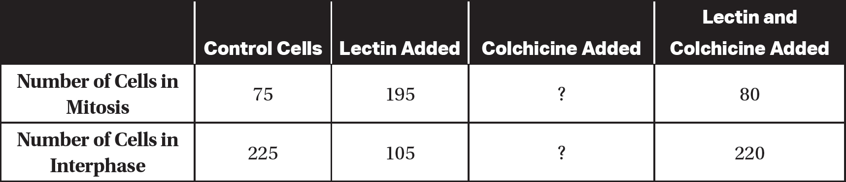
11.Lectin is a molecule commonly found in legumes. Colchicine is a molecule that inhibits the formation of spindle fibers. An experiment was performed to observe the effects of lectin and colchicine on dividing cells. A total of 300 cells were observed for each treatment. Partial results of the experiment are shown in the following table.
(a)Describe the effect of adding lectin to the cells.
(b)Identify the independent variable and the dependent variable in this experiment.
(c)Based on the data presented, predict the most likely effects of adding only colchicine to the cells.
(d)Justify your prediction from part (c).
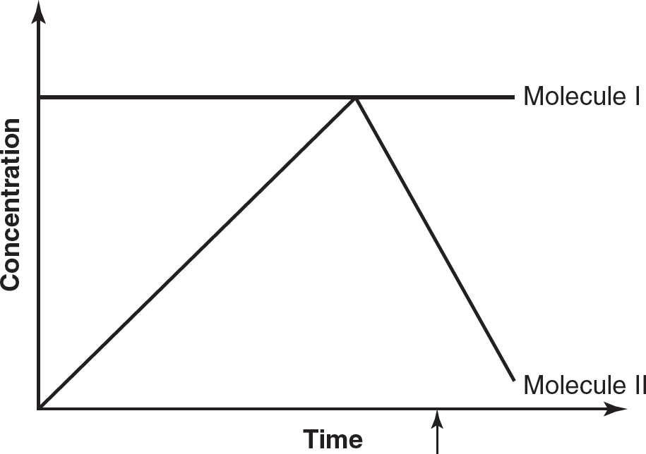
12.The following figure represents the concentrations of two different molecules (cyclin and cyclin-dependent kinase) during the cell cycle, and the arrow indicates the start of mitosis.
(a)Cell division can be described as having three major events: replication of chromosomes, alignment of chromosomes, and separation of chromosomes. Describe the stages of the cell cycle during which each of these three major events occurs.
(b)In the figure, which molecule (cyclin or cyclin-dependent kinase) is represented by the line labeled “Molecule I”? Which molecule (cyclin or cyclin-dependent kinase) is represented by the line labeled “Molecule II”?
(c)Explain why mitosis starts at the point indicated by the arrow in the figure.
(d)A cancer cell has a mutation that results in the constant production of Molecule II. Explain how the process of mitosis might be affected by this mutation.
Long Free-Response
13.A student wants to test whether treating onion root cells with caffeine will significantly increase the number of cells in mitosis. Two groups of onions are grown: one group received distilled water, and the other received distilled water with caffeine. The root tips of both groups are harvested and stained, and the number of cells in interphase and mitosis in each group is counted. The expected and observed data are shown in the following table.
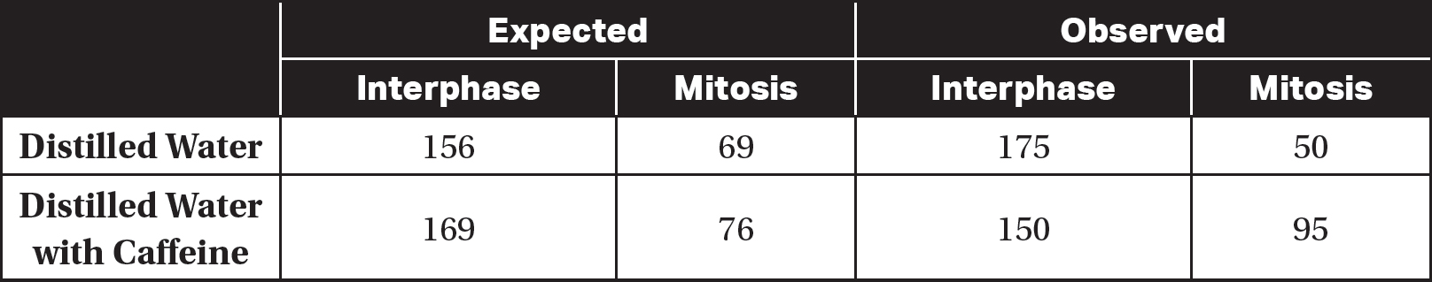
(a)State the null hypothesis for this experiment.
(b)Identify the independent variable and the dependent variable.
(c)Use the chi-square equation and a p-value of 0.05 to analyze the data. Determine if there is a statistically significant difference between the two groups of onions.
(d)Justify your conclusion from part (c).
Answer Explanations
Multiple-Choice
1.(D) DNA is replicated during the S stage of the cell cycle, so a drug that blocks the replication of DNA would have the greatest effect during the S stage. DNA does not replicate during G0, G1, or G2, so choices (A), (B), and (C) are incorrect.
2.(A) During G0, the cell is not dividing and has exited the cell cycle. G1, G2, and S are all stages that prepare the cell to divide, so choices (B), (C), and (D) are incorrect.
3.(C) At the beginning of anaphase, each chromosome consists of two sister chromatids. During anaphase, these chromatids separate, and at the end of anaphase, each of these chromatids is considered a separate chromosome. Thus, anaphase ends with twice as many chromosomes as it had at its beginning. During prophase, chromosomes condense and become visible, but the number of chromosomes does not change. So choice (A) is incorrect. Choice (B) is incorrect because in metaphase, chromosomes line up along the center of the cell but the number of chromosomes does not change. In telophase, two new nuclei are formed and the number of chromosomes per cell at the end of telophase is less than it was at the beginning of telophase. Thus, choice (D) is also incorrect.
4.(B) During metaphase, chromosomes line up in the center of the cell on the metaphase plate. Prophase is when chromosomes condense and become visible, so choice (A) is incorrect. Anaphase is when sister chromatids separate and begin to move to opposite ends of the cell, so choice (C) is incorrect. Telophase is when two new nuclei are formed, so choice (D) is also incorrect.
5.(D) Interphase consists of G1, S, and G2. Therefore, the number of cells in interphase is 73 + 89 + 68 = 230 cells. The total number of cells is 73 + 89 + 68 + 43 = 273. So the percentage of cells in interphase is ![]() . Choice (A) is incorrect because 16% is the approximate percentage of cells that are in mitosis. Choice (B) is incorrect because 33% is the approximate percentage of cells that are in the S stage. Choice (C) is incorrect because 52% is the approximate percentage of cells in G1 and G2.
. Choice (A) is incorrect because 16% is the approximate percentage of cells that are in mitosis. Choice (B) is incorrect because 33% is the approximate percentage of cells that are in the S stage. Choice (C) is incorrect because 52% is the approximate percentage of cells in G1 and G2.
6.(A) Apoptosis is programmed cell death, and it can occur during embryonic development when the soft tissue between the fingers dies to allow the fingers to separate. While choices (B), (C), and (D) also occur during embryonic development, choice (A) is the best answer because it is the most specific to the formation of the fingers during embryonic development.
7.(D) Cytokinesis is the division of the cytoplasm. Choice (A) is incorrect because apoptosis is programmed cell death. DNA replication occurs during the S stage of the cell cycle, so choice (B) is incorrect. Choice (C) is also incorrect because mitosis is the process of evenly distributing replicated chromosomes between the daughter nuclei.
8.(B) Many somatic cells in organs and tissues exhibit density-dependent inhibition. Cancer cells do not exhibit anchorage dependence nor do they exhibit density-dependent inhibition, so choices (A) and (D) are incorrect. Choice (C) is incorrect because noncancerous cells spend most of their time in interphase, not mitosis.
9.(D) Mitosis is the distribution of genetic material between the daughter cells. Cytokinesis, not mitosis, is the distribution of cytosol between the daughter cells, so choice (A) is incorrect. Choice (B) is incorrect because the replication of DNA occurs during the S stage, not mitosis. Replication of cell organelles occurs during G1 and G2, not mitosis, so choice (C) is incorrect.
10.(C) Cytokinesis is the division of the cytoplasm. In animal cytokinesis, a cleavage furrow is formed. In plant cytokinesis, a cell plate is formed. DNA and cellular organelles are replicated during interphase, but the cytoplasm is not yet divided. So choice (A) is incorrect. Choice (B) is incorrect because mitosis is the distribution of genetic material between the daughter cells. G0 is the nondividing phase of the cell cycle, so choice (D) is incorrect.
Short Free-Response
11.(a)Lectin stimulates cell division, as shown by the increased number of cells in mitosis for the treatment with only lectin added (compared to the number of control cells in mitosis).
(b)The independent variable is the presence or absence of lectin and/or colchicine. The dependent variable is the number of cells in mitosis or interphase.
(c)The most likely effects of adding only colchicine to the cells would be that the number of cells in mitosis would be less than that of the control group and the number of cells in interphase would be more than that of the control group.
(d)The addition of lectin alone increased the number of cells in mitosis (compared to that of the control group). The addition of both lectin and colchicine resulted in a number of cells in mitosis that was much closer to that of the control group than it was to that of the group with only lectin added. So colchicine most likely reduces the number of cells in mitosis, and thus a group with only colchicine added would have even fewer cells in mitosis than the control group had.
12.(a)Replication of chromosomes occurs during the S stage of interphase. Alignment of chromosomes occurs during metaphase of mitosis. Separation of chromosomes occurs during anaphase of mitosis.
(b)Molecule I represents the concentration of cyclin-dependent kinases because it is at a constant level throughout the cell cycle. Molecule II represents the concentration of cyclins because its level peaks just before the start of mitosis and then falls off rapidly during mitosis.
(c)Mitosis-promoting factor (MPF) triggers the start of mitosis. Mitosis-promoting factor forms when cyclins bind to cyclin-dependent kinases. The arrow shows the point at which there is enough cyclin to form the MPF needed to trigger the start of mitosis.
(d)If a cancer cell had a mutation that resulted in the constant high production of cyclins (Molecule II), there would be constantly high levels of MPF in the cell. Mitosis would constantly be stimulated, and the cell would divide uncontrollably.
Long Free-Response
13.(a)The null hypothesis is that there is no statistically significant difference between the number of cells in interphase and the number of cells in mitosis in the untreated cells compared to the number of cells in interphase and the number of cells in mitosis in the cells treated with caffeine.
(b)The independent variable is the presence of caffeine. The dependent variable is the number of cells in interphase or mitosis.
(c)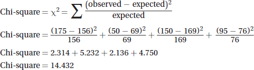
There are four possible outcomes to the experiment, so there are 3 degrees of freedom (df = number of possible outcomes — 1).
Using a p-value of 0.05, the critical value from the chi-square table is 7.81. Based on these calculations, there is likely a statistically significant difference between the two groups of onions.
(d)Since the calculated chi-square value of 14.432 is greater than the critical value of 7.81, the null hypothesis is rejected, and it is possible to say there is likely a statistically significant difference between the treated and untreated groups. The data support the alternative hypothesis that the addition of caffeine increases the mitotic rate in onion root cells.