Biology Premium, 2024: 5 Practice Tests + Comprehensive Review + Online Practice - Wuerth M. 2023
UNIT 2 Cell Structure and Function
5 Cell Organelles, Membranes, and Transport
Learning Objectives
In this chapter, you will learn:
➜Cell Organelles and Their Functions
➜Endosymbiosis Hypothesis
➜The Advantages of Compartmentalization
➜The Importance of Surface Area to Volume Ratios
➜Structure of the Plasma Membrane
➜What Can (and Cannot) Cross the Plasma Membrane
➜Passive Transport
➜Active Transport
Overview
In prokaryotic and eukaryotic organisms (no matter the size), the basic unit of life is the cell. This chapter reviews the functions of different parts of the cell and how cells maintain an internal environment (homeostasis) that supports life.
Cell Organelles and Their Functions
The two major types of cells are prokaryotic cells and eukaryotic cells. (See Figure 5.1.) Prokaryotic cells are simpler in structure. Bacteria are prokaryotic cells. Eukaryotic cells contain membrane-bound organelles and are much more complex than prokaryotic cells. Animals, plants, fungi, and protists are all composed of eukaryotic cells.
All cells (both prokaryotic and eukaryotic) contain genetic material, ribosomes, cytosol, and a plasma membrane. In prokaryotes, the genetic material (DNA) is a circular chromosome located in the center of the cell in an area called the nucleoid region. Bacteria may also contain extra genetic material outside of the chromosome, which is contained in small circular pieces of DNA called plasmids. In eukaryotes, DNA is packaged into linear chromosomes that are contained in a membrane-bound nucleus.
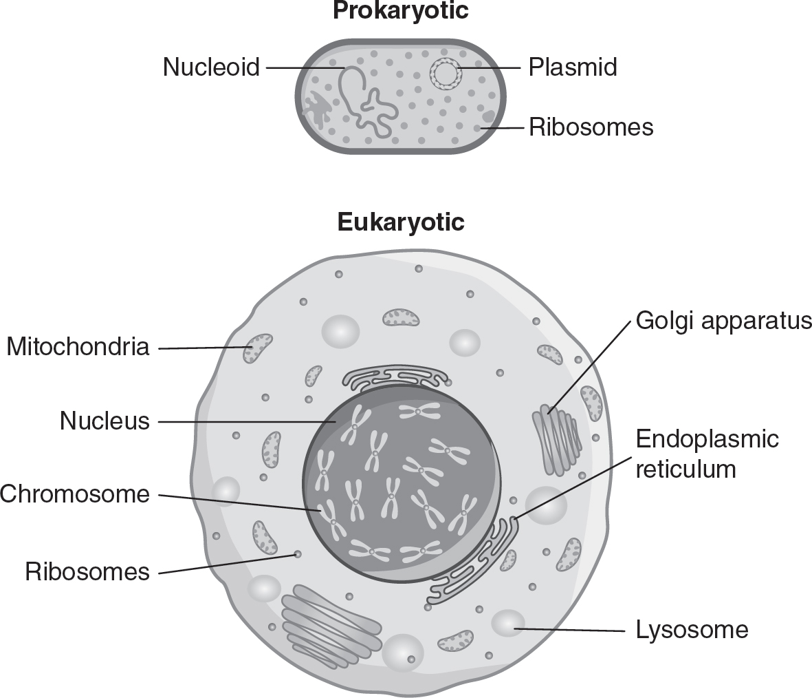
Figure 5.1 Prokaryotic Cell vs. Eukaryotic Cell
Ribosomes function in protein synthesis and are found in both prokaryotic and eukaryotic cells. Ribosomes are made of proteins and ribosomal RNA (rRNA). Both prokaryotic and eukaryotic ribosomes have a large subunit and a small subunit, but the sizes of these subunits differ slightly, as shown in Figure 5.2.
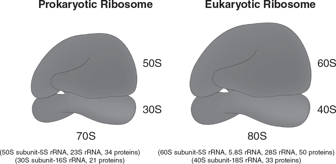
Figure 5.2 Prokaryotic and Eukaryotic Ribosomes
TIP
While it IS important to know that the subunits of prokaryotic ribosomes differ slightly from the subunits of eukaryotic ribosomes, you do NOT need to memorize the specific differences.
During translation, ribosomes assemble amino acids into polypeptide chains according to the mRNA sequence. Free ribosomes are found in the cytosol in both prokaryotes and eukaryotes. In eukaryotes, bound ribosomes are found on the membrane of the rough endoplasmic reticulum.
The endoplasmic reticulum is a series of membrane channels in eukaryotic cells, as shown in Figure 5.3. Rough endoplasmic reticulum has ribosomes bound to its membranes and functions in protein synthesis. Smooth endoplasmic reticulum does not contain ribosomes and functions in the synthesis of lipids and the detoxification of harmful substances in the cell.
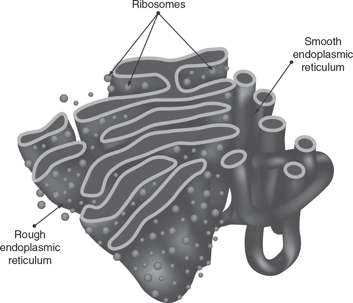
Figure 5.3 Endoplasmic Reticulum
The Golgi complex (also known as the Golgi body) is a stack of flattened membrane sacs (called cisternae), as shown in Figure 5.4. The interior of each cisterna is called the lumen and contains the enzymes necessary for the Golgi complex to function. The Golgi complex controls the modification and packaging of proteins for transport. Proteins made on the free ribosomes of the rough endoplasmic reticulum are sent to the Golgi, which modifies the proteins into their final conformation and packages the finished proteins into vesicles for transport throughout the cell. The Golgi complex is often found near the rough endoplasmic reticulum.
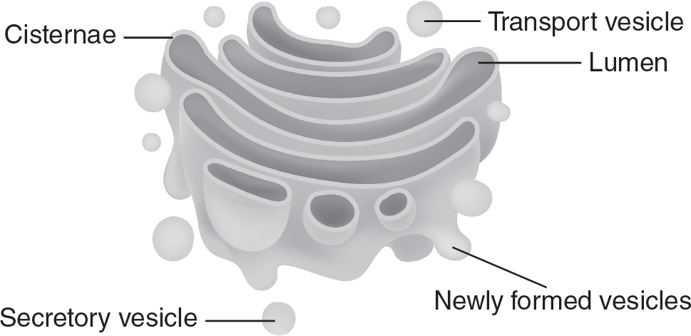
Figure 5.4 Golgi Complex
Lysosomes are membrane-bound sacs, containing hydrolytic enzymes, that function in a variety of cell processes. Lysosomes can help digest macromolecules, break down worn-out cell parts, function in apoptosis, or destroy bacteria and viruses that have entered the cell. (See Figure 5.5.)
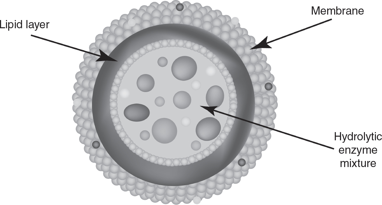
Figure 5.5 Lysosome
Another membrane-bound sac in eukaryotic cells is the vacuole, as shown in Figure 5.6. Vacuoles function in food or water storage, water regulation in a cell, or waste storage until the waste can be eliminated from the cell. In well-hydrated plant cells, vacuoles often occupy the majority of the volume of the cell. By filling up space within the cell, vacuoles provide the plant cell with turgor pressure and support.
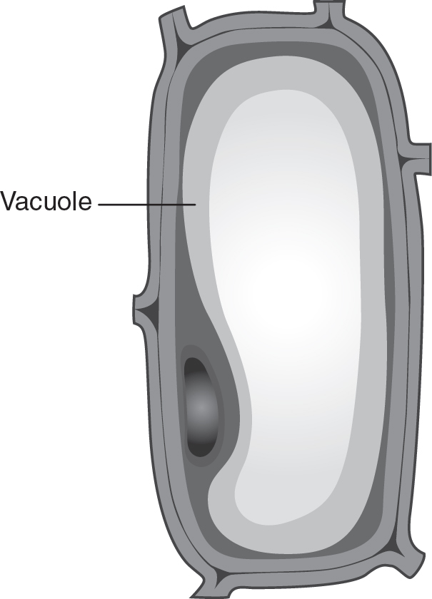
Figure 5.6 Vacuole
Mitochondria produce energy for the cell. The mitochondria have double membranes, with a smooth outer membrane and a folded inner membrane. (See Figure 5.7.) These folds on the inner membrane increase the surface area available for energy production during cellular respiration, which will be discussed in more detail in Chapter 9. The double-membrane structure of the mitochondria also allow them to create the proton gradients that are necessary for ATP production. The center of the mitochondria is an enzyme-containing fluid called the matrix. The reactions of the Krebs cycle (the citric acid cycle) occur in the matrix of the mitochondria. Mitochondria also contain their own mitochondrial DNA (mtDNA). Chapter 14 of this book will explain why mtDNA is inherited in a non-Mendelian fashion. Mitochondria also contain their own ribosomes.
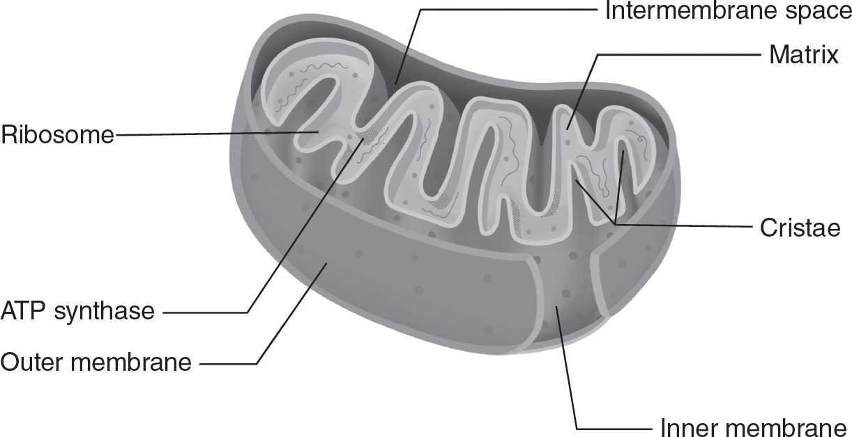
Figure 5.7 Mitochondrion
Chloroplasts are found in plants and algae that carry out photosynthesis. Like the mitochondria, chloroplasts also have a double-membrane structure. Chloroplasts have a smooth outer membrane and pancake-shaped membranous sacs called thylakoids that are stacked into structures called grana (singular: granum). The liquid inside the chloroplast that surrounds the grana is called stroma. (See Figure 5.8.) The membranes of the thylakoids function in the light-dependent reactions of photosynthesis, and the enzymes in the stroma function in the light-independent reactions of photosynthesis. These processes will be discussed in detail in Chapter 8. Like mitochondria, chloroplasts also contain their own chloroplast DNA (cpDNA) and their own ribosomes.
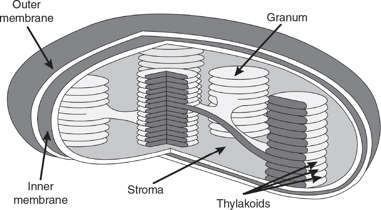
Figure 5.8 Chloroplast
The centrosome, as shown in Figure 5.9, is found in animal cells and helps the microtubules assemble into the spindle fibers needed in cell division. Defects in centrosome function have been associated with dysregulation of the cell cycle in some cancers.
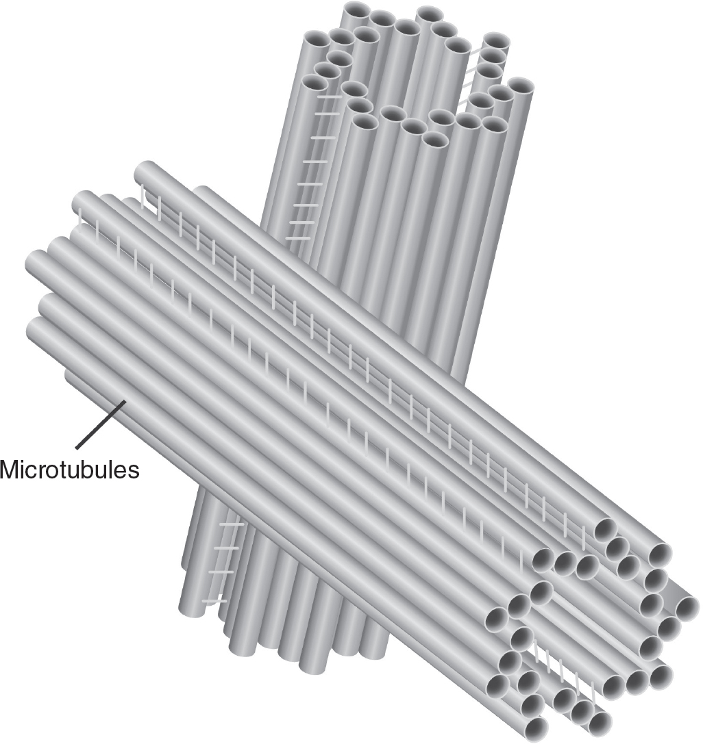
Figure 5.9 Centrosome
Plant cells may contain amyloplasts, as shown in Figure 5.10. Excess glucose produced by photosynthesis is stored as starch molecules in the amyloplasts. Amyloplasts are frequently found in the root and tubers of starchy vegetables, such as potatoes.
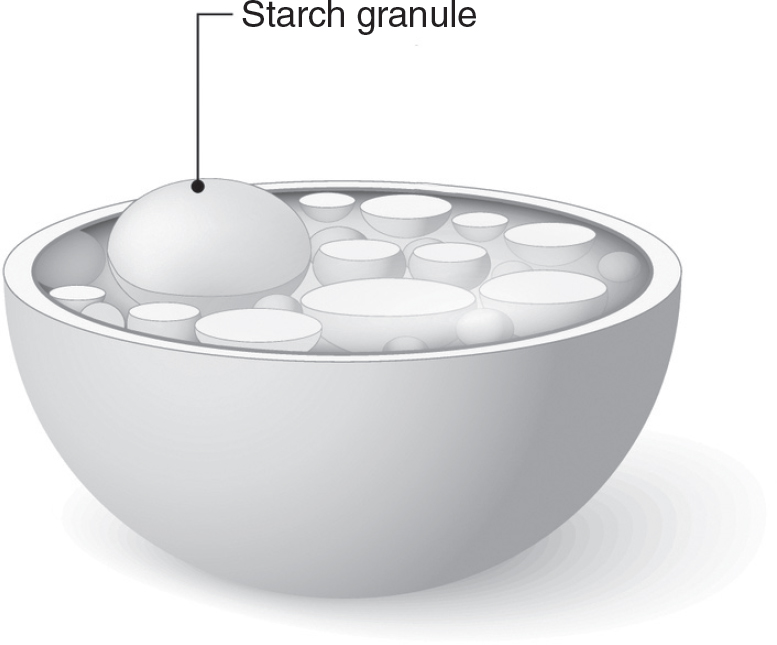
Figure 5.10 Amyloplast
There are several other structures found in both plant and animal cells. The peroxisome helps oxidize molecules and break down toxins in the cell. The nucleolus is not a membrane-bound organelle; that term refers to the region in the nucleus where ribosomes are assembled. Cytoskeleton fibers help give cells their shape and can be used to move items in the cell. Figure 5.11 shows the cellular organelles that are typically found in plant and animal cells.
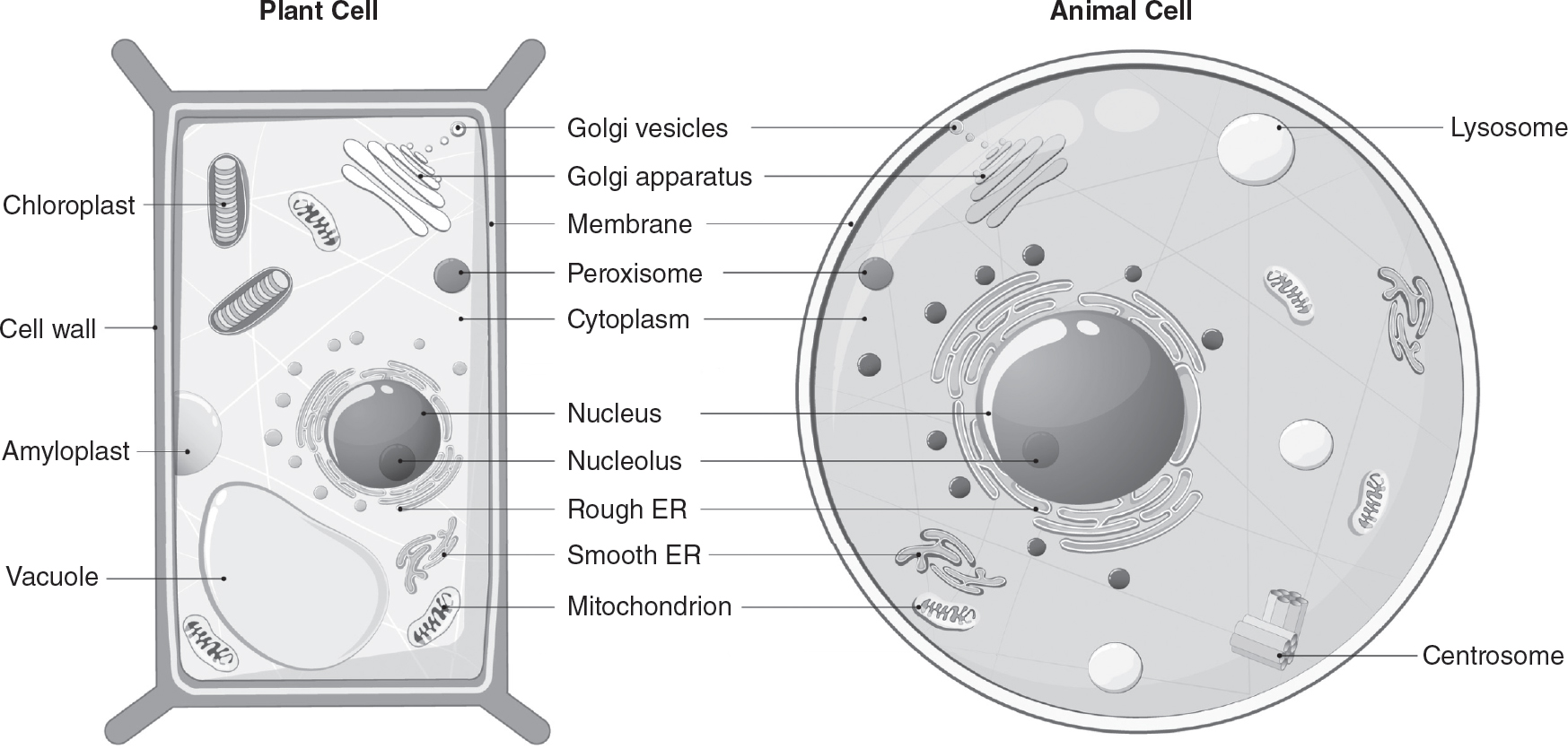
Figure 5.11 Plant and Animal Cells
Endosymbiosis Hypothesis
How did the membrane-bound organelles in eukaryotes originate? The leading theory to explain the origin of membrane-bound organelles is the endosymbiosis hypothesis. The endosymbiosis hypothesis states that membrane-bound organelles, such as mitochondria and chloroplasts, were once free-living prokaryotes that were absorbed into other larger prokaryotes. These prokaryotes became interdependent on each other. The smaller prokaryotes that were engulfed by the larger prokaryotes evolved to become membrane-bound organelles. As with any theory, it is important to understand the evidence for the endosymbiosis hypothesis:
§ Mitochondria and chloroplasts have their own DNA. Mitochondrial and chloroplast DNA is circular, similar to that of prokaryotic DNA.
§ Mitochondria and chloroplasts have their own ribosomes, which are similar in structure to prokaryotic ribosomes.
§ Mitochondria and chloroplasts reproduce by binary fission, similar to how bacteria reproduce.
There are examples of similar relationships in modern organisms. The paramecium Paramecium bursaria is a eukaryote that swallows photosynthetic green algae but does not digest their chloroplasts. Instead, these chloroplasts remain active for months. When P. bursaria swims into the light, the chloroplasts it swallowed perform photosynthesis, supplying food to the paramecium. This process is called kleptoplasty (klepto- means “to steal”).
Next is a discussion of how compartmentalization in cells increases their efficiency.
The Advantages of Compartmentalization
Membrane-bound organelles in eukaryotic cells allow different parts of the cells to specialize their functions, and this specialization allows for greater efficiency within the cell. These membrane-bound organelles form specialized compartments. This compartmentalization allows the cell to separate the enzymes involved in different metabolic processes. By separating these metabolic processes, cells minimize the risk of enzymes and molecules from different processes cross-reacting, which would make these processes less efficient and more difficult to regulate.
Some eukaryotic organelles have internal membranes that are folded (for example, the inner membrane of the mitochondria), which provides greater surface area on which reactions can occur in the organelle. While prokaryotic cells do not have membrane-bound organelles, prokaryotes can fold their plasma membranes to get more surface area on which to perform metabolic processes.
The Importance of Surface Area to Volume Ratios
Living systems need to efficiently eliminate waste products, absorb nutrients, and exchange chemicals and energy with their environment. The ratio of a cell’s surface area to its volume is a key factor in determining the cell’s ability to accomplish these tasks. The larger a cell’s surface area to volume ratio, the more efficiently the cell can accomplish these tasks.
For example, first consider a cell with a spherical shape. The volume of a sphere is proportional to the cube of its radius, as shown in the following formula:
![]()
TIP
You do NOT need to memorize the formulas for surface area or volume. These formulas are on the AP Biology Equations and Formulas sheet that will be provided to you during the AP Biology exam.
The surface area of a sphere is proportional to the square of its radius, as shown in the following formula:
![]()
Dividing the surface area of a sphere formula by the volume of a sphere formula gives you the surface area to volume ratio:
![]()
Note that since r (the radius) is in the denominator, as the radius increases, the surface area to volume ratio will decrease. All materials that are exchanged between a cell and its environment must pass through the surface area of the cell. As the radius of a cell increases, the amount of surface area available per unit volume for exchange of materials gets smaller and smaller. Eventually, this will limit the cell’s ability to efficiently exchange materials with its environment and ultimately impact how big the cell can grow. Larger cells have a lower surface area to volume ratio and a less efficient exchange of materials with the cell’s environment. For this reason, it can be advantageous for an organism to be made of many smaller cells rather than fewer large ones.
One way the surface area to volume ratio can be increased is by folding membranes, as seen in the cristae of the inner mitochondrial membrane. The membrane folds on the inner mitochondrial membrane allow for greater surface area for the electron transport chains that are required for cellular respiration. The lining of the human intestine has many folds called villi that increase the surface area available for nutrient absorption.
Structure of the Plasma Membrane
Plasma membranes are critical in allowing cells to maintain an internal environment that is favorable to life. Plasma (or cell) membranes are selectively permeable, which means that some materials can cross the membrane while other materials cannot. This selective permeability allows the cell to maintain its internal environment.
The plasma membrane is made of a bilayer of phospholipids. Recall from Chapter 4 that phospholipids have a hydrophilic (or polar) phosphate “head” and two hydrophobic (or nonpolar) “tails.” When the bilayer of phospholipids is formed in an aqueous environment, the hydrophobic lipid tails orient themselves away from the water toward the interior of the membrane, while the hydrophilic phosphate heads are oriented facing the aqueous environment on the exterior of the membrane, as shown in Figure 5.12.
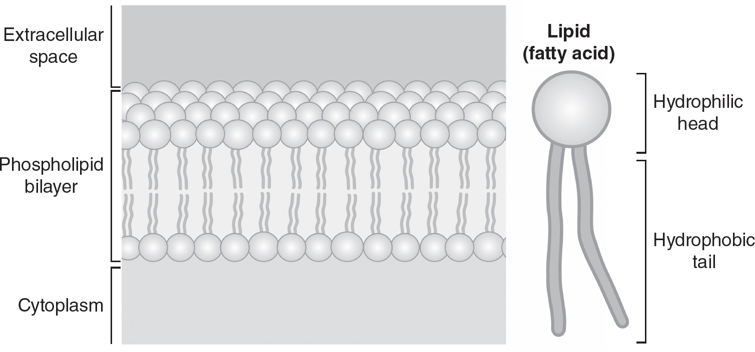
Figure 5.12 Phospholipid
Embedded in this phospholipid bilayer are membrane proteins, modified proteins called glycoproteins, modified lipids called glycolipids, and steroids. These components of the membrane are mobile and can flow throughout the surface of the plasma membrane, allowing the cell to adapt to changing environmental conditions. Proteins in the plasma membrane have many functions, including transporting materials, participating in cell signaling processes, anchoring the cell to its surroundings, and catalyzing chemical reactions. Glycoproteins and glycolipids function in cell recognition. Steroids in the plasma membrane can adjust membrane fluidity in response to changing environmental conditions and the needs of the cell. Individual phospholipids may also move across the surface of the plasma membrane. Because the components of the plasma membrane have mobility, the structure of the membrane is often referred to as a fluid mosaic model. (See Figure 5.13.)
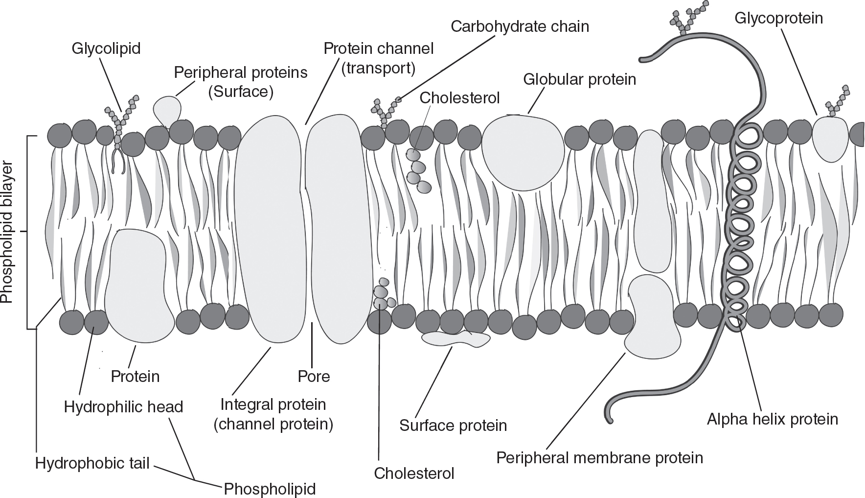
Figure 5.13 Fluid Mosaic Model of a Plasma Membrane
What Can (and Cannot) Cross the Plasma Membrane
The phospholipid bilayer of the cell membrane gives it selective permeability. The hydrophobic lipids of the phospholipids are much larger than the hydrophilic phosphates. Small hydrophobic molecules, such as oxygen (O2), carbon dioxide (CO2), and nitrogen (N2), can easily pass between the phospholipids into and out of the cell. However, large polar molecules and ions cannot cross the cell membrane unassisted. These large polar and charged molecules must use embedded membrane channels or transport proteins to enter or exit the cell. Small polar molecules, like H2O, can pass through the membrane in small quantities. Specialized proteins called aquaporins allow for most of the passage of water in and out of the cell.
Plants, fungi, and prokaryotic cells are surrounded by a cell wall outside of the cell membrane. These cell walls are composed of large carbohydrates (cellulose in plants, glucans in fungi, and peptidoglycan in prokaryotes). The cell walls provide rigidity to the cell and are an additional barrier to substances entering or exiting the cell.
Passive Transport
Passive transport is the movement of molecules from areas of higher concentration to areas of lower concentration. (See Figure 5.14.) Passive transport therefore is said to move molecules “down” their concentration gradient. Passive transport does not require the input of energy. This movement of molecules down their concentration gradient without the input of energy is also called diffusion. The diffusion of water down its concentration gradient across a membrane gets a special name, osmosis. This will be discussed in more detail in Chapter 6.
Molecules that are polar or charged may require a membrane protein in order to diffuse across the cell membrane. The process of passive transport that uses a membrane protein is called facilitated diffusion. One example is the specialized membrane proteins called aquaporins that allow large quantities of water to move down their concentration gradient. Specialized channel proteins can allow the passive transport of ions, such and Ca+2 or Cl—1, down their concentration gradients. Because molecules that use facilitated diffusion require a membrane protein in order to cross the membrane, the rate of facilitated diffusion is limited by the number of membrane proteins available and is said to be saturable.
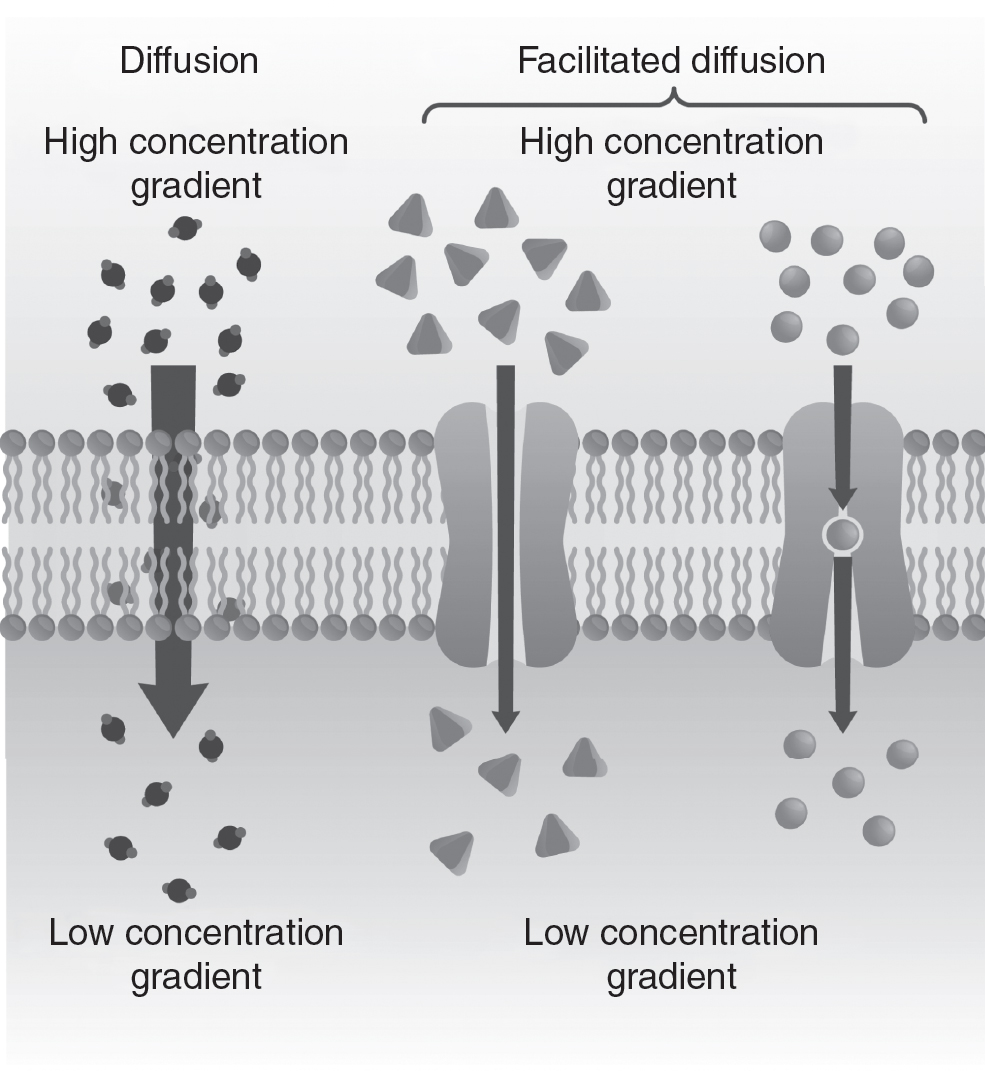
Figure 5.14 Passive Transport
Active Transport
Active transport moves molecules from areas of low concentration to areas of high concentration, as shown in Figure 5.15. This movement of molecules “against” their gradient requires the input of energy.
One example of this is the Na+/K+ pump. This membrane protein requires the input of ATP to pump Na+ ions from their lower concentrations in the cell to an area of higher Na+ concentration outside the cell. This membrane protein also pumps K+ ions from their lower concentrations outside of the cell to an area with higher concentrations of K+ ions inside the cell. For every three Na+ ions pumped out of the cell, two K+ ions are pumped into the cell. This results in a higher concentration of positive ions outside of the cell and helps the cell maintain a membrane potential.
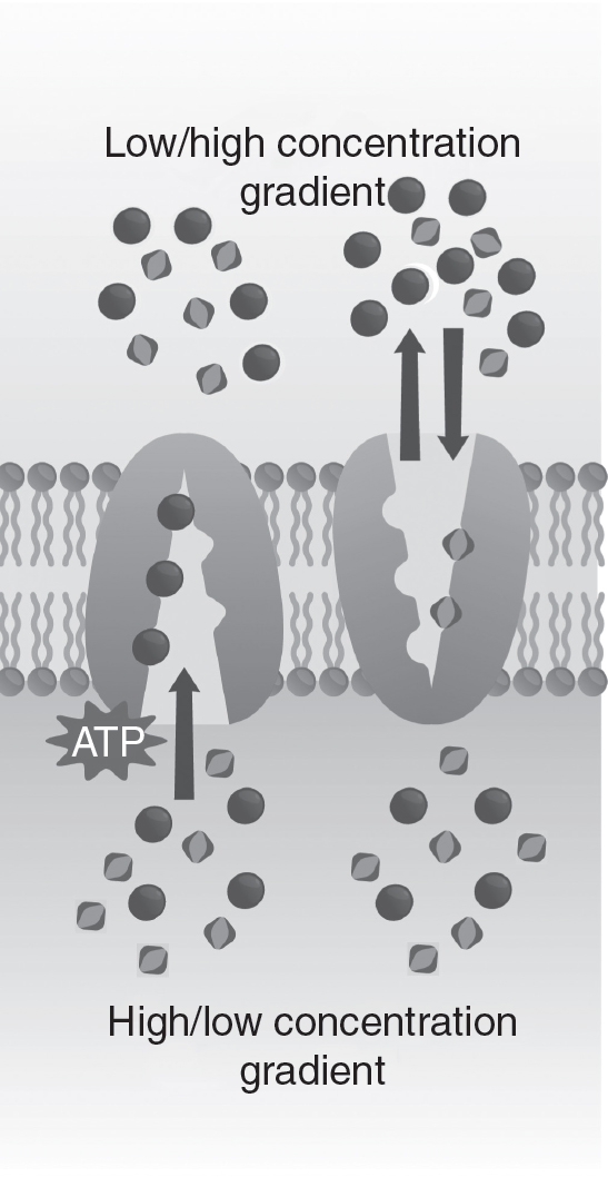
Figure 5.15 Active Transport
Endocytosis and exocytosis also require an input of energy and are forms of active transport. Endocytosis is used by the cell to take in water and macromolecules by enfolding them into vesicles formed from the plasma membrane. In exocytosis, this process is reversed. Vesicles (that contain molecules to be expelled) are fused with the plasma membrane, which then allows these molecules to be expelled from the cell. This movement of large molecules into or out of the cell requires the input of energy. (See Figure 5.16.)
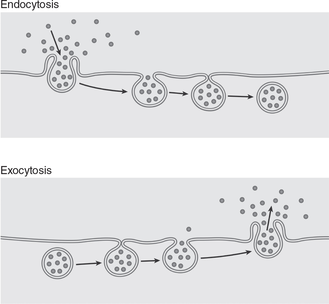
Figure 5.16 Endocytosis and Exocytosis
Practice Questions
Multiple-Choice
1.An animal cell that lacks glycolipids and glycoproteins in its plasma membrane would likely be unable to carry out which of the following functions?
(A)cell recognition
(B)maintaining the fluidity of the phospholipid bilayer
(C)endocytosis
(D)creating a membrane potential
2.When some bacteria are exposed to antibiotics, the bacteria use ATP to try to pump the antibiotics out of their cells. Which of the following processes is most likely used to do this?
(A)osmosis
(B)diffusion
(C)active transport
(D)endocytosis
3.Which of the following is NOT a component of the cell membrane?
(A)phospholipids
(B)cellulose
(C)proteins
(D)glycolipids
4.Muscle cells require large amounts of energy to function. Which cellular organelle is most likely found in high concentrations in muscle cells?
(A)lysosome
(B)Golgi complex
(C)amyloplast
(D)mitochondria
5.Niemann-Pick disease is caused by the cell’s inability to break down large lipid molecules. Which cellular organelle is most likely not functioning properly in an individual with Niemann-Pick disease?
(A)rough endoplasmic reticulum
(B)Golgi complex
(C)lysosomes
(D)ribosomes
6.A student has access to four dyes that stain different components of cells, as shown in the following table.
|
Dye |
Component of Cells Stained |
Nile red |
Lipids |
Hoechst 33342 |
Nuclei |
DRAQ9 |
Cytosol |
Coomassie blue |
Proteins |
Which dye would be the best choice to use to distinguish prokaryotic cells from eukaryotic cells?
(A)Nile red
(B)Hoechst 33342
(C)DRAQ9
(D)Coomassie blue
7.Which types of molecules pass through the cell membrane unassisted most easily?
(A)small and hydrophilic
(B)small and hydrophobic
(C)large and hydrophilic
(D)large and hydrophobic
8.A cell is not able to modify and package proteins for secretion from the cell. Which of the following organelles is most likely not functioning correctly?
(A)ribosomes
(B)lysosomes
(C)vacuoles
(D)Golgi complex
9.Which of the following is NOT evidence that supports the endosymbiosis hypothesis?
(A)Mitochondria and chloroplasts have their own circular DNA.
(B)Mitochondria and chloroplasts have their own ribosomes.
(C)Mitochondria and chloroplasts reproduce by binary fission.
(D)Mitochondria and chloroplasts are found in all eukaryotic cells.
10.A molecule is moving from an area of higher concentration outside of a cell to an area of lower concentration inside the cell. Which process best describes the movement of this molecule?
(A)active transport
(B)diffusion
(C)endocytosis
(D)exocytosis
Short Free-Response
11.A student conducts an experiment to investigate which cell shape would allow for the most efficient exchange of materials with its environment. Agar blocks (containing bromothymol blue dye) are cut into three different shapes to model three differently shaped cells. The agar blocks are then placed in a vinegar solution. As the vinegar diffuses into the agar models, the acid in the vinegar will turn the bromothymol blue dye yellow. The time required for each agar model to turn completely yellow is measured and is an indication of the efficiency of movement of materials into the agar models. The shape, volume, and surface area of each agar block is shown in the table.
|
Shape |
Volume |
Surface Area |
Cylinder |
25.1 cm3 |
50.2 cm2 |
Sphere |
33.5 cm3 |
50.2 cm2 |
Cube |
8 cm3 |
24 cm2 |
(a)Describe the measurement that would best predict the efficiency of each agar block’s exchange of materials with its environment. Calculate that measurement for each agar block.
(b)Identify the independent variable and the dependent variable in this experiment.
(c)Predict which cell would turn completely yellow first.
(d)Justify your prediction from part (c).
12.Apoptosis is programmed cell death. Apoptosis is often triggered by mutations that could cause a cell to form a tumor if the cell continued to grow and multiply.
(a)Describe the organelle in a eukaryotic cell that is most likely to participate in apoptosis.
(b)Explain other functions the organelle from part (a) would have in the cell besides participating in apoptosis.
(c)A mutation causes the enzymes in the organelle from part (a) to become nonfunctional. Predict what effects this would have on the cell.
(d)Justify your prediction from part (c).
Long Free-Response
13.Two different molecules, A and B, can enter a cell using passive transport. A cell that did not initially contain molecule A or molecule B was placed in a beaker with a solution containing equal concentrations of both molecules for 30 minutes. The cell was removed from the beaker, and the concentration of each molecule inside the cell was measured. The experiment was repeated with different, but equal, starting concentrations of molecules A and B in the beaker. The data are shown in the table.
|
Starting Concentration of Molecules A and B in the Beaker (millimolar) |
Concentration of Molecule A Inside the Cell After 30 Minutes (millimolar) ± 2 SEM* |
Concentration of Molecule B Inside the Cell After 30 Minutes (millimolar) ± 2 SEM |
40 |
20 ± 2.1 |
20 ± 2.5 |
80 |
40 ± 1.9 |
40 ± 1.8 |
120 |
60 ± 3.0 |
50 ± 2.9 |
160 |
80 ± 3.2 |
50 ± 2.7 |
*Standard Error of the Mean
(a)Describe two types of passive transport.
(b)Using the axes provided, construct a graph of this data. Include 95% confidence intervals.
(c)Based on the data, make a claim about which molecule (A or B) uses simple diffusion and which molecule uses facilitated diffusion. Justify your claim with evidence from the data.
(d)This experiment is repeated with the addition of a molecule that irreversibly binds to transport proteins in the cell membrane. Make a prediction about what, if any, changes this would lead to in the data. Justify your prediction.
Answer Explanations
Multiple-Choice
1.(A)Glycolipids and glycoproteins are involved in cell recognition, so if a cell lacked these molecules, cell recognition functions would be impaired. Choice (B) is incorrect because cholesterol, not glycoproteins or glycolipids, is involved in maintaining membrane fluidity. Endocytosis is a process that requires energy to bring material into the cell, so choice (C) is incorrect. The Na+/K+ pump uses active transport to create the membrane potential, so choice (D) is also incorrect.
2.(C)Processes that require the use of ATP to move molecules in or out of a cell are called active transport. Choices (A) and (B) are incorrect because osmosis and diffusion are both forms of passive transport. Endocytosis does use energy and is a form of active transport, but that process moves molecules into the cell, not out of the cell. So choice (D) is incorrect.
3.(B)Cellulose is a component of cell walls; it is not a component of the cell membrane. Phospholipids, proteins, and glycolipids are all components of cell membranes, so choices (A), (C), and (D) are all incorrect.
4.(D)Mitochondria are the site of ATP production for the cell. Muscle cells require large amounts of energy, so muscle cells would be expected to have a higher concentration of mitochondria. Choice (A) is incorrect because lysosomes break down macromolecules and cell parts; they do not produce energy. The Golgi complex modifies and packages proteins but is not involved in energy production, so choice (B) is incorrect. Amyloplasts store starches in plant cells, so choice (C) is incorrect.
5.(C)Lysosomes help break down large molecules, so a defect in the lysosomes could be a likely cause of Niemann-Pick disease. The rough endoplasmic reticulum and the ribosomes serve functions in protein synthesis; they do not break down molecules. So choices (A) and (D) are incorrect. Choice (B) is incorrect because the Golgi complex modifies and packages proteins.
6.(B)Eukaryotic cells have nuclei; prokaryotic cells do not. Thus, Hoechst 33342 could distinguish between eukaryotic and prokaryotic cells. Both eukaryotic and prokaryotic cells contain lipids, cytosol, and proteins. Therefore, the dyes that stain these (Nile red, DRAQ9, and Coomassie blue, respectively) would not distinguish between eukaryotic and prokaryotic cells, making choices (A), (C), and (D) incorrect.
7.(B)Small, hydrophobic molecules can slide between the phospholipids in the cell membrane and therefore can cross the cell membrane most easily. Choice (A) is incorrect because hydrophilic molecules could not get past the long hydrophobic lipid tails of the phospholipids that make up the cell membrane’s bilayer. Large molecules cannot pass through the cell membrane unassisted, so choices (C) and (D) are incorrect.
8.(D)The function of the Golgi complex is to modify and package proteins. Ribosomes synthesize proteins, so choice (A) is incorrect. Choice (B) is incorrect because lysosomes contain hydrolytic enzymes, which break down macromolecules and cell parts. Vacuoles store water and other molecules in the cell, so choice (C) is incorrect.
9.(D)Mitochondria and chloroplasts are not found in all eukaryotic cells; chloroplasts are only found in eukaryotic cells that perform photosynthesis. Mitochondria and chloroplasts do have their own circular DNA as well as their own ribosomes, and they both reproduce by binary fission, which are all characteristics they share with prokaryotes. Thus, choices (A), (B), and (C) are all evidence of the endosymbiosis hypothesis.
10.(B)Movement of molecules from an area of higher concentration to an area of lower concentration is diffusion, a passive transport process. Choices (A), (C), and (D) all involve active transport and are therefore incorrect.
Short Free-Response
11.(a)The measurement that would best predict the efficiency of each agar block’s exchange of materials with its environment is the surface area to volume ratio. For the cylinder, the ratio is ![]() . For the sphere, the ratio is
. For the sphere, the ratio is ![]() . For the cube, the ratio is
. For the cube, the ratio is ![]() .
.
(b)The independent variable is the shape of the agar block. The dependent variable is the time it takes for the vinegar to diffuse completely into the cell and turn it yellow.
(c)The cube would turn completely yellow first.
(d)The greater a cell’s surface area to volume ratio, the more efficiently it will exchange materials with its environment. Since the cube has the greatest surface area to volume ratio, it will exchange materials the most efficiently and turn completely yellow first.
12.(a)The lysosome is most likely to participate in apoptosis because it contains hydrolytic enzymes that can break down parts of the cell.
(b)Other functions of the lysosome are to assist in digestion by breaking down large macromolecules and destroying bacteria or viruses that invade the cell.
(c)The hydrolytic enzymes in the lysosome are crucial for the functioning of the lysosome. If those enzymes were not functional, the lysosome could not perform its activities, thus reducing, or even preventing, the cell’s ability to function.
(d)If the hydrolytic enzymes were nonfunctional, the lysosome could not assist in digesting large molecules nor could the lysosome break down waste products in the cell. The cell could simultaneously starve, since it couldn’t digest its food, and be poisoned by its own waste products. In addition, the cell’s ability to defend itself against invading pathogens would be compromised.
Long Free-Response
13.(a)Two types of passive transport are diffusion and facilitated diffusion. In diffusion, a molecule moves from an area of high concentration to an area of low concentration, and no energy is required. In facilitated diffusion, a molecule moves from an area of high concentration to an area of low concentration with the assistance of a transport protein. Again, no energy is required.
(b)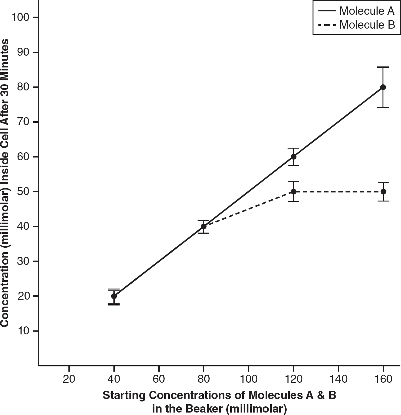
(c)Molecule A is using simple diffusion, and molecule B is using facilitated diffusion. The rate of facilitated diffusion is limited by the number of transport proteins for the molecule in the cell membrane. The rate of simple diffusion is not limited by the number of transport proteins in the cell membrane and will continue to increase as the concentration difference across the cell membrane increases. In the experiment, as the concentration of each molecule in the beaker increases, the amount of molecule A entering the cell continues to increase, but at higher concentrations, the amount of molecule B entering the cell no longer increases. This indicates that molecule B is likely using a transport protein to enter the cell and is therefore using facilitated diffusion.
(d)Since molecule A is simply diffusing across the membrane, the addition of a molecule that binds to transport proteins will likely have no effect on the data for molecule A. However, since molecule B requires a transport protein to enter the cell, the addition of this new molecule would likely prevent molecule B from entering the cell.