CCEA GCSE Biology - Denmour Boyd, James Napier 2017
Unit 2
The circulatory system
Specification points
In GCSE Biology, this chapter covers specification points 2.2.1 to 2.2.8. It covers blood, blood vessels, the heart, and the effects of exercise on pulse rate and the heart.
In Double Award Science, it covers specification points 2.2.1 to 2.2.7 and covers blood, blood vessels, the heart, and the effects of exercise on pulse rate and the heart.
The circulatory system
The circulatory system has three main components: the blood, the blood vessels that carry the blood and the heart that pumps the blood. It has two main functions: transport of blood components and other substances in the blood and protection against disease.
The components of the blood
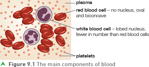
The main components of blood are as follows.
• Red blood cells — the function of these cells is to carry oxygen around the body. Red blood cells are highly specialised for this function. They:
1 contain haemoglobin, which is rich in iron, that carries the oxygen
2 have a biconcave shape that provides a large surface area for diffusion of oxygen
3 have no nucleus and therefore have more space to pack in the haemoglobin.
• White blood cells — the blood contains two types of white blood cell and both are important in defence against disease.
Lymphocytes produce antibodies and phagocytes engulf and digest microorganisms in a process called phagocytosis.
• Platelets — these very small structures are important in blood clotting and the formation of scabs. The platelets work by converting the protein fibrinogen to fibrin. The fibrin forms a mesh network that traps other blood components.
• Plasma — this is the liquid part of the blood. The plasma is responsible for the transport of the blood cells, absorbed food molecules (for example glucose and amino acids), carbon dioxide, hormones and urea.
Tip
You will learn more about the role of white blood cells in defence in Chapter 13.
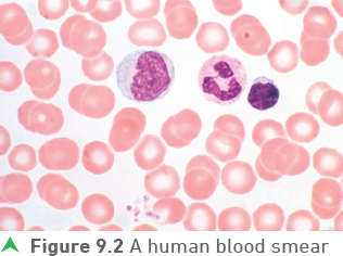
You should examine a blood smear using a microscope. Figure 9.2 shows a typical blood smear as viewed using a microscope.
![]()
Salts and other chemicals in the plasma keep its concentration stable and at a concentration similar to the blood cells. This is important because if red blood cells are placed in water they will take in water by osmosis and burst in a process called cell lysis.
Tip
Red blood cells (like all animal cells) do not have a cell wall, so there is nothing to stop them swelling and bursting if placed in water or a more dilute solution.
Tip
You should be aware that blood contains many more red blood cells than white blood cells.
The blood vessels
There are three main types of blood vessel in the body. The following table shows the main differences between arteries, veins and capillaries.
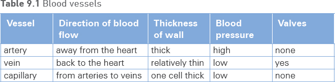
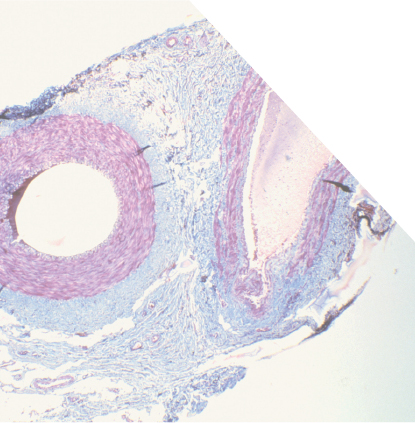
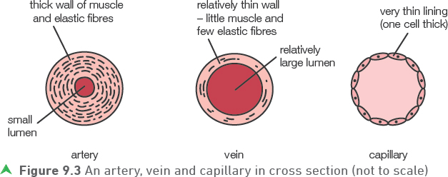
Figure 9.3 represents the three types of blood vessel in cross section.
Tip
The lumen is the space through which the blood flows.
The structure of the blood vessels is closely linked to their function:
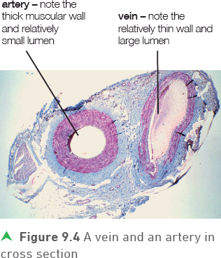
• Arteries normally carry oxygenated blood (rich in oxygen) to the body organs and veins normally carry deoxygenated blood (blood with little oxygen present) back to the heart. The arteries have relatively thick walls as they carry blood at high pressure; the veins have relatively thin walls as they carry blood at low pressure. The walls of the arteries have muscles and elastic fibres which expand as the blood is pulsed through (the elastic fibres give the strength to prevent bursting). After the high pressure pulse, the elastic fibres recoil and muscles contract returning the wall to its original size while maintaining some pressure in the blood. The overall effect is the smoothing out of the blood flow.
• By the time the blood reaches the veins the pressure is low and there is no pulse so the walls need few elastic fibres and muscles. However, valves are necessary to keep the flow in the right direction (unidirectional), preventing backflow. The relatively large lumen of veins reduces friction and further aids the movement of blood.
• Diffusion of oxygen, carbon dioxide, dissolved food and urea takes place between the capillaries and the body cells or vice versa. The one cell thick walls of the capillaries are thin enough to be permeable and allow diffusion to take place.
Test yourself
1 Describe how platelets work.
2 Describe and explain two adaptations of red blood cells.
3 Give one advantage of veins having a large lumen.
Show you can
Explain why veins have much less muscle and fewer elastic fibres than arteries.
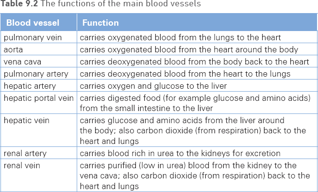
You should be aware of the functions of the blood vessels shown in Figure 9.5.
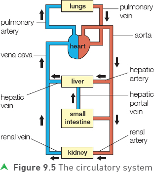
Table 9.2 summaries these functions.
Tip
Figure 9.5 shows that the pulmonary artery and the pulmonary vein are exceptions to the usual rule in that the pulmonary artery carries deoxygenated blood and the pulmonary vein carries oxygenated blood.
The heart
The heart pumps the blood to the lungs and around the body. Figure 9.6 shows why the heart has two sides — the right side pumps the blood to the lungs and the left side pumps the blood that has returned from the lungs around the body.
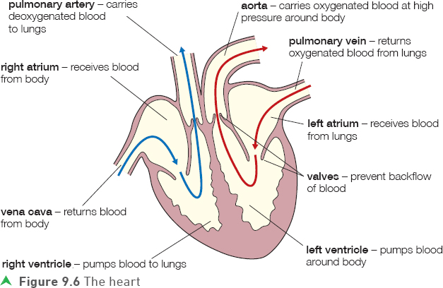
The right atrium receives deoxygenated blood from the body. This passes into the right ventricle where it is pumped out in the pulmonary artery to the lungs. In the lungs, the blood becomes oxygenated and returns to the left atrium of the heart through the pulmonary vein. The oxygen rich blood passes into the left ventricle and is pumped into the aorta and then around the body.
The walls of the ventricles are thicker than the walls of the atria — this is because the ventricles have to pump the blood further than the atria (the atria only pump the blood into the ventricles). The left ventricle wall is thicker (has more muscle) than the right ventricle wall, as it pumps blood around the whole body as opposed to just the lungs.
The heart valves prevent backflow and ensure that the heart acts as a unidirectional pump.
Tip
The heart valves are similar in structure to the valves in the veins. They are ’flap-like’ and will only open one way.
Tip
You need to be able to explain why the walls of the ventricles are thicker than the walls of the atria and also why the wall of the left ventricle is thicker than the right ventricle.
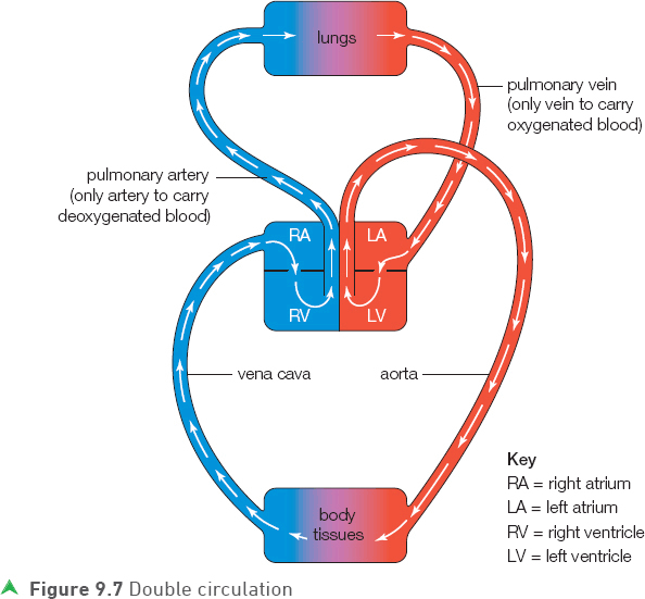
Humans (and other mammals) have a double circulatory system; one circulation system to and from the lungs and one to and from the rest of the body (see Figure 9.7). This means that the blood travels through the heart twice in one circulation of the body.
Figure 9.7 summarises double circulation.
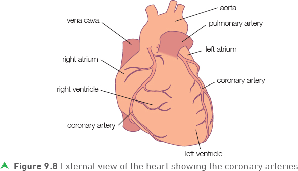
The heart itself receives blood from the coronary arteries, which branch from the aorta almost immediately after it leaves the heart. These are the fine blood vessels that can be seen running over the outer surface of the heart, shown in Figure 9.8.
Test yourself
4 Describe two differences between the structure of the aorta and the vena cava.
5 Describe two differences between the blood in the aorta and in the vena cava.
6 Name two blood vessels that carry blood to the liver.
7 Name the two heart chambers that contain deoxygenated blood.
Show you can
Describe the path of blood from leaving the lungs to returning back to the lungs. Your answer should name the blood vessels and chambers of the heart involved.
Exercise and the circulatory system
Regular exercise benefits the circulatory system in a number of ways. Exercise helps by strengthening the heart muscle (as with any muscle that is exercised). A stronger heart will have an increased cardiac output (pumps more blood per minute) even when not exercising.
Tip
Cardiac output is the volume (amount) of blood pumped by the heart per minute.
You should also investigate the effect of exercise on pulse rate.
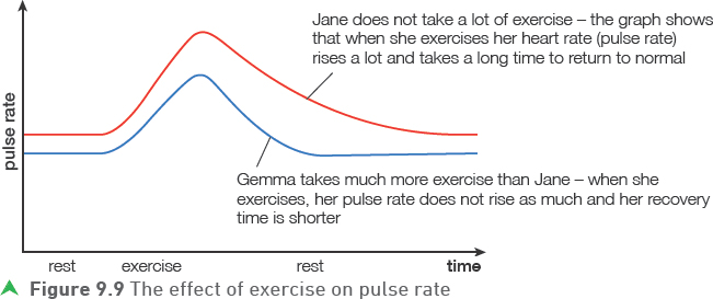
The graph in Figure 9.9 shows the effect of exercise on pulse rate and recovery rate.
The recovery rate is the time it takes for the pulse or heart rate to return to normal after exercise and this will usually be shorter for people who exercise regularly or play a lot of sport.
![]()
You should also know why exercise causes the heart rate to rise. When we exercise, our muscles need more energy as they are contracting more often (and often more vigorously) so the heart has to pump more blood to our muscles so that they get more oxygen and glucose for respiration.
This extra glucose and oxygen is supplied through the increased cardiac output resulting in a higher blood pressure leading to the increased blood flow to the muscles.
Tip
Heart rate and pulse rate will be the same — heart rate is how often the heart beats and the pulse rate how often a ’pulse’ or surge of blood passes round the body — they are the same as each beat causes a new pulse.
Practice questions
1 a) Copy and complete the table below.
(3 marks)
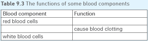
b) Name three things that are transported in the plasma.
(3 marks)
2 a) Give three differences between the structures of arteries and veins.
(3 marks)
b) Figure 9.10 shows how the blood pressure changes as blood flows through the circulatory system.
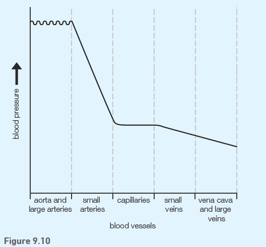
i) In which part of the circulatory system is there the largest fall in blood pressure?
(1 mark)
ii) Suggest what causes the small ripples in blood pressure as the blood flows through the aorta and other large arteries.
(1 mark)
iii) Suggest one reason why it is necessary that blood pressure is low in the capillaries.
(1 mark)
iv) The blood pressure in the veins is low. Name one structure that prevents backflow of blood in the veins.
(1 mark)
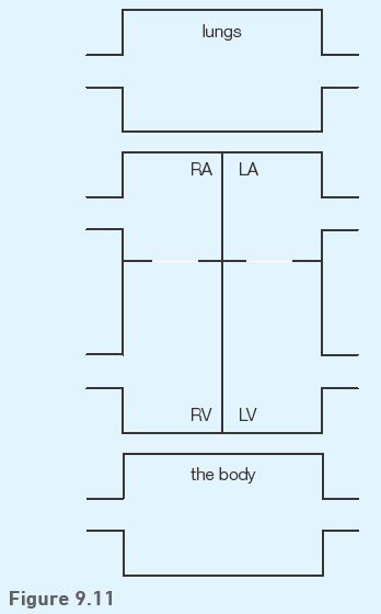
3 Humans are described as having a double circulation system.
a) Describe what is meant by a double circulation system.
(1 mark)
b) The partially completed Figure 9.11 represents the human circulation. Copy the diagram and use arrows to complete it to show the direction of blood flow between the structures shown.
(4 marks)
4 a) Figure 9.12 shows the effect of exercise on the pulse rate of two girls.
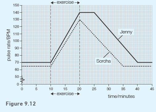
i) Calculate the increase in Sorcha’s pulse rate during exercise.
(1 mark)
ii) Calculate the percentage increase in Sorcha’s pulse rate during exercise.
(1 mark)
b) Use the graph to suggest which girl was the fitter. Explain your choice giving two pieces of evidence from the graph.
(2 marks)
c) If the girls continued to exercise for a period of time, suggest the effect this would have, if any, on recovery time.
(1 mark)