CCEA GCSE Biology - Denmour Boyd, James Napier 2017
Unit 2
The genome, chromosomes, DNA and genetics
Specification points
This chapter covers sections 2.4.1 to 2.4.13 (Double Award Science 2.4.1 to 2.4.12) of the specification. It is about the genome, chromosomes, genes, DNA, mitosis and meiosis, genetics, genetic conditions, genetic screening and genetic engineering.
The genome, chromosomes, genes and DNA
Most living cells contain a nucleus (control centre). The nucleus is the control centre because it contains chromosomes that are subdivided into smaller sections called genes. There are hundreds of genes in each chromosome. Figure 10.1 shows a single chromosome. Normally they occur in functional pairs (except sex cells) — we will see why later in this chapter.
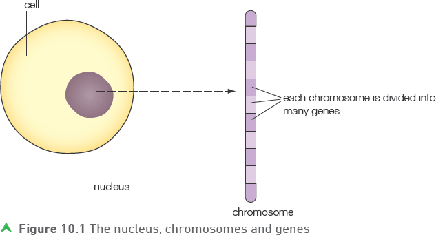
It is the genes in our bodies that control characteristics such as eye and hair colour — the features that make us who we are. Therefore, genes are sections of chromosomes that code for particular characteristics. Inside genes and chromosomes there is a very important molecule that gives them their properties. This molecule is DNA (deoxyribonucleic acid). In effect, genes are short lengths of DNA that code for a particular protein or characteristic.
All the DNA in an individual is referred to as their genome.
Tip
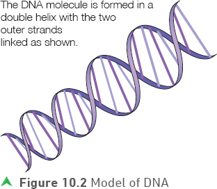
It might be easier to remember this diagram if you think of a ladder with interlinking rungs that is twisted round on itself.
Tip
The genome includes all the DNA in all the genes in all the chromosomes in an individual (all the genetic material).
The structure of DNA
DNA consists of three smaller units, which are regularly repeated throughout the length of the molecule. These units are deoxyribose sugar, phosphate and bases. There are four different types of base: adenine, guanine, cytosine and thymine. In the double helix (Figure 10.2), the rungs of the ’ladder’ are the bases and the sides are alternating units of deoxyribose sugar and phosphate.
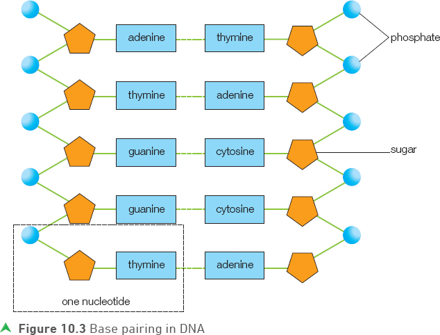
Each repeating unit of DNA (consisting of a phosphate, a sugar and a base) is called a nucleotide. Figure 10.3 shows that bases link the two sides of the molecule together in such a way that adenine only combines with thymine and guanine only combines with cytosine. This arrangement is known as base pairing. The arrangement of the bases along the length of the DNA is what determines how a gene works.
Tip
The letters A, G, C and T are often used to identify the four bases. A only combines with T and C with G.
Test yourself
1 Define the term gene.
2 Name the four DNA bases.
3 What is meant by the unique nature of an individual’s DNA?
Show you can
If 20% of the DNA bases in an individual are adenine, what percentage will there be of the other bases?
If we map the sequence of bases along each individual’s chromosomes we will find that while there will be similarities among different individuals, no two people have the same sequence of bases along the entire length of all their chromosomes (except for identical twins.)
Tip
The sequence of bases in everyone’s DNA is unique, apart from identical twins.
![]()
How does DNA work?
The DNA works by providing a code to allow the cell to make the proteins that it needs. The DNA determines which proteins, and in particular which enzymes, are made. Enzymes are extremely important proteins that control the cell’s reactions. Therefore, by controlling the enzymes, the DNA controls the development of the cell and in turn the entire organism.
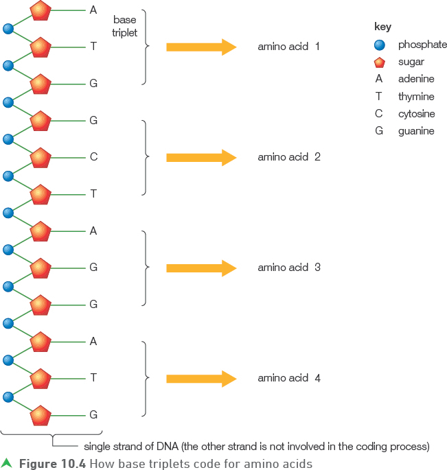
The bases along one side (strand) of the DNA molecule — the coding strand — form the genetic code. Each sequence of three bases (a triplet) along this coding strand codes for a particular amino acid — the building blocks of proteins. The sequence of three bases that codes for an amino acid is called a base triplet. As a protein consists of many amino acids linked together it is important that the correct base triplets are arranged in the correct sequence along the coding strand.
Figure 10.4 shows how base triplets code for particular amino acids. In Figure 10.4 the first and fourth base triplets have the same code and this means that amino acid 1 and amino acid 4 are also the same. The model only shows a small section of a gene and a small section of the protein that it produces codes for.
Cell division
Most living organisms grow by increasing their number of cells. Cells double in number by splitting in half. It is important when cells divide during growth that the two new cells (daughter cells) end up with exactly the same genetic makeup as each other and the parent cell — they are clones of the parent cell. This means that every cell in the growing organism has the same number and type of genes and chromosomes, and that these are the same as they were in its very first cell, the zygote. This type of cell division is called mitosis.
Mitosis
Mitosis can be defined as cell division in which the exact duplication of chromosomes takes place to produce daughter cells that are genetically identical to each other and to the parent cell.
Figure 10.5 summarises the process of mitosis and shows that the two daughter cells contain exactly the same chromosomes as each other and also as the parent cell from which they were produced.
Tip
As new (daughter) cells formed by mitosis grow to ’normal’ cell size before dividing again, growth of an organism usually involves both cell division and cell growth.
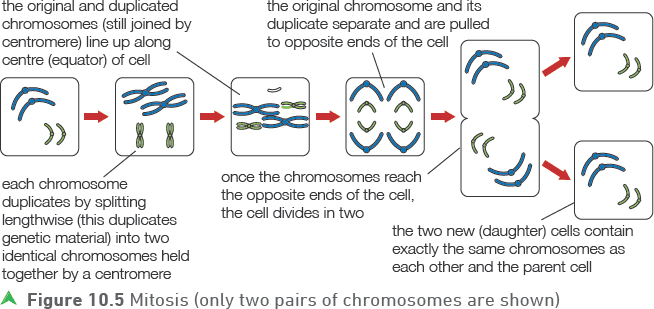
Mitosis is a type of cell division used in growth, to replace worn out cells and to repair damaged tissue.
Meiosis
Meiosis is another type of cell division. It only takes place in the sex organs (the testes and ovaries) during the production of gametes (sperm or eggs). The purpose of meiosis is to produce gametes with half the number of chromosomes of all the other (non-gamete) cells in the body. As meiosis halves the chromosome numbers in the daughter cells it is also known as reduction division.
![]()
Most human cells have 46 chromosomes, arranged in 23 pairs, but the sperm and eggs that are produced by meiosis have only 23 chromosomes. It is not just any 23 chromosomes from the 46 but one chromosome from each pair that passes into each gamete. It could be either chromosome of a particular pair that passes into a particular gamete, and as there are 23 pairs of chromosomes in total, there are millions of potential chromosome combinations in any gamete: 223 possibilities.
This random independent assortment of chromosomes in meiosis at gamete formation gives unique gametes (the chance of any two gametes being identical is so small that it is virtually impossible) and so helps to produce variation in offspring.
Figure 10.6 summarises the role of meiosis and the random nature of fertilisation itself in producing variation in living organisms.
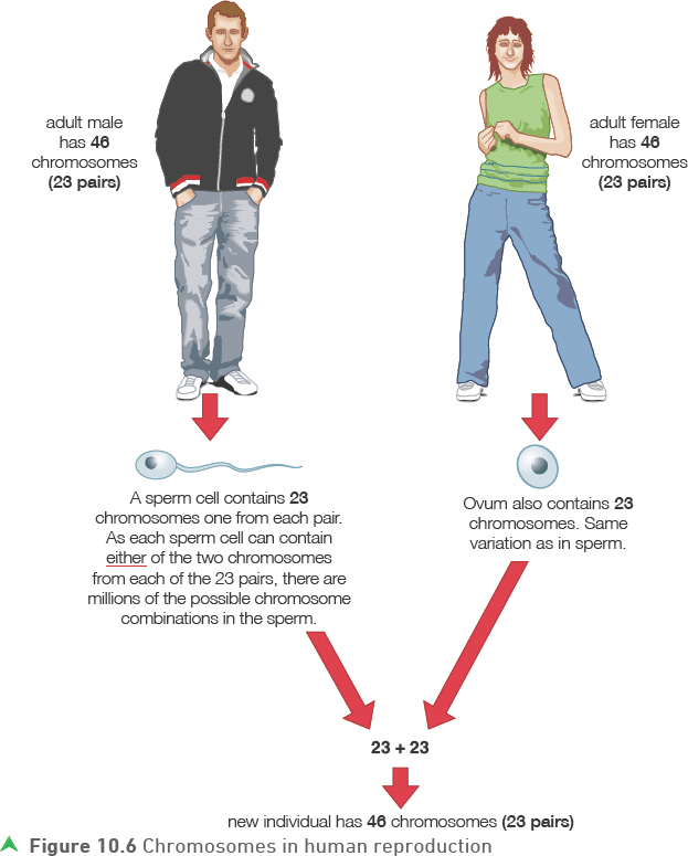
The chromosome number in the gametes is referred to as the haploid number (23 in humans). The normal number in an organism is called the diploid number (46 in humans). The roles of fertilisation include restoring the diploid number in the offspring and combining the different arrangements of chromosomes produced during the process of meiosis.
The differences between mitosis and meiosis
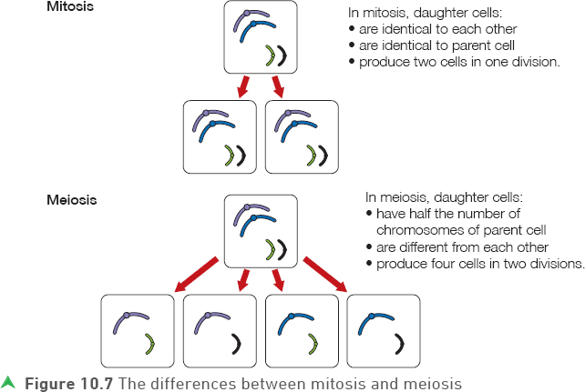
Figure 10.7 summarises the differences between mitosis and meiosis.
Test yourself
4 Give the three functions of mitosis.
5 What is meant by the term clone?
6 Meiosis produces haploid cells. What is meant by the term haploid?
7 Where in the human body does meiosis occur?
Show you can
Describe the function of meiosis and explain why it is important that it takes place.
Tip
Remember that mitosis produces identical cells (clones) — diploid cells produce other identical diploid cells. In meiosis a diploid cell produces haploid cells that are genetically different from each other (and from their parent cell).
Genetics
The science of genetics explains how characteristics pass from parents to offspring.
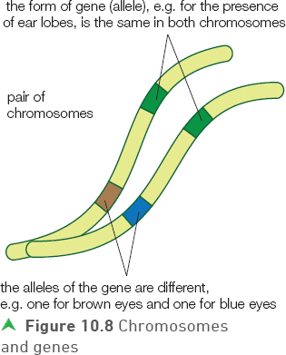
Figure 10.8 shows the following.
• Chromosomes are arranged in pairs — humans have 23 pairs, which is 46 chromosomes in total.
• Genes are sections of chromosomes that carry the code for particular characteristics such as eye colour.
• Similar genes occupy the same position on both chromosomes in a pair.
• Genes exist in different forms, called alleles, and the alleles may be homozygous (the same), or heterozygous (different) on the two chromosomes of a pair.
Genetic crosses
A monohybrid genetic cross is a cross between two individuals where the genetics of one characteristic (for example height in peas or eye colour in humans) is considered.
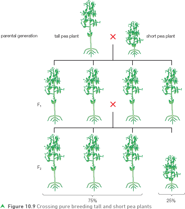
Pea plants occur in their normal tall form or in a much shorter variety. One genetic cross that can be carried out is a cross between tall and short plants. Before carrying out this cross, the tall plants can be allowed to breed with each other for a period of time to ensure they always produce tall plants. The same can be done with the short plants by allowing only short plants to breed together until it is certain they could only produce short offspring. The parent plants to be used are then referred to as pure breeding.
Tip
Remember that alleles are different forms of the same gene.
When pure breeding tall plants are crossed with pure breeding short plants (the parental generation), all the plants in the first, or F1 generation, (the offspring) are tall. However, if these F1 plants are crossed with other F1 plants, their offspring (the second or F2 generation) are a mixture of tall and short plants in the ratio of 75% tall to 25% small.
Explanation of this monohybrid cross
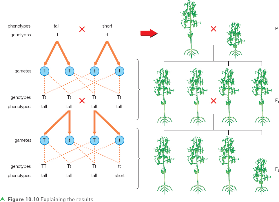
The results of this (and other) genetic crosses can be explained by understanding the gene involved — in this case, the gene that determines height. There are two forms of the height gene — a tall and a short gene. The different forms of a gene are called alleles. In the example of height in peas, there is an allele for tallness and an allele for shortness. In genetic crosses, alleles are represented by a single letter, for example T for tall and t for short.
As the parental plants were pure breeding, the tall plants only carried the tall alleles and short plants only carried the short alleles. These plants containing only one type of allele are homozygous (TT or tt). When both types of allele are present, the individual is heterozygous (Tt).
The paired symbols (TT, Tt or tt) used in genetics are referred to as the genotype and the outward appearance (tall or short) is the phenotype.
When gametes are produced, only one allele (from each gene) from each parent passes on to the offspring. This is fully explained by our understanding of meiosis, as we know that only one chromosome, and therefore one allele, of each pair can pass into a gamete.
The F1 plants in our cross must have received one T allele from their tall parent and one t allele from the short parent. The F1 plants were therefore Tt (heterozygous). Although all these plants contained both the T and the t allele, they were tall. This can be explained by considering the T allele as being dominant over the recessive t allele. The recessive condition will only be expressed, or visible, in the phenotype when only recessive alleles are present in the genotype (tt).
Figure 10.10 shows that when the F1 plants were interbred a ratio of 3 : 1 (tall : short) was produced. This ratio was achieved because the two alleles (T and t) of one parent were produced in equal numbers during meiosis and they had an equal chance of combining with the T or the t allele produced by the other parent during fertilisation.
When completing genetic diagrams, it is helpful to use a small grid called a Punnett square.
Figure 10.11 shows how to use a Punnett square. This is a way of setting out a genetic cross in table format. In this example, using height in peas as before, a heterozygous pea (Tt) is crossed with a homozygous recessive pea (tt). There are extra notes on the diagram to help your understanding of genetic crosses.
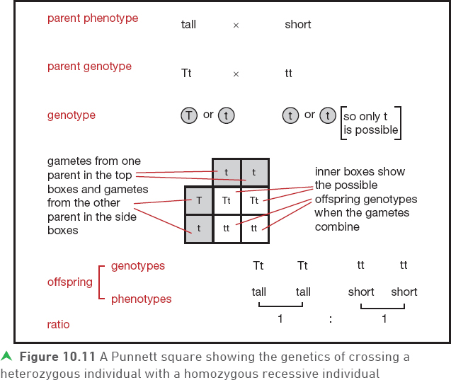
Figure 10.12 uses Punnett squares to show examples of some monohybrid crosses that can occur in humans. In these examples of eye colour, brown eye colour (B) is dominant to blue eye colour (b).
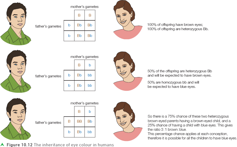
Some important points about genetic crosses
• Ratios will only be accurate when large numbers of offspring are produced. This is because it is totally random which gametes, and therefore alleles, fuse during fertilisation.
• It is common practice to use the same letter for both the dominant and recessive alleles, with the dominant allele being the capital and the recessive allele written in lower case.
• If a 3 : 1 ratio is present in the offspring of a particular cross, both of the parents involved will be heterozygous for the characteristic being considered.
• If a 1 : 1 ratio is produced in a cross, one parent will be heterozygous and the other homozygous recessive.
Tip
It is useful to learn the parental genotypes that produce 3 : 1 and 1 : 1 offspring ratios as these genetic ratios are common in exam questions.
![]()
The test cross (back cross)
A tall pea plant can be either homozygous (TT) or heterozygous (Tt). Both genotypes give exactly the same phenotype. Sometimes, in agriculture or in the breeding of domestic animals, it is important to know the genotype of a particular animal or plant that is showing the dominant phenotype. To identify the unknown genotype a test or back cross is carried out, like the one shown in Figure 10.13.
The animal or plant in question is crossed with a homozygous recessive individual. If the offspring are produced in sufficient numbers it is possible to identify the unknown genotype.
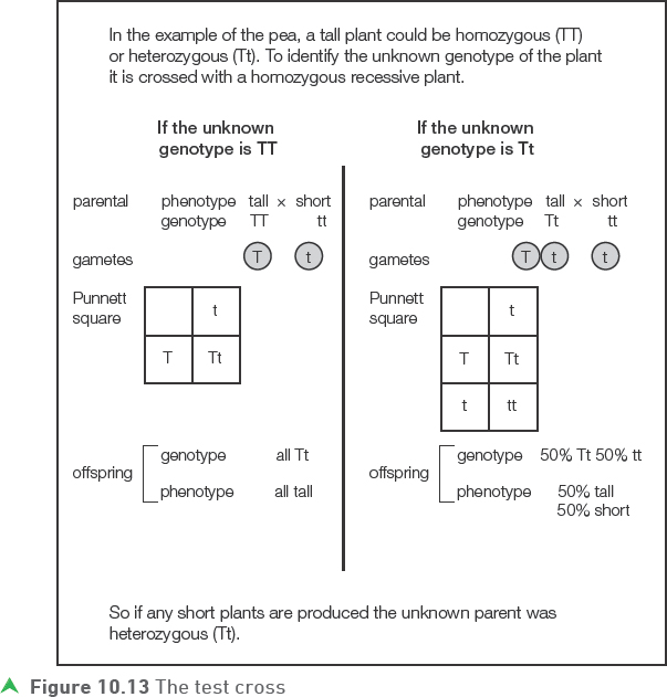
Tip
In a test cross if any offspring are homozygous recessive then the unknown parent must have been heterozygous.
![]()
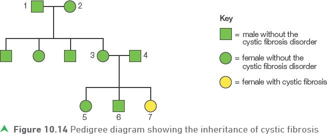
Pedigree diagrams
A pedigree diagram shows the way in which a genetic condition is inherited in a family or group of biologically related people. Figure 10.14 is an example of a pedigree diagram showing how the condition cystic fibrosis is inherited. Cystic fibrosis is a medical condition caused by having two recessive alleles of a particular gene.
Test yourself
Use Figure 10.14 and your knowledge to answer the following questions. Let F = normal allele; f = cystic fibrosis allele (note: the normal allele is dominant and the cystic fibrosis allele recessive).
8 What is the genotype of the child (7) with cystic fibrosis?
9 What are the genotypes of the parents of child 7 (3 and 4)?
10What is the probability that the next child of these parents will have cystic fibrosis?
In this diagram, one of the grandchildren (7) has cystic fibrosis. It is possible to use the information provided to work out the probability of other children having the condition. Genetic counsellors often construct pedigree diagrams and use them to advise parents who have a genetic condition or who may be carriers.
Pedigree diagrams can be used in any type of genetic cross but they are obviously very valuable in tracing and predicting harmful genetic conditions.
Show you can
What can you say about the genotypes of the grandparents of child 7 (1 and 2) in terms of carrying the cystic fibrosis allele?
![]()
Gregor Mendel — the founder of genetics
Much of our understanding of genetics is based on the work carried out by Gregor Mendel. Mendel was born in 1822 in what is now the Czech Republic and as a young man joined the church. As a monk in a large monastery he developed an interest in the breeding of the garden pea, plants that were common in the monastery garden.
Mendel noticed that garden pea plants had many characteristics that varied from plant to plant. These characteristics included pea shape and pea colour. The pea seeds produced by the garden plants were either green or yellow and they could be round or wrinkled. Mendel carried out a range of breeding experiments in which he crossed (mated) plants carrying particular contrasting characteristics that he was interested in. By careful observation of the offspring produced, he was able to draw conclusions about the nature of inheritance.
Mendel carried out the monohybrid crosses on peas described earlier in this chapter. Although he did not know about chromosomes and genes he was able to deduce a number of things, including:
• that certain traits (characteristics) in living organisms are determined by factors within the organism (we now call these factors genes)
• that the factors (genes) for a particular characteristic can be present in two different forms (we now call these different forms alleles)
• that the two factors (alleles) in an individual separate during gamete formation (meiosis)
• an understanding of the classic monohybrid ratios such as 3 : 1 and 1 : 1.
Tip
It is important to appreciate that modern genetics builds on Mendel’s work, rather than contradicting it.
Sex determination in humans
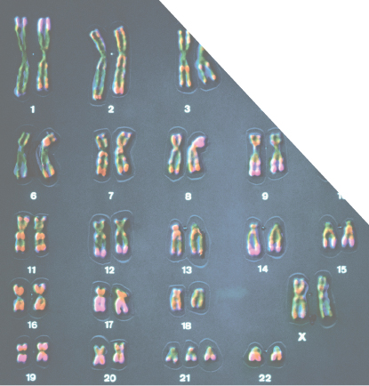
Sex in humans is another characteristic that is genetically determined. Humans have 46 chromosomes in each cell (except gametes) consisting of 22 pairs of normal chromosomes and one pair of sex chromosomes. The sex chromosomes determine the sex of each individual. Males have one X and one Y sex chromosome whereas females have two X chromosomes.
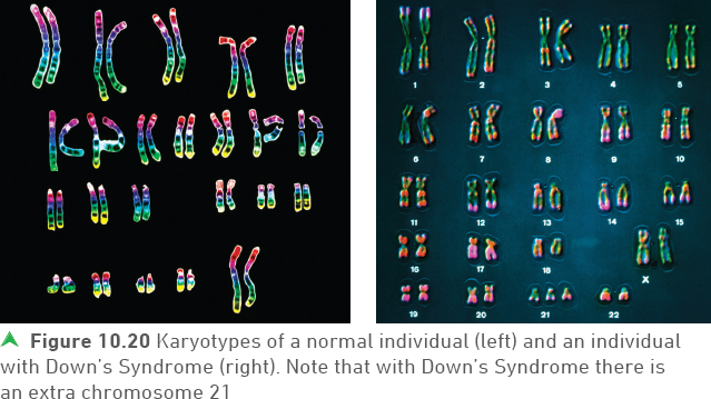
An image of a complete set of chromosomes is known as a karyotype. Figure 10.16 shows the karyotypes of the human male and female.
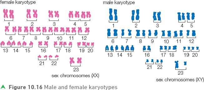
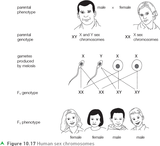
During meiosis the female will provide one X chromosome for each egg (ovum), but half the male’s sperm will have an X chromosome and half will have a Y chromosome. As there will be an equal chance of an X or a Y chromosome from the male being involved in fertilisation there will be equal numbers of males and females produced. Again, the random nature of fertilisation must be emphasised.
![]()
Sex linkage
The X and Y chromosomes are not only responsible for sex determination, they also have genes that code for a number of body functions. Each of the 22 normal (non-sex) pairs of chromosomes has the same gene present on both chromosomes and in the same position. The alleles may be different (alleles for blue or brown eyes) but the gene (gene for eye colour) is present on both. However, in the sex chromosomes the X is much larger than the Y and carries genes that are not present on the Y.
This is particularly important in males as they only have one X chromosome. Therefore any recessive allele carried on an X chromosome in a male will show its effect in the phenotype — there is no dominant allele to mask its effect, as is the situation with females who have two X chromosomes.
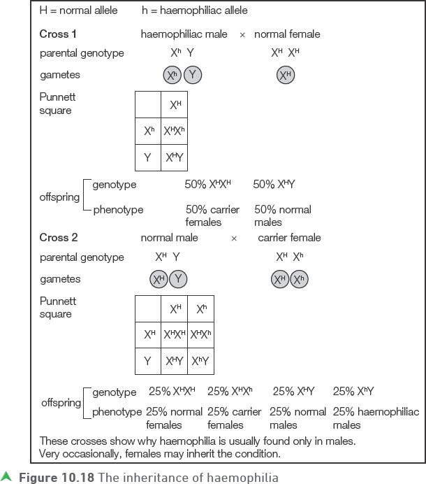
Haemophilia is a sex-linked condition that is almost exclusively found in males. Females seldom show sex-linked conditions but they are often carriers. In sex-linked conditions, carriers are females who have one dominant and one recessive allele on their X chromosomes. In the female the recessive allele usually does not affect the phenotype as it is masked by the dominant allele.
Haemophilia is a condition whereby individuals who are only carrying recessive alleles are unable to make all the products required to clot their blood.
Tip
In genetic conditions caused by a recessive allele, individuals who are heterozygous for the condition (they ’carry’ the harmful allele but don’t show the condition) are referred to as carriers.
Tip
When carrying out crosses involving sex linkage, it is important to use symbols that represent both the type of sex chromosome (X or Y) and any allele carried, for example XhY.
![]()
Test yourself
11 What are the sex chromosomes in a human male?
12 On which types of chromosomes are sex-linked conditions such as haemophilia found?
Show you can
Although haemophilia is almost always found in males only, it is possible for females to have haemophilia. Use a Punnett square to show how this is possible.
Genetics and health
Genetic conditions
Genetic conditions are caused by a fault with genes or chromosomes (a genetic fault). Some genetic conditions (but not all) are inherited; this means they are passed from parent to child.
Haemophilia, cystic fibrosis, Huntington’s disease and Down’s Syndrome are all genetic conditions that affect humans. However, they are each caused by a different type of genetic problem.
![]()
• Haemophilia — this condition is caused by a problem with the blood-clotting mechanism. Sufferers are at risk of excessive bleeding even from very small wounds or bruising. It is a sex-linked inherited condition caused by a recessive allele on the X chromosome.
• Cystic fibrosis — individuals with cystic fibrosis have frequent and serious lung infections and problems with food digestion. It is caused by a recessive allele, so affected individuals must be homozygous recessive.
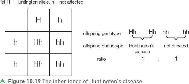
• Huntington’s disease — Huntington’s disease affects nerve cells in the brain, leading to brain damage, which usually becomes apparent in middle age. It is fatal and there is no cure. It is caused by the presence of a dominant allele.
Example
If one parent is heterozygous for Huntington’s disease and the other parent does not have the Huntington’s disease allele, then each child has a 50% chance of developing Huntington’s disease. Explain.
Answer
• Down’s Syndrome — this condition is caused by the presence of an extra chromosome, so that affected individuals have 47 rather than 46. Humans normally have 23 chromosomes in each gamete (sperm or egg). Occasionally gametes are formed with 24 chromosomes so if one of these gametes is involved in fertilisation with a ’normal’ gamete then the child produced will have 47 chromosomes. Individuals with Down’s Syndrome have reduced muscle tone and reduced cognitive development.
Tip
Haemophilia, cystic fibrosis and Huntington’s disease are inherited (the faulty genes are passed from parent to child) and can pass through many generations. Down’s Syndrome is a mistake during gamete formation — it is not inherited as such (it is not the passing of a faulty gene or chromosome from parent to child).
Genetic screening
Genetic screening may be used to reduce the incidence of diseases or conditions caused by problems with our chromosomes or genes. It involves testing people for the presence of a particular allele or other genetic abnormality. Whole populations can be tested, or testing can be targeted at selected groups or individuals where the probability of having (or passing on) a particular condition is high.
One type of genetic screening is amniocentesis. This involves inserting a needle into the amniotic fluid surrounding the foetus and withdrawing some of the fluid. Foetal cells in the fluid are then examined for the presence of genetic abnormalities.
Figure 10.21 shows amniocentesis taking place. The structure to the left of the needle shows ultrascanning taking place (with the image of the foetus in the background).
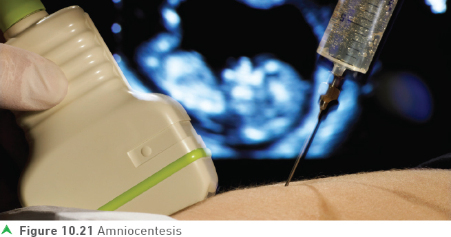
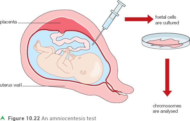
Amniocentesis tests can be carried out for a range of genetic conditions including Down’s Syndrome and cystic fibrosis (see Figure 10.22). However, as amniocentesis carries a risk of miscarriage (around 1%), it is usually only routinely used with those pregnant women who have a greater risk of having a child with a genetic abnormality.
Mothers with a greater risk include:
• those who have previously carried a foetus with a genetic abnormality
• those who have a family history of a genetic condition
• those where possible problems have been identified in an earlier medical examination, for example a blood test
• older mothers.
When screening for Down’s Syndrome, pregnant mothers are offered a blood test between 10—14 weeks of pregnancy. The blood test will determine if the possibility of having a Down’s Syndrome child is raised. The amniocentesis is normally then offered to those mothers who appear to have a higher risk based on the earlier screening. While not as accurate as amniocentesis, it carries no risk to the foetus or the mother, but it can help identify those women who may wish to take the riskier amniocentesis procedure.
Tip
The blood test for Down’s Syndrome is less precise than amniocentesis testing but it poses no risk.
Genetic screening — ethical and moral issues
If a foetus is diagnosed with a genetic condition the potential parents have some very difficult decisions to make and this creates a real dilemma for many. Is abortion the best thing to do?
Many parents will argue ’yes’, as it prevents having a child who could have a poor quality of life; a lot of time may need to be spent caring for the child with the abnormality at the possible expense of time with their other children.
Many parents will argue ’no’ as the unborn child doesn’t have a say, or they will argue that it is not morally right to ’kill’ a foetus. Additionally, abortion is banned in some religions and in some countries.
Some other issues arising from genetic screening are listed below.
• Who decides on who should be screened?
• Is there an acceptable risk associated with genetic screening? For example, amniocentesis for Down’s Syndrome screening has a small risk of miscarriage.
• Costs of screening compared to the costs of treating individuals with a genetic condition — should cost be a factor?
• Should genetic screening be extended to more than just serious genetic conditions? What if it can predict life expectancy?
It is now possible to screen everyone (whether before birth, in childhood or as an adult) for many different alleles. The information obtained is referred to as a genetic profile. Should this information be available to life insurance companies and employers?
If someone is identified with a genetic condition that is not obvious as yet, but may shorten their life span, then life insurance companies may not insure them and if they do then the insurance could be very expensive.
There are arguments for making genetic information publically available — it could help with medical research for example.
Tip
It is possible for anyone to pay for their genetic profile. Many companies carry out genetic profiles and provide a wide range of feedback including the risk of developing a range of diseases.
Test yourself
13 Which of the following conditions is caused by the presence of a dominant allele?
haemophilia
cystic fibrosis
Huntington’s disease
14 Give the genotype of someone who is a carrier for cystic fibrosis (use F for the dominant allele and f for the recessive allele).
15 Which of the following conditions is caused by a chromosome (rather than a gene) abnormality?
cystic fibrosis
Down’s Syndrome
Huntington’s disease
Show you can
When screening for Down’s Syndrome explain why the blood test is given before taking an amniocentesis test, rather than the other way around.
Genetic screening is the process of identifying genetic issues that are present in someone’s genome. Genetic engineering is the process of actually manipulating the genome in an organism for medical or other reasons.
Genetic engineering
Genetic engineering involves taking a piece of DNA, usually a gene, from one organism (the donor) and adding it to the genetic material of another organism (the recipient).
Genetic engineering is defined as a process that modifies the genome of an organism to introduce desirable characteristics.
Commonly, DNA that codes for a desired product is incorporated into the DNA of bacteria. This is because bacterial DNA is easily manipulated and also because bacteria reproduce so rapidly that large numbers can quickly be produced with the new gene.
As a result the bacteria will produce a valuable product coded for by the added gene, such as a drug or hormone that may be difficult or expensive to produce by other means. Once the new genetic material is built into them, the bacteria are added to special fermenters or bioreactors where they reproduce rapidly in suitable growing conditions that maximise the production of the desired product.
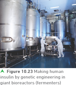
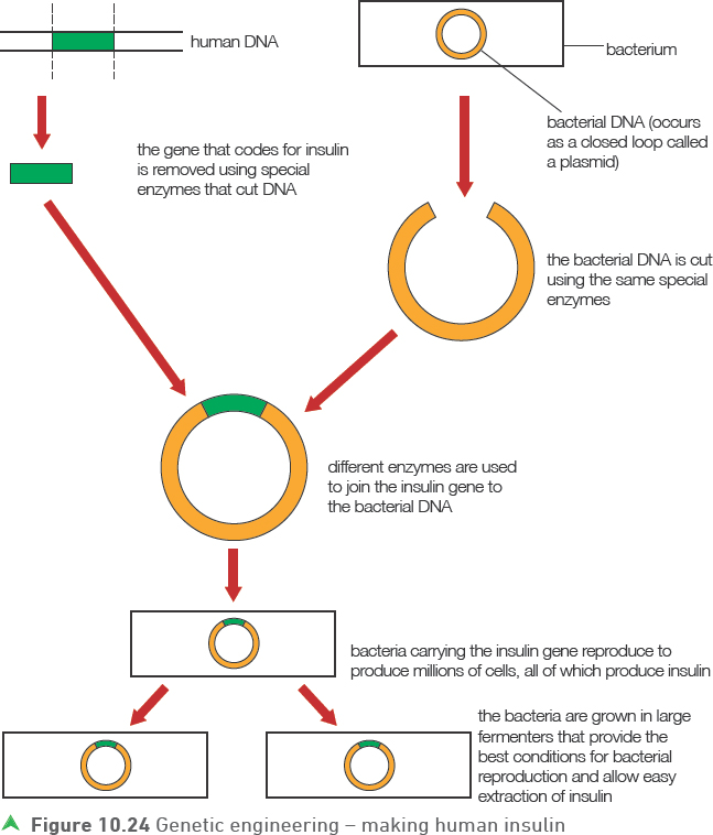
One of the best examples of genetic engineering providing essential products for humans is the production of genetically engineered human insulin, as shown in Figure 10.24. Diabetes is becoming increasingly common and as a result many more people require insulin than in the past. Before the development of genetic engineering, insulin was obtained from the pancreases of domestic animals such as pigs and cattle.
![]()
Tip
Restriction enzymes are often described as ’molecular scissors’ as their function is to cut the molecule of DNA in a particular way.
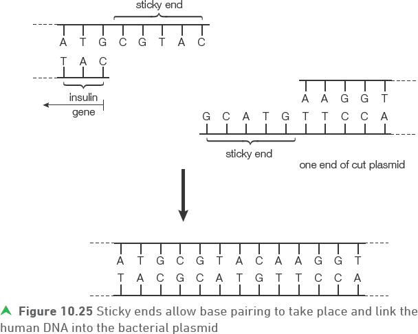
The enzymes that cut and isolate the human insulin gene and cut the bacterial plasmid are called restriction enzymes. These cut the DNA in such a way that one of the two strands extends further than the other one. The longer strand will have ’free’ exposed bases that are not paired. The key is that each restriction enzyme will leave complementary sections of exposed bases in both the plasmid and the human insulin gene so that they can join by base pairing between each other. Not surprisingly, the exposed strands of DNA and their bases are called ’sticky ends’ (Figure 10.25).
![]()
Following the production of the insulin by genetic engineering (also called recombinant DNA technology) the insulin needs to be extracted from the fermenter, purified and packaged before it can be used for medical purposes. These processes that take place after the insulin is produced by the genetically engineered bacteria are referred to as downstreaming.
Many other medical and non-medical products are now made by genetic engineering. In general, pure forms can be produced more quickly, more cheaply and in greater quantities than by older extraction methods.
Advantages of producing human insulin by genetic engineering include:
• before genetically engineered insulin, the amount of insulin available was limited by the number of animals brought to the abattoirs for slaughter
• the extraction process was time consuming and there was the risk of transferring infections
• using animal insulin creates ethical issues for some people
• an additional complication is the fact that non-human insulin differs in structure to human insulin and is therefore not quite as effective for humans.
Practice questions
1 a) Figure 10.26 represents an animal cell. Only one pair of chromosomes is shown.
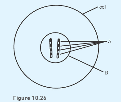
i) Name the structures labelled A on the chromosome.
(1 mark)
ii) Name the part of the cell labelled B where chromosomes are found.
(1 mark)
b) Chromosomes are formed of DNA.
i) Name the two components in the DNA backbone.
(2 marks)
ii) Explain what is meant by the ’unique nature of an individual’s DNA’.
(1 mark)
![]()
c) Figure 10.27 outlines the role of DNA in cells.
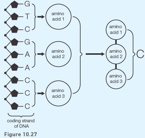
i) How many base triplets are present in the diagram?
(1 mark)
ii) How many bases would be needed to code for a sequence of 45 amino acids?
(1 mark)
iii) What evidence is there that the three amino acids coded for in the diagram are different from each other?
(1 mark)
iv) Name structure C.
(1 mark)
2 a) Figure 10.28 represents the process of mitosis.
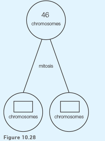
i) Copy and complete the diagram to show the number of chromosomes in each daughter cell.
(1 mark)
ii) Name one function of mitosis.
(1 mark)
b) i) Figure 10.29 below represents the process of meiosis. Only two pairs of chromosomes are shown.
Copy and complete the diagram to show the four possible chromosome arrangements in the daughter cells.
(2 marks)
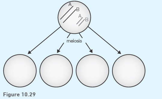
ii) Name one part of the human body where meiosis occurs.
(1 mark)
iii) What name is given to the cells that are produced by meiosis?
(1 mark)
3 a) The allele that causes albinism is recessive to the normal allele.
i) Copy and complete the Punnett square below to show the offspring of a cross between one parent who is heterozygous for albinism and the other parent who has albinism. (Let A = normal; a = albinism)
(2 marks)
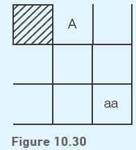
ii) From the Punnett square, what is the probability of these parents having a child with albinism?
(1 mark)
![]()
b) Figure 10.31 shows a pedigree diagram for the inheritance of albinism in a family. (Let A = normal; a = albinism)
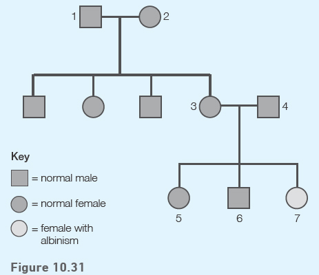
i) What are the genotypes of the parents of child 7 (3 and 4)?
(1 mark)
ii) What are the possible genotypes for the brother and sister of the child with albinism (5 and 6)?
(1 mark)
4 Presence or absence of a widow’s peak in humans is genetically determined by the two alleles of one gene.
The allele for having a widow’s peak is dominant. Let W = widow’s peak; w = widow’s peak absent.
a) State the genotype(s) that will result in the presence of a widow’s peak.
(1 mark)
b) Use a Punnett square to show the possible genotypes of children from two heterozygous parents.
(2 marks)
c) What proportion of the offspring are homozygous?
(1 mark)
d) What is the probability of these heterozygous parents’ next child being a girl with a widow’s peak?
(1 mark)
![]()
5 Figure 10.33 represents a human insulin gene and a plasmid from a bacterium.
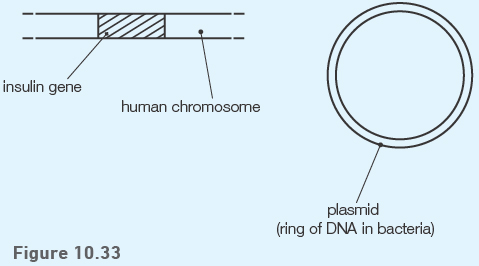
a) Describe how the insulin gene can be incorporated into the plasmid.
(3 marks)
b) Once the plasmid is placed back into the bacterium, describe the stages which follow to enable the bacteria to produce large quantities of insulin.
(2 marks)
c) State the term that is used to refer to the extraction and purification of the insulin.
(1 mark)
d) Give two advantages of producing insulin by this method.
(2 marks)