CCEA GCSE Biology - Denmour Boyd, James Napier 2017
Unit 1
Coordination and control
Specification points
This chapter covers specification points 1.6.1 to 1.6.17. It covers the Central Nervous System, the eye, neurones, synapses, voluntary and reflex actions, homeostasis, hormones, insulin and diabetes, excretion and osmoregulation and plant hormones.
In Double Award Science it covers specification points 1.6.1 to 1.6.12 and covers the central nervous system, voluntary and reflex actions, homeostasis, hormones, insulin and diabetes, excretion and osmoregulation and plant hormones.
Responding to the environment — the nervous system
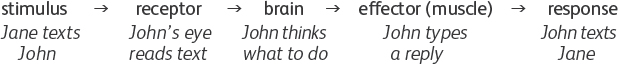
We can respond to the environment and anything that we respond to is called a stimulus. In animals, each type of stimulus affects a receptor in the body. There are many types of receptors, each sensitive to a particular type of stimulus or sense (for example sight, sound, touch, taste and smell). If a receptor is stimulated it may cause a different part of the body, an effector (for example muscles) to produce a response.
![]()
The flowchart above is a simplification because it suggests that we will automatically produce a response when we are stimulated. For example, if we hear a sound (the stimulus) we might respond or not, depending on what the sound is.
Coordination
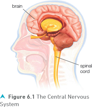
In reality, the receptors and effectors are linked by a coordinator. This coordinator is usually the brain but may also be the spinal cord. Together the brain and the spinal cord are known as the Central Nervous System (CNS), as shown in Figure 6.1.
Nerve cells or neurones link the receptors and the effectors to the coordinator. A neurone carries information as small electrical charges called nerve impulses. The brain acts as a filter and determines which receptors link up with which effectors and whether or not a particular stimulus brings about a response.
A more complete flowchart is:
The overall total of our responses to the environment around us is described as our behaviour.
Sometimes our receptors are grouped together into complex sense organs. Examples include the nose (smell), the ear (sound) and the eye (sight).
![]()
The eye
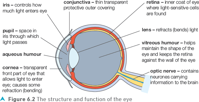
The eye is a specialised sense organ containing receptors that are sensitive to light. Figure 6.2 shows the main parts of the eye and their functions.
The eye is filled with two fluids which allow light to pass through. The fluids also push out against the wall and so maintain the round shape of the eye. In front of the lens the fluid (aqueous humour) is water-like while behind the lens a jelly-like liquid (vitreous humour) keeps the retina pressed against the wall of the eye.
Focusing the eye
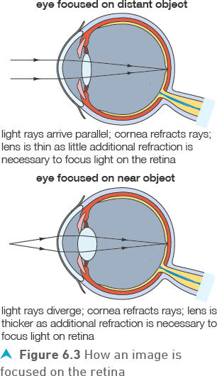
As light rays enter and pass through the cornea, some bending (refraction) of the light takes place. Further bending takes place as the light passes through the lens. By adjusting the thickness of the lens, light can be focused on the retina, irrespective of its angle as it enters the eye. Figure 6.3 shows the role of the lens in focusing light rays from distant and near objects on the retina.
Tip
Most of the refraction happens as the light passes through the cornea, with the lens doing the ’fine tuning’ to focus on the retina.
![]()
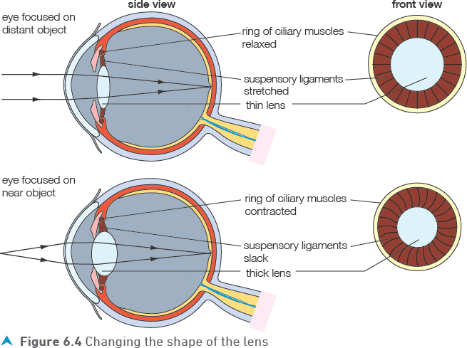
How does the lens change shape (accommodation)?
The ciliary muscle is a ring of muscle that surrounds the lens of the eye. The lens is attached to the ciliary muscle by suspensory ligaments that resemble small pieces of thread. If the ciliary muscle relaxes, it springs out to give a bigger diameter. When this happens, the suspensory ligaments tighten and pull on the lens and it becomes thinner. The opposite happens to make the lens thicker; the ciliary muscle contracts to form a tighter circle with a smaller diameter, causing the suspensory ligaments to slacken, reducing pressure on the lens and allowing it to spring back to its original thicker shape.
![]()
Controlling the amount of light entering the eye
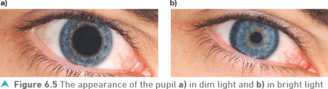
It is important that the correct amount of light enters the eye and reaches the retina. Too little or too much light can damage the sensitive light receptor cells in the retina. Dim light produces a large pupil to allow as much light as possible to enter the eye. In bright light, the pupil is reduced to a small size to restrict the amount of light entering. This is shown in Figure 6.5.
Test yourself
1 What is a receptor?
2 Explain the role of the central nervous system.
3 Where is the cornea in the eye?
4 What is the role of the iris?
![]()
Show you can
Describe how muscles and ligaments in the eye can change the shape of the lens causing light to be focused on the retina.
![]()
Neurones
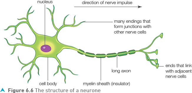
Receptors such as the eye and effectors such as muscles are connected to the Central Nervous System by nerve cells, the neurones. The structure of neurones is adapted for the function of transmitting electrical impulses very quickly across the nervous system. Figure 6.6 shows the typical structure of a neurone.
![]()
The adaptations include:
• The nucleus, which controls the activities of the neurone is surrounded by most of the cell’s cytoplasm in the cell body.
• The axon is a long extension of the cytoplasm which can be up to one metre in length so that nerve impulses can be transmitted from the central nervous system to the extremities of the body by one cell.
• The myelin sheath is a fatty layer around the axon produced by special cells. It acts as an insulator and helps to speed up the nerve impulses.
• The branched ends of the axon and the small branching extensions from the cell body allow the neurone to make junctions with many other nerve cells.
Tip
Although neurones have the same basic structures as all animal cells they are one of the most specialised types of cell in the body.
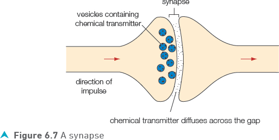
Synapses
The junction between two neurones is called a synapse.
![]()
As shown in Figure 6.7 there is always a small gap between the two neurones.
Since there is a gap between adjoining neurones, the nerve impulse cannot pass from one neurone to the next as an electrical impulse. When an electrical impulse reaches the end of the axon leading into the synapse, it causes the axon to release a special chemical.
The chemical, known as the transmitter substance, quickly diffuses across the gap. If the concentration of transmitter substance is high enough on the other side of the synapse it triggers an electrical impulse in the axon leaving the junction and the nerve impulse continues on its way.
Voluntary and reflex actions
Many of our actions are voluntary. This means we deliberately choose to do them and they involve conscious thought. However, there is another type of action that does not involve conscious thought — these are reflex actions.
![]()
Reflex actions
If you accidently touch a very hot object you respond immediately by rapidly withdrawing your hand from the danger area. The advantage of this is that you move your hand away before it can get burned too badly. This type of action does not involve any ’thinking’ time, as the time taken to consider the response would cause unnecessary damage to the body. All reflex actions have two main characteristics in common:
• they occur very rapidly
• they do not involve conscious control (thinking time).
What makes a reflex action so rapid? In a reflex pathway, the total length of the pathway is kept as short as it possibly can be, with the minimum number of neurones involved. In addition, there are relatively few gaps between the neurones (synapses), as they are the places where the nerve impulses travel relatively slowly.
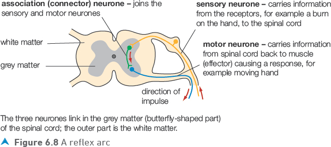
Figure 6.8 shows the nerve pathway when a hand touches a hot object.
There are three types of neurone involved in this response.
The diagram shows that both the association and the motor neurones begin with the cell body. The diagram also shows that only two synapses (short gaps between neurones that slow nervous communication) are involved in this pathway. This system of structures involved in a reflex action is called a reflex arc.
Test yourself
5 How do the branch ends of a neurone adapt it to its function?
6 Explain the difference between a voluntary and a reflex action.
7 What is a synapse?
![]()
8 Where is a transmitter substance found?
![]()
Show you can
Draw a flow chart to summarise the pathway of a reflex arc.
Hormones
Another type of communication system is controlled by hormones. Hormones are chemical messages produced by special glands which release them into the blood. Although the hormones travel all around the body in the blood, they can only affect certain organs called target organs.
Tip
Each hormone affects different target organs. Some hormones affect many organs while some organs are affected by several hormones.
Hormones usually act more slowly than the nervous system and over a longer period. Good examples to illustrate these points are the sex hormones: oestrogen and testosterone. The changes brought about by testosterone in males and oestrogen in females come about over many years.
Hormones also have an important role in maintaining the internal environment of the body in a relatively constant state in response to changes outside and inside the body. The maintenance of this constant state is referred to as homeostasis. Two examples of the homeostatic role of hormones are controlling the concentration of glucose in the blood by the hormone insulin and controlling the water content of the body, referred to as osmoregulation.
Tip
While nervous responses are fast and short term, hormone responses are slow and continue over long periods of time.
Insulin and blood glucose
Insulin is a hormone that prevents the concentration of glucose (sugar) in the blood becoming too high. Glucose is constantly needed by all cells for respiration and therefore must always be present at a sufficient concentration. However, if there is too much glucose in the blood this can damage body cells due to water loss by osmosis (see Chapter 8).
Tip
Insulin lowers blood glucose concentration — it is not enough to say that it controls blood glucose concentration.
Insulin is produced and released into the blood by special cells in the pancreas in response to increasing or high blood glucose concentrations. This usually occurs after a meal, especially if the meal is rich in carbohydrates.
The main target organ for insulin is the liver where it causes:
• increased absorption of glucose from the blood, so reducing the blood glucose concentration
• the conversion of excess glucose into glycogen, which is stored in the liver and to a lesser extent in muscle cells
• increased respiration.
Tip
Meals rich in carbohydrates are digested into sugars which can then be absorbed into the blood stream.
Figure 6.9 summarises how insulin controls the blood glucose concentration.
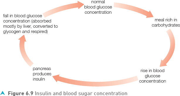
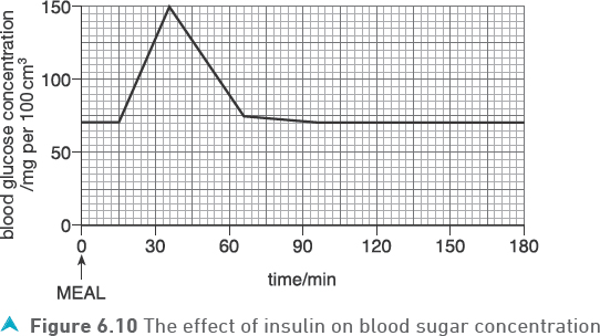
Figure 6.10 shows how the concentration of blood glucose typically varies after eating a meal.
When blood glucose concentration is low, less insulin is produced. This means that the above processes do not take place or take place at a slower rate, helping to raise the concentration of glucose in the blood.
![]()
Negative feedback
In general, negative feedback is a mechanism to ensure that the concentration or level of something does not deviate too far from the normal value. The mechanism usually involves at least one hormone. Such mechanisms include feedback because the body constantly monitors the concentration or level of what it controls. In the case of blood glucose concentration, the pancreas fulfils this role, responding by adjusting the amount of insulin it releases into the blood. The mechanism is negative feedback because increasing the hormone has the opposite (negative) effect on what is being monitored — increasing the amount of insulin causes the blood glucose concentration to fall back towards normal.
Diabetes — when blood glucose regulation fails
Diabetes is a fairly common condition in which the body does not produce enough insulin to keep the blood glucose at the normal concentration. Individuals who develop diabetes are unable to control the concentration of their blood glucose without treatment and the following symptoms are often present:
• There is glucose in the urine. This happens because their blood glucose concentration is so high that some is filtered out by the kidneys and passed into the urine.
• Affected individuals are often thirsty and because they drink so much they need to go to the toilet a lot.
• Lethargy may result.
There are two kinds of diabetes, Type 1 and Type 2. Type 1 diabetes normally develops in childhood. Type 2 diabetes usually only develops in older people, but is becoming increasingly common in young people.
Type 1 diabetes is usually treated by the injection of insulin and by a carefully controlled diet where the intake of carbohydrates is carefully monitored.
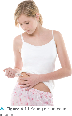
Figure 6.11 shows a young girl injecting herself with insulin. Even with the use of insulin injections and a carefully controlled diet, it is difficult for people with diabetes to control their blood sugar concentration very accurately. Problems may arise if too much insulin is injected or if not enough food is eaten at regular intervals. If the blood sugar concentration drops too low, a hypoglycaemic attack (a hypo) may occur and unconsciousness will result. If blood sugar concentrations remain too high for a long period serious medical complications can result.
Type 2 diabetes has a slightly different cause in that insulin is produced but stops working effectively. Type 2 diabetes is often associated with poor diet, obesity and lack of exercise. The treatment of Type 2 diabetes therefore includes changes to diet and exercise to achieve weight loss along with medication in the form of tablets and injections. The increasing number of people with these characteristics largely explains the increase in the number of people with diabetes.
Test yourself
9 What are the differences between a hormone response and nervous response?
10 What is homeostasis?
11 Why is it important to control the blood glucose concentration?
12 Give three symptoms of diabetes.
Long-term effects and future trends
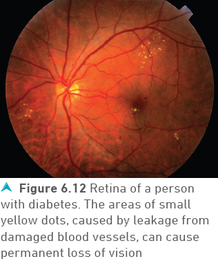
People who have had diabetes for a long time (in some cases undiagnosed and unknown) and whose blood sugar concentration is not tightly controlled run the risk of developing long term complications. These include eye damage (see Figure 6.12), or even blindness, heart disease, strokes and kidney damage. These complications are usually due to the high blood sugar concentration damaging the capillaries that supply the part of the body involved.
Show you can
Describe how insulin controls the blood sugar concentration.
Osmoregulation
Osmoregulation is another homeostatic process in the body. It controls the amount of water in the blood and other body fluids. Water, like the sugars in the blood, has the potential to damage cells due to osmosis causing excessive movements of water.
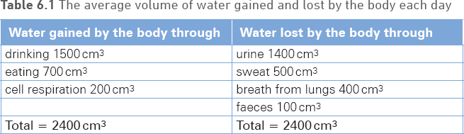
The body gains water mainly by drinking and from the food we eat. A small amount also comes from cell respiration which produces water as a by-product. At the same time, water is lost from the body through evaporation in the lungs, evaporation while sweating and the production of urine by the kidneys and in faeces. Table 6.1 lists the average volume of water gained and lost by the body each day.
In normal conditions the volume of water gained balances the volume lost but if conditions change then osmoregulation brings the volumes back into balance.
For example, in very warm weather or during vigorous exercise the body will lose more water as sweat. This can be partially balanced by increasing the amount we drink but the kidneys also produce a more concentrated urine containing less water. If we drink larger volumes of liquid than normal the kidneys again act to bring the volumes back into balance by producing large volumes of dilute urine.
Tip
Osmoregulation is the way the body balances the water it gains (drinking, eating and respiration) with the water it loses (urine, sweat, breathing and faeces).
Example
Table 6.2 shows the water gained and lost by a boy who has played a game of football on a warm summer day.
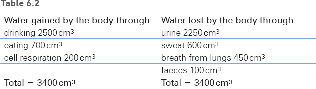
In this situation the volumes of water gained and lost have changed because:
• Playing football has increased the water lost as sweat and by breathing more often.
• During the game, he has drunk extra water to compensate for these losses.
• That has increased his total gained to 3400 cm3.
• To maintain the water balance in the body the total lost must also increase to 3400 cm3.
• The kidneys produce a larger volume of dilute urine.
Kidneys — how they affect the volume of urine
The kidneys are part of the excretory system, which functions to remove wastes from the body and to carry out osmoregulation. Figure 6.13 shows the excretory system.
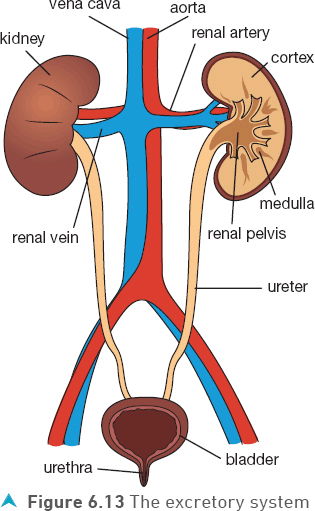
Blood passes into each kidney through the renal artery. When it reaches the cortex of the kidney much of the liquid portion of the blood and many of the substances dissolved in it are filtered out. Then in the medulla region water and other substances are reabsorbed back into the blood in a controlled way until normal concentrations are reached.
The excess water and dissolved substances pass into the urine, which collects in the renal pelvis, and passes down the ureter into the bladder. There it is stored before being passed out via the urethra.
Tip
It is important to learn the correct spelling of ureter and urethra.
![]()
Anti-diuretic hormone (ADH)
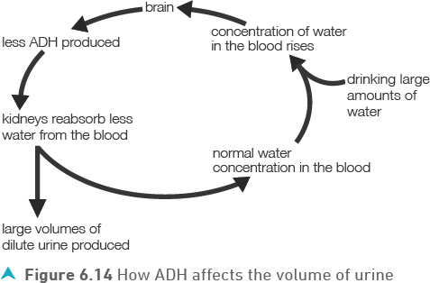
The process of reabsorption of water back into the blood by the kidneys is controlled by a hormone, the anti-diuretic hormone (ADH).
ADH is produced by the part of the brain where the amount of water in the blood is monitored. The ADH is released into the blood and travels to its target organ, the kidneys. In the kidney medulla, ADH allows more water to pass from the urine back into the blood (be reabsorbed). As a result, the urine has a lower volume and is more concentrated.
As an example of how ADH works, consider a person who has drunk a lot of water on a hot day. The blood reaching their brain will contain much more water than normal. In response, the brain stops or reduces the production of ADH. In the kidney, low or no ADH means much less water is reabsorbed into the blood leaving large volumes of dilute urine to be produced. Figure 6.14 shows how ADH affects the volume of urine.
Test yourself
13 List three ways the body gains water.
14 Name the blood vessel that transports blood to the kidney.
15 Name the tube that carries urine from the kidney to the bladder.
16 Where is ADH produced?
![]()
Show you can
After drinking a litre of water, explain how ADH would cause a large volume of dilute urine to be produced.
Sensitivity in plants
Plants, like animals, respond to changes in the environment. However, they respond to fewer types of stimuli and in general the response is slower. Plants respond to the environmental stimuli that have the greatest effect on their growth. Roots grow towards water when a moisture gradient exists and shoots tend to grow away from the effects of gravity (they grow upwards). Reasons for these responses are fairly obvious, as they ensure that the plants react in such a way that they receive the best conditions for growth. The response of a plant to light is called phototropism and this response has been investigated in detail to establish how it occurs.
Phototropism — responding to light
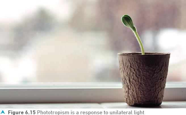
Most of you will have observed that plants grow towards a light source. Plants left on a window sill or against the wall of a house usually do not grow straight up, but bend towards the Sun. You can also probably guess that this response ensures that the plant stem and leaves receive more light than they otherwise would do if there was no response. This means that more photosynthesis takes place and there will be more growth.
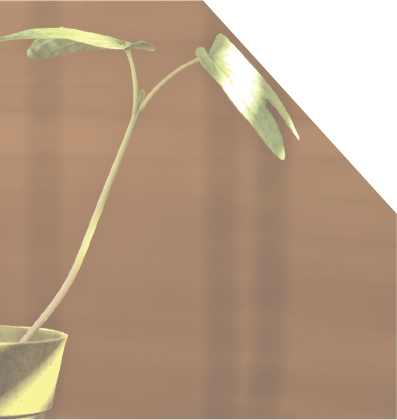
Although it is easy to observe the effect of phototropism, what causes it to occur? Figure 6.16 shows the growth of young seedlings in unilateral light (light coming from one side or source only) and highlights some of the features of phototropism. Can you use the diagram to identify what part of the plant is sensitive to the light source?
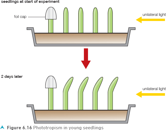
Figure 6.16 shows that it is the shoot tip that is sensitive to light, as when it is covered the phototropic response does not occur. This and many other similar experiments have led to our understanding that the response of phototropism is controlled by a plant hormone called auxin. When the stem is illuminated from one side this hormone tends to accumulate more on the non-illuminated side. The effect of the hormone is to increase the growth of the non-illuminated side of the stem more rapidly than the side receiving most light. This differential growth that occurs when one side of the stem grows more than the other side leads to the stem bending in the direction of the light.
![]()
The auxin is actually produced at the tip of the shoot and diffuses downwards. As it does so, light on one side causes the auxin to accumulate on the non-illuminated side. The main effect of this high concentration of auxin is to make each cell grow by elongation (become longer) more than it normally would. The cells in the non-illuminated side of the stem therefore increase in length more than those in the illuminated side, causing the stem to bend towards the light. Figure 6.17 shows how auxin causes a stem to bend towards light.

Practice questions
1 The word equation summarises a nervous system response.
![]()
a) What is a stimulus?
(1 mark)
b) i) Name the parts of the Central Nervous System.
(2 marks)
ii) What is the role of the Central Nervous System?
(1 mark)
c) Give one example of an effector.
(1 mark)
d) Some responses are voluntary while others are reflex.
Explain the difference between a voluntary and a reflex response.
(2 marks)
2 a) What effect would eating chocolate have on blood glucose concentration?
(1 mark)
b) Name the organ which produces insulin.
(1 mark)
c) Describe and explain the effect insulin has on blood glucose concentration.
(3 marks)
d) Name the condition caused by the failure of the body to produce insulin.
(1 mark)
e) Describe how this condition can be treated.
(1 mark)
![]()
3 Figure 6.18 shows the front of an eye in bright light.
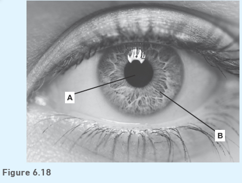
a) Name parts A and B.
(2 marks)
b) The bright light is switched off.
i) Describe what happens to part B.
(1 mark)
ii) Explain why this change is necessary.
(2 marks)
![]()
4 Figure 6.19 shows a section through an eye.
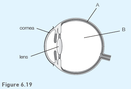
a) Name and give the function of parts A and B.
(4 marks)
b) i) The lens and the cornea work together to carry out one function. Describe this function.
(1 mark)
![]()
ii) Describe in detail how the muscles and ligaments in the eye allow the lens to carry out this function.
(3 marks)
5 Describe how an electrical nerve impulse crosses a synapse.
(4 marks)
![]()
6 Figure 6.20 shows a reflex arc.
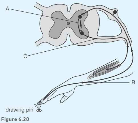
a) Name neurones A, B and C.
(3 marks)
![]()
b) Describe how neurone B is adapted to its function.
(1 mark)