CONCEPTS IN BIOLOGY
PART V. THE ORIGIN AND CLASSIFICATION OF LIFE
23. The Animal Kingdom
23.4. Body Plans
Although animals come in a variety of sizes and shapes, you can see certain evolutionary trends and a few basic body plans.
Symmetry
Symmetrical objects have similar parts that are arranged in a particular pattern. For example, the parts of a daisy flower and a bicycle are arranged symmetrically.
Asymmetry is a condition in which there is no pattern to the individual parts. Asymmetrical body forms are rare and occur only in certain species of sponges, which are the simplest kinds of animals.
Radial symmetry occurs when a body is constructed around a central axis. Any division of the body along this axis results in two similar halves. Although many animals with radial symmetry are capable of movement, they do not always lead with the same portion of the body; that is, there is no anterior, or head, end. Starfish and jellyfish are examples of organisms with radial symmetry.
Bilateral symmetry exists when an animal is constructed with equivalent parts on both sides of a plane. Animals with bilateral symmetry have a head and a tail region. There is only one way to divide bilateral animals into two mirrored halves. Animals with bilateral symmetry move head first, and the head typically has sense organs and a mouth. The feature of having an anterior head end is called cephalization (cephal = head). It appears that bilateral symmetry was an important evolutionary development since most animals have bilateral symmetry (figure 23.4).
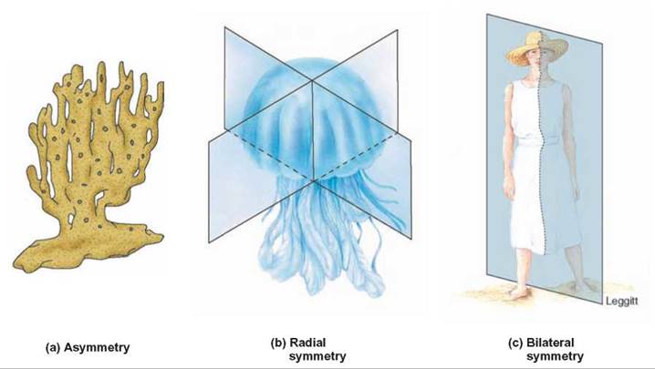
FIGURE 23.4. Kinds of Symmetry
(a) This sponge has a body that cannot be divided into symmetrical parts and is therefore asymmetrical. (b) In animals such as this with radial symmetry, any cut along the central body axis results in similar halves. (c) In animals with bilateral symmetry, only one one plane results in similar halves.
Embryonic Cell Layers
Animals differ in the number of layers of cells of which they are composed. When we look at the development of embryos we find that the embryos of the simplest animals (sponges) do not form distinct, tissuelike layers. However, jellyfishes and their relatives have embryos that consist of two layers. The ectoderm is the outer layer and the endoderm is the inner layer. Because their embryos are composed of two layers, these animals are said to be diploblastic. In adults, these embryonic cell layers give rise to an outer, protective layer and an inner layer that forms a pouch and is involved in processing food.
All the other major groups of animals have embryos that are triploblastic. Triploblastic animals have three layers of cells in their embryos. Sandwiched between the ectoderm and endoderm is a third layer, the mesoderm. In the adult body, the ectoderm gives rise to the skin or other surface covering, the endoderm gives rise to the lining of the digestive system, and the mesoderm gives rise to muscles, connective tissue, and other organ systems involved in the excretion of waste, the circulation of material, the exchange of gases, and body support (figure 23.5).
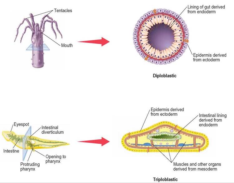
FIGURE 23.5. Embryonic Cell Layers
Diploblastic organisms have two embryonic cell layers. The outer ectoderm becomes the epidermis and the inner endoderm becomes the lining of the gut. Triploblastic organisms have three embryonic cell layers: the ectoderm, endoderm, and mesoderm. The mesoderm forms most of the tissues and organs of the body.
Body Cavities
A coelom is a fluid-filled body cavity that separates the outer body wall of the organism from the gut and internal organs. The development of a coelom was an important step in animal evolution.
Simple animals, such as jellyfish and flatworms, are acoelomate, which means that they have no space separating their outer surface from their internal organs. However, most animals have some form of a coelom. Because organs such as the gut and heart are not embedded in a mass of cells but are suspended in a space (the coelom), they have a greater freedom of movement than the organs of acoelomate animals. Organs are not loose in the coelom; they are held in place by sheets of connective tissue called mesenteries.
Mesenteries also support the blood vessels connecting the various organs.
It is often difficult to visualize the presence of a coelom as a cavity, because the cavity is filled with organs and a small amount of fluid. Perhaps a common example will help.
The coelom in a turkey is the cavity where you stuff the dressing. In the living bird this cavity contains a number of organs, including those of the digestive, excretory, and circulatory systems.
Some animals do not have a true coelom but have a similar space called a pseudocoelom. A pseudocoelom differs from a true coelom in that it is located between the lining of the gut and the outer body wall. In other words, animals with a pseudocoelom do not have muscles around their digestive system. In addition, there are no mesenteries suspending the gut from the outer body wall. Nematode worms and several related groups of animals have a pseudocoelom (figure 23.6).
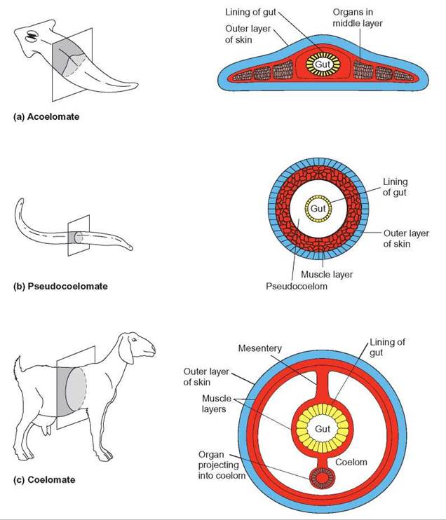
FIGURE 23.6. Body Cavities
(a) Acoelomate animals, such as flatworms, have no open space between the gut and outer body layer. (b) Roundworms, commonly found in soil, have a body cavity called a pseudocoelom. It contains some cells, and there are no muscles surrounding the gut. (c) Other animals, including all vertebrates, have a coelom, which is a fluid-filled space that separates internal organs from the outer body wall. In addition, the coelom is lined with connective tissue of mesodermal origin. Organs project into the coelom and are held in place by thin sheets of connective tissue called mesenteries.
Segmentation
Many kinds of bilaterally symmetrical organisms have segmented bodies.
Segmentation is the separation of an animal’s body into a number of recognizable units from its anterior to its posterior end. Segmentation is associated with the specialization of certain parts of the body. Three common groups of animals show segmentation: annelid worms, arthropods, and chordates.
Annelid worms have a series of very similar segments with minor differences between them. Segmentation in arthropods is modified so that several segments are specialized as a head region and more posterior segments are less specialized. Many of the posterior segments have legs and other appendages. Among the arthropods, insects show a great deal of specialization of segments. In chordates, the segmentation is of a different sort but is obvious in the arrangement of muscles and the vertebral column (figure 23.7). Studies of the genes that control development show that all bilaterally symmetrical animals have essentially the same genes controlling their development and how different regions or segments of the body develop (How Science Works 23.1).
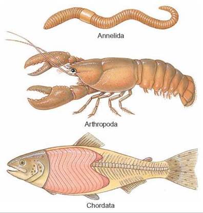
FIGURE 23.7. Segmentation
Segmentation is associated with the specialization of certain parts of the body. Annelid worms show many segments with little specialization. Arthropods show a highly developed head region, with the more posterior segments less specialized. Chordates show the segmentation of muscles and skeletal structures.
HOW SCIENCE WORKS 23.1
Genes, Development, and Evolution
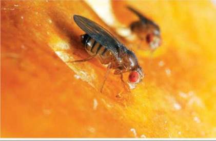
One of the important discoveries of modern molecular genetics is the remarkable similarity in the kinds of genes found in all organisms. This has important implications for understanding the evolution of organisms. It appears that once a new, valuable gene is created through the process of mutation, it is preserved in evolutionary descendants. One example is a group of genes known as homeotic genes. These genes regulate how an organism's body is formed by helping to define which end of the developing embryo is the head and which is the tail. As the embryo develops and regular body segments form, the homeotic genes also help define what each segment becomes. In insects, one segment might give rise to antennae, while another gives rise to wings or legs. Homeotic genes were first discovered in the fruit fly (Drosophila melanogaster), which has been a favorite species for students of animal genetics for 100 years (see photo). Fruit flies are ideal for genetic studies for several reasons: They are easy and inexpensive to raise in the lab, a new generation can be produced every 10 days, and large numbers of offspring are produced.
It is now known that homeotic genes control the same developmental processes in all organisms that are bilaterially symmetrical (their left side mirrors their right side). This trend is so overwhelming that some scientists have suggested that the presence of one type of homeotic genes, the Hox genes, should be used to define the Animal kingdom.
Essentially the same genes with the same functions can be found in widely different animals, such as fruit flies, earthworms, sea urchins, tapeworms, and humans. This means that the study of fruit flies can be used to discover how the same genes function in humans and other animals. Because homeotic genes are involved in regulating embryonic development and cellular differentiation, these studies can be used to help identify the causes of human embryonic development abnormalities and other diseases like cancer.
Skeletons
A skeleton is the part of an organism that provides structural support. Most animals have a skeleton. It serves as strong scaffolding, to which other organs can be attached. In particular, the skeleton provides places for muscle attachment and, if the skeleton has joints, the muscles can move one part of the skeleton with respect to others. Some aquatic organisms, such as sea anemones and many kinds of worms, are generally supported by the dense medium in which they live and lack well-developed skeletons. However, most aquatic animals have a skeleton. Most terrestrial animals have a strong structure that supports them in the thin medium of the atmosphere.
There are two major types of skeletons: internal skeletons (endoskeletons) and external skeletons (exoskeletons) (figure 23.8). The vertebrates (fish, amphibians, reptiles, birds, mammals), echinoderms (starfish, sea urchins, etc.), and some other groups have internal skeletons. The various organs are attached to and surround the skeleton, which grows in size as the animal grows. Arthropods (crustaceans, spiders, insects, millipedes, centipedes), nematodes, and some other groups have an external skeleton that surrounds all the organs. It is generally hard and has joints. These animals accommodate growth by shedding the old skeleton and producing a new, larger one. This period in the life of an arthropod is dangerous, because for a short period it is without its hard, protective outer layer. Many other animals have structures that have a supportive or protective function (such as clams, snails, and corals) and these are sometimes called skeletons, but they do not have joints.
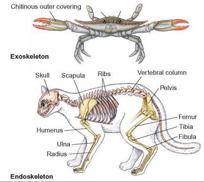
FIGURE 23.8. Skeletons
There are two major types of skeletons; endoskeletons and exoskeletons. Endoskeletons are typical of vertebrates and echinoderms, and exoskeletons are typical of arthropods and their relatives.
Some organisms use water as a kind of supportive skeleton. Annelid worms and some other animals have fluid-filled coeloms. Because water is not compressible, but it is movable, compressive forces by muscles can cause the animal’s shape to change. This is similar to what happens with a water-filled balloon; compression in one place causes it to bulge out somewhere else.
23.4. CONCEPT REVIEW
6. Describe body forms that show asymmetry, radial symmetry, and bilateral symmetry.
7. Give an example of an animal that has a coelom and one with a pseudocoelom.
8. Give an example of an animal with an exoskeleton, and one with an endoskeleton.
9. How does an animal with an exoskeleton grow?
10. How do diploblastic and triploblastic animals differ?
11. What is one advantage of segmentation?