CONCEPTS IN BIOLOGY
PART VI. PHYSIOLOGICAL PROCESSES
24. Materials Exchange in the Body
24.5. Obtaining Nutrients: The Digestive System
The digestive system is the organ system responsible for the processing and distribution of nutrients; it consists of a muscular tube and glands that secrete digestive juices into the tube. Four kinds of activities are involved in getting nutrients to the cells that need them: mechanical processing, chemical processing, nutrient uptake, and chemical alteration. Figure 24.10 shows the various structures of the digestive system.

FIGURE 24.10. The Digestive System
The teeth, tongue, and enzymes from the salivary glands modify the food before it is swallowed. The stomach adds acid and enzymes and further changes the food’s texture. The food is eventually emptied into the duodenum, where the liver and pancreas add their secretions. The small intestine also adds enzymes and is involved in absorbing nutrients. The large intestine is primarily involved in removing water.
Mechanical and Chemical Processing
The purpose of the digestive system is to break down large chunks of food into small molecules, which the circulatory system can distribute to the body’s cells. The first step in the digestive process is mechanical processing.
It is important to grind large particles into small pieces by chewing to increase their surface areas and allow for more efficient chemical reactions. It is also important to add water to the food, which further disperses the particles and provides the watery environment needed for these chemical reactions. Materials also must be mixed so that all the molecules that need to interact with one another have a good chance of doing so. The oral cavity and the stomach are the major body regions involved in reducing the size of food particles. The teeth are involved in cutting and grinding food to increase its surface area. The watery mixture that is added to the food in the oral cavity is called saliva, and the three pairs of glands that produce saliva are known as salivary glands. Saliva contains the enzyme salivary amylase, which begins the chemical breakdown of starch. Saliva also lubricates the oral cavity and helps bind food before swallowing.
In addition to having taste buds that help identify foods and potentially toxic materials, the tongue performs the important service of helping position the food between the teeth and pushing it to the back of the throat for swallowing. The oral cavity is very much like a food processor, in which mixing and grinding take place.
Once the food has been chewed, it is swallowed and passes down the esophagus to the stomach. The process of swallowing involves a complex series of events. First, a ball of food, known as a bolus, is formed by the tongue and moved to the back of the oral cavity. There, it stimulates the walls of the throat, also called the pharynx. Nerve endings in the lining of the pharynx are stimulated, causing a reflex contraction of the walls of the esophagus, which transports the bolus to the stomach. Because both food and air pass through the pharynx, it is important to prevent food from getting into the lungs. During swallowing, the larynx is pulled upward, causing a flap of tissue, called the epiglottis, to cover the opening to the trachea and preventing food from entering the trachea.
In the stomach, additional liquid, called gastric juice, is added to the food. Gastric juice contains enzymes and hydrochloric acid. The major enzyme of the stomach is pepsin, which initiates the chemical breakdown of protein. The pH of gastric juice is very low, generally around pH 2. Consequently, very few kinds of bacteria or protozoa emerge from the stomach alive. Those that do survive have characteristics that protect them from the gastric juice as they pass through the stomach. The entire mixture is churned by the contractions of the three layers of muscle in the stomach wall. The combined activities of enzymatic breakdown, chemical breakdown by hydrochloric acid, and mechanical processing by muscular movement result in a thoroughly mixed liquid. This liquid eventually leaves the stomach through a valve known as the pyloric sphincter and enters the small intestine (How Science Works 24.1).
The first part of the small intestine is called the duodenum. In addition to producing enzymes, the duodenum secretes several kinds of hormones that regulate the release of food from the stomach and the release of secretions from the pancreas and liver. The pancreas produces a number of digestive enzymes and secretes large amounts of bicarbonate ions, which neutralize the acids that enter from the stomach so that the pH of the duodenum is about pH 8. The liver, a large organ in the upper abdomen, performs several functions, one of which is the secretion of bile. When bile leaves the liver, it is stored in the gallbladder prior to being released into the duodenum. When bile is released from the gallbladder, it assists mechanical processing by breaking large fat globules into smaller particles, much as soap breaks up fat particles into smaller globules that are suspended in water and washed away. This process is called emulsification. Emulsification is important because fats are not soluble in water, yet the reactions of digestion must take place in a water solution.
Along the length of the small intestine, additional watery juices are added until the mixture reaches the large intestine or colon. The 1.5-meter-long large intestine is primarily involved in reabsorbing the water that has been added to the food tube when saliva, gastric juice, bile, pancreatic secretions, and intestinal juices are introduced into the digestive system. The large intestine is also home to a variety of bacteria. Most live on food that was not absorbed in the small intestine. Some provide additional benefit by producing vitamins that can be absorbed from the large intestine. A few are capable of causing disease.
Several kinds of enzymes have been mentioned in this section. Each is produced by a specific organ and has a specific function. Chapter 5 introduced the topic of enzymes and how they work. Some enzymes, such as those involved in glycolysis, the Krebs cycle, and protein synthesis, are produced and used inside cells; others, such as the digestive enzymes, are produced by cells and secreted into the digestive tract. Digestive enzymes are simply a class of enzymes; they have the same characteristics as all other enzymes. They are protein molecules that speed up chemical reactions and are sensitive to changes in temperature or pH. The various digestive enzymes, the sites of their production, and their functions are listed in table 24.2.
TABLE 24.2. Digestive Enzymes and Their Functions
Enzyme |
Site of Production |
Molecules Altered |
Molecules Produced |
Salivary amylase |
Salivary glands |
Starch |
Smaller polysaccharides |
Pepsin |
Stomach lining |
Proteins |
Peptides |
Gastric lipase |
Stomach lining |
Fats |
Fatty acids and glycerol |
Chymotrypsin |
Pancreas |
Polypeptides (long chains of amino acids) |
Peptides |
Trypsin |
Pancreas |
Polypeptides |
Peptides |
Carboxypeptidase |
Pancreas |
Peptides (several amino acids) |
Smaller peptides and amino acids |
Pancreatic amyla |
se Pancreas |
Polysaccharides (many sugar molecules attached together) |
Disaccharides |
Pancreatic lipase |
Pancreas |
Fats |
Fatty acids and glycerol |
Nuclease |
Pancreas |
Nucleic acids |
Nucleotides |
Aminopeptidase |
Intestinal lining |
Peptides |
Smaller peptides and amino acids |
Dipeptidase |
Intestinal lining |
Dipeptides |
Amino acids |
Lactase |
Intestinal lining |
Lactose |
Glucose and galactose |
Maltase |
Intestinal lining |
Maltose |
Glucose |
Sucrase |
Intestinal lining |
Sucrose |
Glucose and fructose |
Nuclease |
Intestinal lining |
Nucleic acids |
Nucleotides |
HOW SCIENCE WORKS 24.1
An Accident and an Opportunity
On the morning of June 6, 1822, on Mackinac Island in northern Lake Huron, a 19-year-old French-Canadian fur trapper named Alexis St. Martin was shot in the stomach by the accidental discharge from a shotgun. The army surgeon at Fort Mackinac, Michigan, Dr. William Beaumont, was called to attend to the wounded man. Part of St. Martin's stomach and body wall had been shot away, and parts of his clothing were embedded in the wound. Although Dr. Beaumont did not expect St. Martin to live, he quickly cleaned the wound, pushed portions of the lung and stomach that were protruding back into the cavity, and dressed the wound. Finding St. Martin alive the next day, Beaumont was surprised and encouraged to do what he could to extend his life. In fact, Dr. Beaumont cared for St. Martin for 2 years. When the wound was completely healed, the stomach had fused to the body wall and a hole allowed Dr. Beaumont to look into the stomach. Fortunately for St. Martin, a flap of tissue from the lining of the stomach closed off the opening, so that what he ate did not leak out.
Dr. Beaumont found that he could look through the opening and observe the activities in the stomach and recognized that this presented an opportunity to study the stomach's function in a way that had not been done before. He gathered gastric juice, had its components identified, introduced food into the hole with a string attached so that he could retrieve the food particles that were partially digested for examination, and observed the effect of emotion on digestion. He discovered many things that were new to science and contrary to the teachings of the time. He recounted many of his observations and experiments in his journal: "I consider myself but a humble inquirer after truth—a simple experimenter. And if I have been led to conclusions opposite to the opinions of many who have been considered luminaries of physiology, and, in some instances, from all the professors of this science, I hope the claim of sincerity will be conceded to me, when I say that such difference of opinion has been forced upon me by the convictions of experiment, and the fair deductions of reasoning."
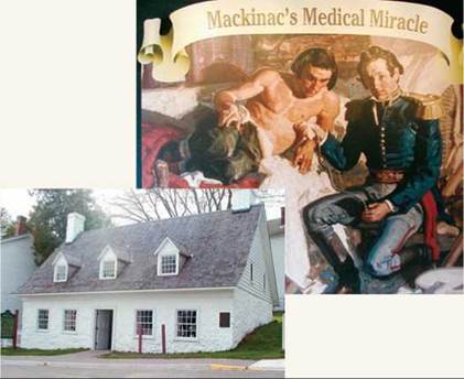
House where Dr. Beaumont treated Alexis St. Martin
Following are some of his important discoveries.
1. He measured the temperature of the stomach and found that it does not heat up when food is introduced, as was thought at the time: "But from the result of a great number of experiments and examinations, made with a view to asserting the truth of this opinion, in the empty and full state of the organ, ... I am convinced that there is no alteration of temperature. ..."
2. He found that pure gastric juice contains large amounts of hydrochloric acid. This was contrary to the prevailing opinion that gastric juice was simply water: "I think I am warranted, from the result of all the experiments, in saying, that the gastric juice, so far from being "inert as water," as some authors assert, is the most general solvent in nature, of alimentary matter—even the hardest bone cannot withstand its action."
3. He observed that gastric juice is not stored in the stomach but, rather, is secreted when food is eaten: "The gastric juice does not accumulate in the cavity of the stomach, until alimentary matter is received, and excite its vessels to discharge their contents, for the immediate purpose of digestion."
4. He realized that digestion begins immediately when food enters the stomach. The prevailing opinion of the day was that nothing happened for an hour or more: "At 2 o'clock p.m.—twenty minutes after having eaten an ordinary dinner of boiled, salted beef, bread, potatoes, and turnips, and drank [sic] a gill of water, I took from his stomach, through the artificial opening, a gill of the contents ... Digestion had evidently commenced, and was perceptually progressing, at the time."
5. He discovered that food in the stomach satisfies hunger even though it is not eaten: "To ascertain whether the sense of hunger would be allayed without food being passed through the oesophagus [sic], he fasted from breakfast time, til 4 o'clock, p.m., and became quite hungry. I then put in at the aperture, three and a half drachms of lean, boiled beef. The sense of hunger immediately subsided, and stopped the borborygmus, or croaking noise, caused by the motion of the air in the stomach and intestines, peculiar to him since the wound, and almost always observed when the stomach is empty."
St. Martin did not take kindly to these probings and twice ran away from Dr. Beaumont's care back to Canada, where he married, had two children, and resumed his former life as a voyager and fur trapper. He did not die until the age of 83, having lived over 60 years with a hole in his stomach.
Nutrient Uptake
Digestion results in a variety of simple organic molecules that are available for absorption from the tube of the gut into the circulatory system. As simple sugars, amino acids, glycerol, and fatty acids move into the circulatory system, surface area again becomes important. The amount of material that can be taken up is limited by the surface area available. There are several ways in which the structure of the intestinal tract provides a large surface area. First, the small intestine is a very long tube; the longer the tube, the greater the internal surface area. In a typical adult human, it is about 6 to 8 meters long. In addition to length, the lining of the small intestine consists of millions of fingerlike projections, called villi, which increase the surface area. The cells that make up the villi also have folds in their surface membranes. All of these characteristics increase the surface area available for the transport of materials from the gut into the circulatory system. Scientists estimate that the cumulative effect of all these features produces a total intestinal surface area of about 250 square meters. That is equivalent to about the area of a basketball court.
The surface area by itself would be of little value if it were not for the intimate contact of the circulatory system with the intestinal lining. Each villus contains several capillaries and a branch of the lymphatic system called a lacteal. The close association between the intestinal surface and the circulatory and lymphatic systems allows for the efficient uptake of nutrients from the cavity of the gut into the circulatory system (figure 24.11).
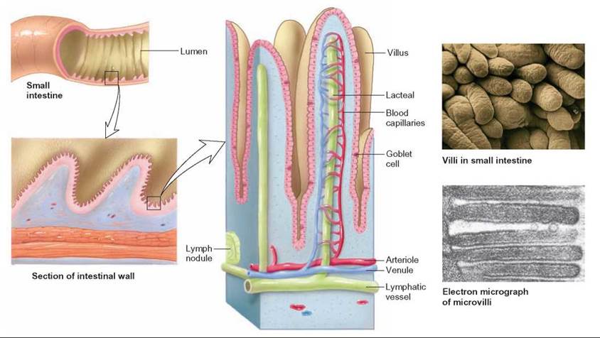
FIGURE 24.11. The Exchange Surface of the Intestinal Tract
The surface area of the intestinal lining is increased by the many fingerlike projections known as villi. Within each villus are capillaries and lacteals. Most kinds of materials enter the capillaries, but most fat-soluble substances enter the lacteals, part of the lymphatic system, giving them a milky appearance. Because the lymphatic system empties into the circulatory system, fat-soluble materials also eventually enter the circulatory system. The close relationship between the vessels and the epithelial lining of the villi allows for the efficient exchange of materials from the intestinal cavity to the circulatory system.
Several processes are involved in the transport of materials from the small intestine to the circulatory system. Some molecules, such as water and many ions, simply diffuse through the wall of the small intestine into the circulatory system. Other materials, such as amino acids and simple sugars, are assisted across the membrane by carrier molecules. Fatty acids and glycerol are absorbed into the intestinal lining cells, where they are resynthesized into fats and enter lacteals in the villi. Because the lacteals are part of the lymphatic system, which eventually empties its contents into the circulatory system, fats also are transported by the blood. They just reach the blood by a different route.
Chemical Alteration: The Role of the Liver
When the blood leaves the small intestine, it flows directly to the liver through the hepatic portal vein. Portal veins are blood vessels that collect blood from capillaries in one part of the body and deliver it to a second set of capillaries in another part of the body without passing through the heart. Thus, hepatic portal veins collect nutrient-rich blood from the small intestine and deliver it directly to the liver. As the blood flows through the liver, enzymes in the liver cells modify many of the molecules and particles that enter them. One of the functions of the liver is to filter any foreign organisms from the blood that might have entered through the intestinal cells.
The liver also detoxifies many dangerous molecules that might have entered with the food. Many foods contain toxic substances that could be harmful if not destroyed by the liver. Ethyl alcohol is one obvious example. Many plants contain various kinds of toxic molecules that are present in small quantities and could accumulate to dangerous levels if the liver did not perform its role of detoxification.
In addition, the liver is responsible for modifying nutrient molecules. The liver collects glucose molecules and synthesizes glycogen, which can be stored in the liver for later use. When glucose is in short supply, the liver can convert some of its stored glycogen back into glucose. Although amino acids are not stored, the liver can change the relative numbers of various amino acids circulating in the blood. It can remove the amino group from one kind of amino acid and attach it to a different carbon skeleton, generating a different amino acid. The liver can also take the amino group off amino acids, so that what remains of the amino acid can be used in aerobic cellular respiration. The toxic amino groups are then converted to urea by the liver. Urea is secreted back into the bloodstream for disposal in urine.
24.5. CONCEPT REVIEW
10. Describe three ways in which the digestive system increases its ability to absorb nutrients.
11. List three functions of the liver.
12. Name five digestive enzymes and describe their functions.
13. What is the role of bile, saliva, enzymes, and hydrochloric acid in digestion?
14. How is fat absorption different from the absorption of carbohydrate and protein?