CONCEPTS IN BIOLOGY
PART VI. PHYSIOLOGICAL PROCESSES
27. Human Reproduction, Sex, and Sexuality
27.7. Oogenesis, Ovulation, and Menstruation
Oogenesis is the production of egg cells. This process starts before a girl is born, during prenatal development of the ovaries. This occurs when diploid cells in each ovary cease dividing by mitosis and enlarge in preparation to divide by meiosis. These are the potential egg cells. All of these cells form in the embryonic ovaries before a female is born. At birth, they number approximately 2 million, but that number has been reduced by cell death to between 300,000 and 400,000 cells by puberty. These cells stop their development in an early stage of meiosis and remain in the ovary inside a follicle.
At puberty and on a regular monthly basis thereafter, the sex hormones stimulate one of these cells to continue meiosis. However, in telophase I, the two cells that form receive unequal portions of cytoplasm, a kind of lopsided division (figure 27.12). The smaller of the two cells is called a polar body, and the larger haploid cell is commonly referred to as an egg or ovum, although technically it is not. Just prior to ovulation, the follicle of the soon-to-be-released egg grows and moves near the surface of the ovary. When this maturation is complete, ovulation occurs when the follicle ruptures and the egg is released. It is swept into the oviduct (fallopian tube) by ciliated cells and travels toward the uterus (figure 27.13). Because of the action of luteinizing hormone, the follicle from which the egg was ovulated develops into a glandlike structure, the corpus luteum. This gland produces hormones (progesterone and estrogen), which prevent the release of other eggs. If the egg is fertilized, it completes meiosis with the sperm DNA inside and the haploid egg and sperm nuclei unite to form the zygote. If the egg is not fertilized, it passes through the vagina to the outside of the body during menstruation (figure 27.14).
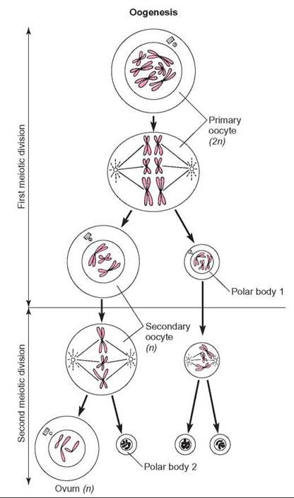
FIGURE 27.12. Oogenesis
This diagram illustrates the process of oogenesis in human females. Not all of the 46 chromosomes are shown. Carefully follow the chromosomes as they segregate, recalling the details of the process of meiosis.
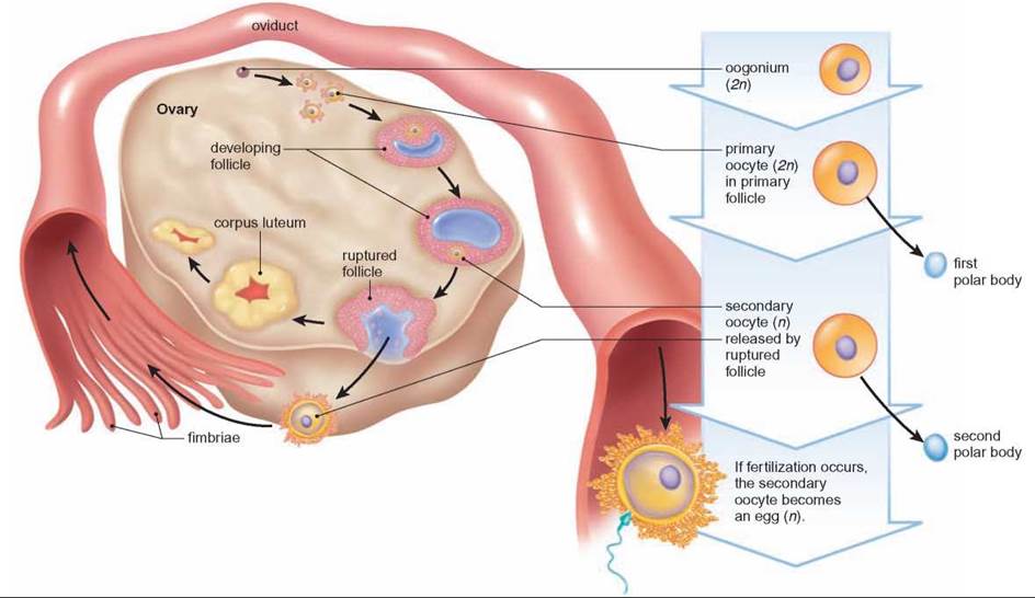
FIGURE 27.13. Ovarian Cycle and Ovulation
After puberty the ovary goes through a regular monthly cycle that involves growth of follicles, ovulation, and the development of a corpus luteum.
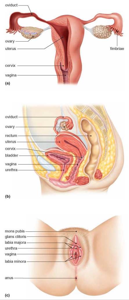
FIGURE 27.14. The Human Female Reproductive System
(a) After ovulation, the cell travels down the oviduct to the uterus. If it is not fertilized, it is shed when the uterine lining is lost during menstruation. (b) The human female reproductive system, side view. (c) External view of female reproductive structures.
One distinguishing characteristic between eggs and sperm is their relative age. In males, sperm production continues throughout the life span. Sperm do not remain in the tubes of the male reproductive system for very long; they are either released shortly after they form or they die and are harmlessly absorbed. In females, meiosis begins before birth, but the oogenesis process is not completed, and an egg cell is not released for many years. An egg released when a woman is 37 years old began meiosis 37 years earlier. During that time, the cell was exposed to many influences, a number of which may have damaged the DNA or interfered with the meiotic process. The increased risk for abnormal births in older mothers may be related to the age of their eggs. Damaged DNA in sperm is less likely to be a problem because new sperm are being produced daily.
Hormones control the cycle of changes in breast tissue, the ovaries, and the uterus. In particular, estrogen and progesterone stimulate milk production by the breasts and cause the uterine lining to become thicker and filled with blood vessels prior to ovulation. This ensures that, if fertilization occurs, the resultant embryo will be able to attach itself to the uterine wall and receive nourishment. If the cell is not fertilized, the lining of the uterus, known as the endometrium, is shed. This is known as menstruation, menstrual flow, the menses, or a period. Once the endometruim has been shed, it begins to build up again (figure 27.15).
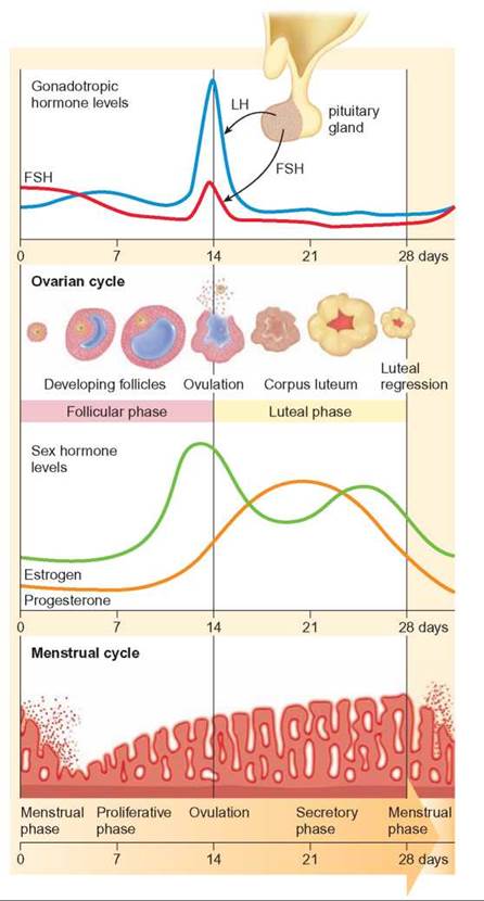
FIGURE 27.15. The Ovarian and Uterine Cycles in Human Females
The release of an egg (ovulation) is timed to coincide with the thickening of the lining of the uterus. The uterine cycle in humans involves the preparation of the uterine wall to receive the embryo if fertilization occurs. Knowing how these two cycles compare, it is possible to determine when pregnancy is most likely to occur by noting when menstruation begins.
The activities of the ovulatory cycle and the menstrual cycle are coordinated. During the first part of the menstrual cycle, increased amounts of FSH cause the follicle to increase in size. Simultaneously, the follicle secretes increased amounts of estrogen, which cause the uterine lining to thicken. When ovulation occurs, the remains of the follicle are converted into a corpus luteum by LH. The corpus luteum begins to secrete progesterone and the nature of the uterine lining changes as a result of the development of many additional blood vessels. This is organized so that, if an embryo arrives in the uterus shortly after ovulation, the uterine lining is prepared to accept it. If pregnancy does not occur, the corpus luteum degenerates, resulting in a reduction in the amount of progesterone needed to maintain the uterine lining, and it is shed. At the same time that hormones are regulating ovulation and the menstrual cycle, changes are taking place in the breasts. The same hormones that prepare the uterus to receive the embryo also prepare the breasts to produce milk. These changes in the breasts, however, are relatively minor unless pregnancy occurs.
27.7. CONCEPT REVIEW
15. What structures are associated with the human female reproductive system? What are their functions?
16. What are the differences between oogenesis and spermatogenesis in humans?