CONCEPTS IN BIOLOGY
PART II. CORNERSTONES: CHEMISTRY, CELLS, AND METABOLISM
4. Cell Structure and Function
4.3. The Structure of Cellular Membranes
One feature common to all cells is the presence of cellular membranes, thin sheets composed primarily of phospholipids and proteins. The current model of how cellular membranes are constructed is known as the fluid-mosaic model. The fluid-mosaic model, considers cellular membranes to consist of two layers of phospholipid molecules and that the individual phospholipid molecules are able to move about within the structure of the membrane (How Science Works 4.1). Many kinds of proteins and some other molecules are found among the phospholipid molecules within the membrane and on the membrane surface. The individual molecules of the membrane remain associated with one another because of the physical interaction of its molecules with its surroundings. The phospholipid molecules of the membrane have two ends, which differ chemically. One end, which contains phosphate, is soluble in water and is therefore called hydrophilic (hydro = water; phile = loving). The other end of the phospholipid molecule consists of fatty acids, which are not soluble in water, and is called hydrophobic (phobia = fear).
In diagrams, phospholipid molecules are commonly represented as a balloon with two strings (figure 4.6). The balloon represents the water-soluble phosphate portion of the molecule and the two strings represent the 2 fatty acids. Consequently, when phospholipid molecules are placed in water, they form a double-layered sheet, with the water-soluble (hydrophilic) portions of the molecules facing away from each other. This is commonly referred to as a phospholipid bilayer (figure 4.7). If phospholipid molecules are shaken in a glass of water, the molecules automatically form double-layered membranes. It is important to understand that the membranes formed are not rigid but, rather, resemble a heavy olive oil in consistency. The component phospholipid molecules are in constant motion as they move with the surrounding water molecules and slide past one another. Other molecules found in cell membranes are cholesterol, proteins, and carbohydrates.
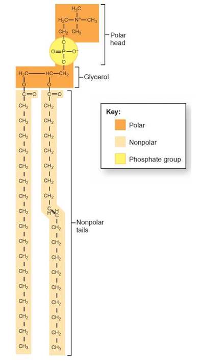
FIGURE 4.6. A Phospholipid Molecule
Phospholipids have a hydrophobic (water-insoluble) portion and a hydrophilic (water-soluble) portion. The hydrophilic portion contains phosphate and is represented as a balloon in many diagrams. The fatty acids are represented as two strings on the balloon.
Because cholesterol is not water-soluble, it is found in the middle of the membrane, in the hydrophobic region. It appears to play a role in stabilizing the membrane and keeping it flexible. There are many different proteins associated with the membrane. Some are found on the surface, some are partially submerged in the membrane, and others traverse the membrane and protrude from both surfaces. These proteins serve a variety of functions, including:
1. helping transport molecules across the membrane,
2. acting as attachment points for other molecules, and
3. functioning as identity tags for cells.
Carbohydrates are typically attached to the membranes on the outside of cells. They appear to play a role in cell-to- cell interactions and are involved in binding with regulatory molecules.
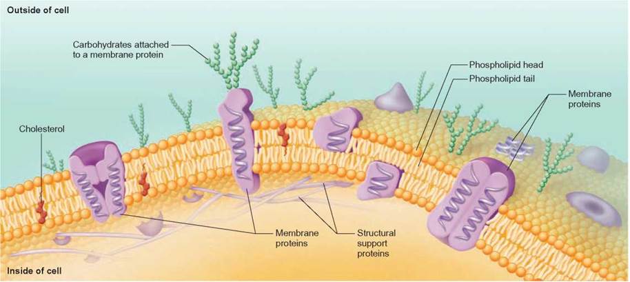
FIGURE 4.7. The Nature of Cellular Membranes
The membranes in all cells are composed primarily of protein and phospholipids. Two layers of phospholipid are oriented so that the hydrophobic fatty acid tails extend toward each other and the hydrophilic phosphate-containing heads are on the outside. Proteins are found buried within the phospholipid layer and are found on both surfaces of the membrane. Cholesterol molecules are also found among the phospholipid molecules. Carbohydrates are often attached to one surface of the membrane.
HOW SCIENCE WORKS 4.1
Developing the Fluid-Mosaic Model
The fluid-mosaic model describes the current understanding of how cellular membranes are organized and function. As is typical during the development of most scientific understandings, the fluid-mosaic model was formed as a result of the analysis of data from many experiments. We will look at three characteristics of cellular membranes and how certain experiments and observations about these characteristics led scientists to develop the fluid-mosaic model.
1. What is the chemical nature of cellular membranes and how do they provide a barrier between the contents of the cell and the cell's environment?
In 1915, scientists isolated cellular membranes from other cellular materials and chemically determined that they consisted primarily of lipids and proteins. The scientists recognized that, because lipids do not mix with water, a layer of lipid could serve as a barrier between the watery contents of a cell and its watery surroundings.
2. How are the molecules arranged within the membrane?
Nearly 10 years after it became known that cellular membranes consist of lipids and proteins, two scientists reasoned from the chemical properties of lipids and proteins that cellular membranes probably consist of two layers of lipid. This arrangement became known as a bilayer. They were able to make this deduction because they understood the chemical nature of lipids and how they behave in water. But this model did not account for the proteins, which were known to be an important part of cellular membranes because proteins were usually isolated from cellular membranes along with lipids. Also, artificial cellular membranes—made only of lipids—did not have the same chemical properties as living cellular membranes.
The first model to incorporate proteins into the cellular membrane was incorrect. It was called the sandwich model, because it placed the lipid layers of the cellular membrane between two layers of protein, which were exposed to the cell's watery environment and cytoplasm. Although incorrect, the sandwich model was very popular into the 1960s, because it was supported by images from electron microscopes, which showed two dark lines, with a lighter area between them.
One of the biggest problems with the sandwich model was that the kinds of proteins isolated from the cellular membrane were strongly hydrophobic. A sandwich model with the proteins on the outside required these hydrophobic proteins to be exposed to water, which would have been an unstable arrangement.
In 1972, two scientists proposed that the hydrophobic proteins are actually made stable because they are submerged in the hydrophobic portion of the lipid bilayer. This hypothesis was supported by an experimental technique called freeze-fracture.

Freeze-fracture experiments split a frozen lipid bilayer, so that the surface between the two lipid layers could be examined by electron microscopy. These experiments showed large objects (proteins) sitting in a smooth background (phospholipids), similar to the way nuts are suspended in the chocolate of a flat chocolate bar. These experiments supported the hypothesis that the proteins are not on the surface but, rather, are incorporated into the lipid bilayer.
3. How do these protein and lipid molecules interact with one another within the cellular membrane?
The answer to this question was provided by a series of hybrid-cell experiments. In these experiments, proteins in a mouse cell and proteins in a human cell were labeled differently. The two cells were fused, so that their cellular membranes were connected. At first, one-half of the new hybrid cell contained all mouse proteins. The other half of the new hybrid cell contained all human proteins. However, over several hours, the labeled proteins were seen to mix until the mouse and human proteins were evenly dispersed. Seeing this dispersion demonstrated that molecules in cellular membranes move. Cellular membranes consist of a mosaic of protein and lipid molecules, which move about in a fluid manner.
4.3. CONCEPT REVIEW
5. What are the prime molecules that make up cell membranes?
6. Describe the structure of cellular membranes based on the fluid-mosaic model.