CONCEPTS IN BIOLOGY
PART II. CORNERSTONES: CHEMISTRY, CELLS, AND METABOLISM
4. Cell Structure and Function
4.5. Nonmembranous Organelles
Suspended in the cytoplasm and associated with the membranous organelles are various kinds of structures that are not composed of phospholipids and proteins arranged in sheets. These are referred to as nonmembranous organelles.
Ribosomes
Ribosomes are nonmembranous organelles responsible for the synthesis of proteins from amino acids. They are composed of RNA and protein. Each ribosome is composed of two subunits—a large one and a small one (figure 4.16). Ribosomes assist in the process of joining amino acids together to form proteins. Many ribosomes are attached to the endoplasmic reticulum. Because ER that has attached ribosomes appears rough when viewed through an electron microscope it is called rough ER. Areas of rough ER are active sites of protein production. Many ribosomes are also found floating freely in the cytoplasm wherever proteins are being assembled. Cells that are actively producing protein (e.g., liver cells) have great numbers of free and attached ribosomes. The details of how ribosomes function in protein synthesis will be discussed in chapter 8.
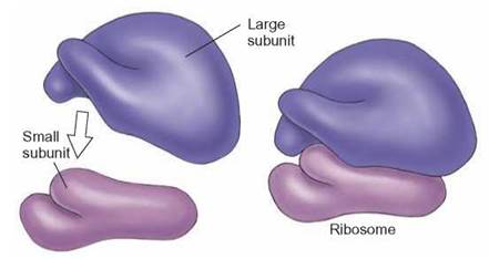
FIGURE 4.16. Ribosomes
Each ribosome is constructed of two subunits. Each of the subunits is composed of protein and RNA. These globular organelles are associated with the construction of protein molecules from individual amino acids. The 2009 Nobel Prize in Chemistry was awarded to Drs. Venkatraman Ramakrishan, Thomas A. Steitz, and Ada E. Yonath for determining the structure and function of ribosomes.
Microtubules, Microfilaments, and Intermediate Filaments
The interior of a cell is not simply filled with liquid cytoplasm. Among the many types of nonmembranous organelles found there are elongated protein structures known as microtubules, microfilaments (actin filaments), and intermediate filaments. All three types of organelles interconnect and some are attached to the inside of the plasma membrane, forming the cytoskeleton of the cell (figure 4.17). These cellular components provide the cell with shape, support, and the ability to move.
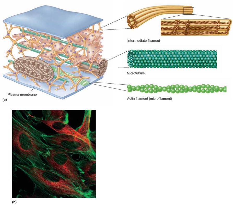
FIGURE 4.17. The Cytoskeleton
Microtubules, microfilaments (actin filaments), and intermediate filaments are all interconnected within the cytoplasm of the cell.
(a) These structures, along with connections to other cellular organelles, form a cytoskeleton for the cell. The cellular skeleton is not a rigid, fixed-in-place structure but, rather, changes as the actin and intermediate filaments and microtubule component parts are assembled and disassembled. (b) The elements of the cytoskeleton have been labeled with a fluorescent dye to make them visible. The microtubules have fluorescent red dye, and actin filaments are green.
Think of the cytoskeleton components as the internal supports and cables required to construct a circus tent. The shape of the flexible canvas cover (i.e., the plasma membrane) is determined by the location of internal tent poles (i.e., microtubules) and the tension placed on them by attached wire or rope cables (i.e., intermediate filaments and microfilaments). Just as in the tent analogy, when one of the microfilaments or intermediate filaments is adjusted, the shape of the entire cell changes. For example, when a cell is placed on a surface to which it cannot stick, the internal tensions created by the cytoskeleton components can pull together and cause the cell to form a sphere.
During cell division, microtubules and microfilaments are involved in moving the chromosomes that contain the DNA and making other adjustments needed to make two cells from one. Microfilaments and microtubules of the cytoskeleton also transport organelles from place to place within the cytoplasm. In addition, information can be transported through the cytoskeleton. Enzymes attached to the cytoskeleton are activated when the cell is touched. Some of these events even affect gene activity.
Centrioles
An arrangement of two sets of microtubules at right angles to each other makes up a structure known as a centriole. Each set of microtubules is composed of nine groups of short microtubules arranged in a cylinder (figure 4.18). The centrioles of many cells are located in a region called the centrosome. The centrosome is often referred to as the microtubule organizing center and is usually located close to the nuclear membrane.
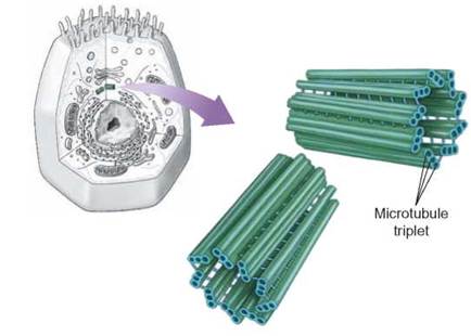
FIGURE 4.18. The Centriole
These two sets of short microtubules are located just outside the nuclear membrane in many types of cells.
During cell division, centrioles are responsible for organizing microtubules into a complex of fibers known as the spindle. The individual microtubules of the spindle are called spindle fibers. The spindle is the structure to which chromosomes are attached, so that they can be separated properly during cell division. The functions of centrioles and spindle fibers in cell division will be referred to again in chapter 9. One curious fact about centrioles is that they are present in most animal cells but not in many types of plant cells, although plant cells do have a centrosome. Other structures, called basal bodies, resemble centrioles and are located at the base of cilia and flagella.
Cilia and Flagella
Many cells have microscopic, hairlike structures known as cilia and flagella, projecting from their surfaces (figure 4.19). These structures are composed of microtubles and are covered by plasma membrane. In general, flagella are long and few in number and move with an undulating whiplike motion; cilia are short and more numerous and move back and forth like oars on a boat. Both function to move the cell through its environment or to move the environment past the cell. Both cilia and flagella are constructed of a cylinder of nine sets of microtubules similar to those in the centriole, but they have an additional two microtubules in the center. This is often referred to as the 9 + 2 arrangement of microtubules.
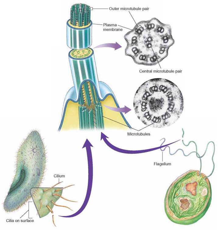
FIGURE 4.19. Cilia and Flagella
Cilia and flagella have the same structure and function. They are composed of groups of microtubules in a 9 + 2 arrangement, are surrounded by plasma membrane, and function like oars or propellers that move the cell through its environment or move the environment past the cell. Flagella are less numerous and longer than cilia.
The cell can control the action of these microtubular structures, enabling them to be moved in a variety of ways. The protozoan Paramecium is covered with thousands of cilia, which move in a coordinated, rhythmic way to move the cell through the water. A Paramecium can stop when it encounters an obstacle, reverse its direction, and then move forward in a new direction. Similarly, the cilia on the cells that line the human trachea beat in such a way that they move mucus and particles trapped in the mucus from the lungs. Many single-celled algae have flagella that beat in such a way that the cells swim toward a source of light.
Some kinds of Bacteria and Archaea also have flagella. However, their structure and the way they function are quite different from those of eukaryotic cells.
Inclusions
Inclusions are collections of materials that do not have as well defined a structure as the organelles we have discussed so far. They might be concentrations of stored materials, such as starch grains, sulfur, or oil droplets, or they might be a collection of miscellaneous materials known as granules. Unlike organelles, which are essential to the survival of a cell, inclusions are generally only temporary sites for the storage of nutrients and wastes.
Some inclusion materials are harmful to other cells. For example, cells of the rhubarb plant contain an inclusion composed of oxalic acid, an organic acid. If you eat rhubarb leaves, the oxalic acid dissolves and later recrystalizes in the kidneys, contributing to kidney stones. The crystals might also cause harm to the glomeruli in the kidneys. Eating the stalks is unlikely to cause these problems since the concentration of oxalic acid is less in the stalks than in the leaves. Similarly, certain bacteria store, in their inclusions, crystals of a substance known to be harmful to insects. Spraying plants with these bacteria is a biological method of controlling the insect pest population while not interfering with the plant or with humans.
In the past, cell structures such as ribosomes, mitochondria, and chloroplasts were also called granules because their structure and function were not clearly known. As scientists learn more about inclusions and other unidentified particles in the cells, they, too, will be named and more fully described.
4.5. CONCEPT REVIEW
10. List the nonmembranous organelles of the cell and describe their functions.