Biology For Dummies
Part IV Systems Galore! Animal Structure and Function
Chapter 14
Moving and Shaking: Skeletal and Muscular Systems
In This Chapter
Seeing how animals move from one place to another
Surveying the various skeletal systems
Understanding what makes muscles so valuable
The coordinated efforts of muscles and skeletons are what make animal movements possible. Muscles pull or push, and skeletons give the muscles something to pull or push against. Prepare to find out all about how animals move from place to place as you discover the different types of skeletons and the fundamentals of muscle function in this chapter.
Doing the Locomotion, Animal-Style
Fish swim, dogs run, frogs jump, worms crawl, and birds fly. Each of these types of locomotion, movement from one place to another, requires animals to use energy to overcome the forces of friction and gravity that would otherwise hold them to the Earth.
Each different animal is adapted for the environment it lives in and the type of locomotion it performs.
Swimming animals, such as fish and whales, have bodies that are shaped to minimize resistance as they move through the water. They’re often coated with water-resistant mucus and may have structures that make them more buoyant.
Birds have hollow bones and wings that are shaped like those of an airplane to create added lift during flight.
Animals that walk or run on land, such as lions and elephants, have a strong skeleton and muscles to support them against the force of gravity.
Rabbits, kangaroos, and other animals that jump or hop have extralarge leg muscles and strong tendons to help put some spring in their hop.
Animals that slither or crawl, think worms and snakes, have smooth, tubular bodies to lessen resistance due to friction as they move over or through soil.
The Types of Skeletal Systems
Skeletons support animals, give their bodies shape, and protect their internal organs, but not all animals have the same type of skeleton. Following are the three different kinds of skeletons you may see in your study of biology:
Hydrostatic skeletons: Found in creatures such as worms and jellies, hydrostatic skeletons are basically chambers filled with water. Animals with this skeleton type move and change their shape by squeezing their water-filled chambers — just like what happens when you squeeze a water balloon.
Exoskeletons: These are exactly what they sound like — skeletons on the outside of the body. You’re probably quite familiar with these hard exterior coverings because they’re found on crabs, lobsters, and many insects. Exoskeletons are rigid and can’t expand as animals grow, so animals must molt, or shed, their exoskeletons periodically. After an animal molts, its new exoskeleton is soft — as in a soft-shelled crab.
Endoskeletons: The most familiar of all skeleton types is the endoskeleton. After all, it’s the kind of skeleton you have. An endoskeleton exists within an animal’s body. The human endoskeleton is hard because it’s partially constructed of the mineral calcium. The endoskeletons of other animals may be more flexible — for example, the endoskeleton of a shark is made of cartilage, the same material that makes up the soft parts of your nose.
 Animals with hydrostatic skeletons and exoskeletons are considered invertebrates, meaning they don’t have a backbone. Animals with endoskeletons, like you, are considered vertebrates because they have a backbone.
The following sections not only break down the parts of a vertebrate animal’s skeleton but they also get you more familiar with the important components of your skeleton — bones and joints.
Splitting apart vertebrate skeletons
All vertebrate skeletons— whether they belong to humans, snakes, bats, or whales — developed from the same ancestral skeleton (which explains why you may notice similarities between your skeleton and that of your pet dog or cat). Today, these animals show their relationship to each other in part due to homologous structures — structures that are equivalent to each other in their origin (see Chapter 12 for more on homologous structures and their importance to the study of evolution).
All vertebrates’ skeletons, whether yours (see Figure 14-1), a whale’s, or a cat’s, have two main parts:
The axial skeleton: This part supports the central column, or axis, of the animal. The axial skeleton includes the skull, the backbone (also called the vertebral column), and the rib cage. The skull protects the brain, the backbone protects the spine, and the rib cage protects the lungs and heart.
The appendicular skeleton: This part extends from the axial skeleton out into the arms and legs (which are also known as appendages). It includes the shoulders, pelvis, and bones of the arms and legs.
 In some vertebrates, such as snakes, the appendicular skeleton has become extremely reduced or nonexistent.
Boning up on bones
If you’ve ever watched an old Western movie, you’ve probably seen images of bones bleached white by the Sun and scattered alongside a pioneer trail. The dry white bones of these images are very different from the living bones that are in your body right now. Bone is actually a moist, living tissue that contains different layers and cell types.
Fibrous connective tissue covers the exterior of bones and helps heal breaks in an injured bone by forming new bone.
Bone cells, which are embedded in a bone matrix, give cells their hard nature. The cells actually make the matrix, which consists of collagen that has been hardened by the attachment of calcium and phosphate crystals.
Cartilage covers the ends of bones and protects them from damage as they rub against each other.
The tissues found within living bone fall into two categories:
Spongy bone tissues are filled with little holes, similar to those you see in volcanic rocks. These holes are filled with red bone marrow, which is the tissue that produces your blood cells.
Compact bone tissues are hard and dense. A cavity within compact bone is filled with yellow bone marrow, which is mostly stored fat. If the body suddenly loses a large amount of blood, it converts the yellow bone marrow to red bone marrow so that blood cell production can be increased.
Figure 14-1: The human skeleton.
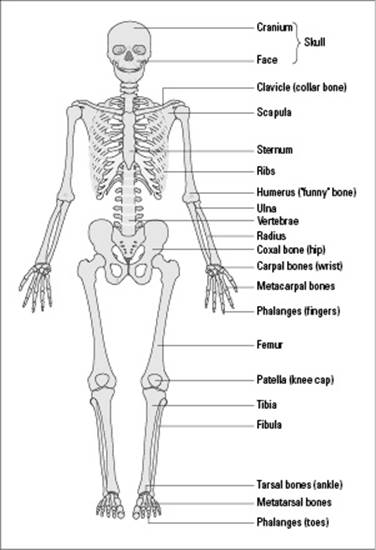
From LifeART®, Super Anatomy 1, © 2002, Lippincott Williams & Wilkins
Got broken bones?
The number of broken bones in American children is on the rise, and doctors think this increase may be due to kids exercising less and drinking less milk. Exercise is important because it puts weight on your bones, which encourages bone growth and helps keep bones dense. Milk is important because the calcium and vitamin D found within it are vital for the development of healthy, strong bones. Doctors recommend milk as an excellent source of concentrated calcium that’s easily absorbed by the body. To get your daily dose, drink four 8-ounce glasses per day, or take a calcium supplement (with your doctor’s permission, of course). (Note: Bone mass stops increasing after age 20, so get your calcium in before then!)
Joining the movement fun
Joints are structures where two bones are attached so that bones can move relative to each other. Bones are held together at joints by ligaments, which are strong, fibrous, connective tissues.
Three different types of joints enable the many movements of animals:
Ball and socket joints consist of one bone, with a rounded, ball-like end, that fits into another bone, which has a smooth, dishlike surface. Your arms and legs fit into your skeleton with ball and socket joints, which is why you’re able to rotate your arms and legs in all directions.
Pivot joints allow you to swivel a bone. When you rotate your arm so that your palm faces up, then down, then up again, you’re using a pivot joint.
Hinge joints allow you to bring two bones close together or move them farther apart, much like you open and close a book. Your elbows and knees have pivot joints that allow you to extend and contract your arms and legs.
Why Muscles Are So Essential
Muscles are extremely important to your body — and not just because toned ones make your body look better and grow stronger. Without muscles, you couldn’t walk, run, or play sports. You couldn’t even obtain nutrients from your food or send blood to all the organs and tissues throughout your body.
Following is a rundown of all the things muscles do for you:
Muscles allow you to stand upright. The force of gravity is strong; without your muscles, it’d keep you pinned to the ground. Your muscles contract so you can push against the surface of the Earth and stand upright and assume different positions.
Muscles make it possible for you to move. Every little movement that your body performs, including blinking and smiling (or frowning), is controlled by your muscles.
Muscles allow you to digest. Muscles all along your digestive tract keep food moving downward and outward. Peristalsis, the squeezing of food down through the esophagus, stomach, and intestines, is due to the contraction of these muscles. If they didn’t contract to squeeze food throughout your digestive system, you’d never be able to obtain the nutrients you need to survive.
Muscles affect the rate of blood flow. Blood vessels contain muscle tissue that allows them to dilate so blood can flow faster or to contract so blood flow slows down. Muscle contraction is also responsible for the movement of blood through your veins. (Not to mention that your heart is actually a muscle; without it, your blood wouldn’t be flowing anywhere, period!)
Muscles help you maintain a normal body temperature. Muscles give off heat when they contract, and your body uses that heat to maintain your body temperature because you continually lose some heat through your skin. This fact explains why you shiver when you’re cold; shivering is your body’s way of trying to generate heat.
Muscles hold your skeleton together. The ligaments and tendons at the ends of your muscles wrap around joints, holding together the joints — and therefore the bones of your skeleton.
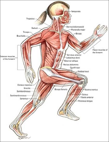
Figure 14-2 gives you an up-close view of the human muscular system. To find out about the specific types of muscle tissue in your body and how your muscles contract, check out the next sections.
Muscle tissue and physiology
Muscle tissues are made up of muscle fibers, and muscle fibers contain many, many myofibrils — the parts of the muscle fiber that contract. Myofibrils are perfectly aligned, which makes muscles look striped, or striated. The repeating unit of these striations — containing light and dark bands — is called a sarcomere.
Figure 14-2: The human muscular system.
Three types of muscle tissue exist within your body:
Cardiac muscle is found in the heart. The fibers of cardiac muscle have one nucleus (so they’re uninucleated), are striated (so they have light and dark bands), and are cylindrical in shape and branched. The fibers interlock so contractions can spread quickly through the heart. Between contractions, cardiac fibers relax completely so the muscle doesn’t get fatigued. Cardiac muscle contraction is totally involuntary, meaning it occurs without nervous stimulation and doesn’t require conscious control.
Smooth muscle is found in the walls of internal organs that are hollow, such as the stomach, bladder, intestines, or lungs. The fibers of smooth muscle tissue are uninucleated, shaped like spindles, and arranged in parallel lines; they form sheets of muscle tissue. Smooth muscle contraction occurs involuntarily and more slowly than skeletal muscle contraction, which means smooth muscle can stay contracted longer than skeletal muscle and not fatigue as easily.
Skeletal muscle is probably what you think of when you picture a muscle. The fibers of skeletal muscle have many nuclei (so they’re multinucleated) and are both striated and cylindrical; they run the length of the muscle. Skeletal muscle is controlled by the nervous system (which we describe in Chapter 18). The movement and contraction of skeletal muscle can be stimulated consciously, which means you consciously decide that you’re going to stand up and walk across the room, an action that requires the use of muscle. Therefore, skeletal muscle contraction is said to be voluntary.
Muscle contraction
Muscle contractions rely on the movements of the filaments that make up myofibrils. Basically, the different filament types slide over each other to cause a muscle contraction; this general theory is referred to as the sliding-filament theory.
The two filament types found in myofibrils are
Actin (thin) filaments: An actin, or thin, filament is made up of two strands of actin, which is a protein that’s wound in a double helix (just like DNA). It has molecules of troponin and tropomyosin at binding sites along its actin double helix.
Myosin (thick) filaments: A myosin, or thick, filament contains groups of myosin, a type of protein with a bulbous end. Multiple strands of myosin are mixed together in opposite directions in muscle tissue, so it appears that both ends of myosin filaments are bulbous.
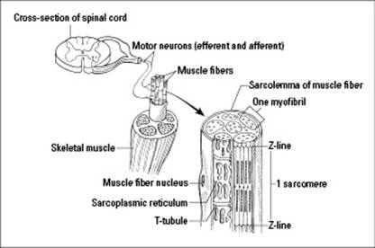
The actin filaments are attached to something called a Z-line, and the myosin filaments lie between the actin filaments, unattached to Z-lines. From Z-line to Z-line is one sarcomere, which is a unit of contraction. You can see all of these parts and more in Figure 14-3, which shows you how skeletal muscle is connected to the nervous system and how it contracts.
Figure 14-3:Structure and function of a skeletal muscle.
The other element necessary for muscle contractions is adenosine triphosphate, or ATP for short (we cover this energy-storing molecule in greater detail in Chapter 5). A muscle fiber contains only enough ATP to sustain a contraction for about one second. After muscle cells use up their available ATP, they get more by
Using energy from stored phosphocreatine molecules: Phosphocreatine, which is made up of ATP and creatine, forms during periods of no contraction. It’s quickly broken down to release more ATP as the low amounts of ATP in a muscle cell are used up.
Increasing the rate of cellular respiration: Muscle cells are loaded with mitochondria, organelles that perform cellular respiration. During cellular respiration, the mitochondria use oxygen to break down food molecules and transfer their energy to ATP (described in Chapter 5). As your muscle cells use up their ATP, you breathe harder to supply them with more oxygen so they can do more cellular respiration.
Recycling ADP molecules into ATP: Every time a muscle cell uses an ATP molecule for energy, a phosphate is removed, producing adenosine diphosphate, or ADP. Human muscle cells take every two molecules of ADP produced during contraction and recombine them to make a new ATP molecule plus a molecule of adenosine monophosphate, or AMP.
Resorting to lactic acid fermentation: Lactic acid fermentation, which produces a small amount of ATP through the partial breakdown of glucose, is a worst-case scenario for muscle cells. Cells make a lot more ATP for each glucose molecule they break down by cellular respiration than they do by lactic acid fermentation. So, cells resort to lactic acid fermentation only when ATP can’t be obtained through cellular respiration, a situation that can occur when the body’s oxygen stores are depleted.
According to the sliding-filament theory, muscle contraction occurs via the following process:
1. ATP binds to the bulbous end of a myosin filament and splits into a molecule of ADP plus a molecule of inorganic phosphate (Pi).
The ADP and Pi stay attached to the myosin.
2. Calcium binds to the troponin in the actin filament, which causes the tropomyosin in the actin filament to move out of the way so the binding sites on the actin filament can open.
3. After the actin filament’s binding sites are exposed, myosin binds to the actin, which causes myosin to release the ADP and Pi.
4. When myosin releases the ADP and Pi so it can link to actin, the shape of the bulbous end of the myosin filament changes and the actin filament slides toward the middle of the sarcomere, pulling the Z-lines at the end of the sarcomere closer together.
The result? A shortening, or contraction, of the muscle fiber.
5. The connection between the actin and myosin filaments breaks when another ATP molecule attaches to the bulbous end of the myosin filament.