Must Know High School Biology - Kellie Ploeger Cox 2019
PART FIVE Forms of Life
Animal Digestion
MUST ![]() KNOW
KNOW
![]() Digestion is based on a combination of mechanical (chewing, churning, bile) and chemical (digestive enzymes, HCl) processes.
Digestion is based on a combination of mechanical (chewing, churning, bile) and chemical (digestive enzymes, HCl) processes.
Unlike the autotrophs who make their own food, heterotrophs have to eat. Once the food goes into your mouth (ingestion), the process of digestion takes over. In its simplest form, digestion is taking large bits of food and breaking them up into smaller bits. The catabolic process of digestion is then followed by absorption, when those small food bits are brought into the circulatory system, where the organic molecules and essential nutrients will be used for building and energy. Finally, any indigestible material is passed out of the body (elimination).
Your diet consists of a delicious array of macromolecules: carbohydrates, proteins, fats, and nucleic acids. As we learned earlier, macromolecules are composed of small subunits. A gigantic starch molecule is made up of thousands of individual glucose monomers. Proteins are huge polymers made up of amino acids linked together. A fat molecule is made of three fatty acids attached to a glycerol molecule. And nucleic acid (DNA or RNA) is composed of repeated nucleotides.
![]()
IRL
Weird concept, right? You actually eat quite a bit of DNA. Think about how much of what you eat is actually made of cells. Meat is muscle (cells), salad is plant tissues (cells), bread is made of ground wheat (cells). On average, you ingest about 0.5 grams of DNA a day … that works out to be tens of millions of miles of the macromolecule!
Digestion is the process of dismantling these big macromolecules into their smaller components. When the macromolecules were synthesized in the first place (i.e., starch created by a plant basking in the sun), it was most likely through a dehydration reaction.
Don’t forget how important dehydration reactions are in the creation of the four macromolecules (carbohydrates, lipids, proteins, and nucleic acids):
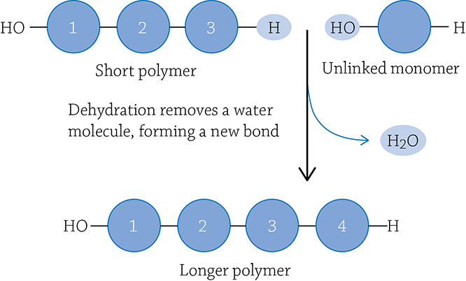
It makes sense, then, to break them down. The body employs the opposite reaction, hydrolysis:
A hydrolysis reaction will use a water molecule to break apart a covalent bond linking subunits together in a larger macromolecule. As we go through the stages of digestion, we will often see a hydrolysis reaction do the work of dismantling your lunch.
The Steps of Digestion
Most animals have an alimentary canal, a one-way tube which food passes into, through, and out of the body. Food is oftentimes pushed along by peristalsis, rhythmical contractions of the smooth muscle lining the canal. Along the way, food is broken up in two manners (and here is our must know concept for this chapter): mechanical digestion and chemical digestion. Mechanical digestion is the inelegant process of physically breaking up big chunks into smaller chunks, thus increasing the surface area for chemical digestion. Chemical digestion is when specific enzymes get to work catalyzing the hydrolysis reactions to liberate the monomers.
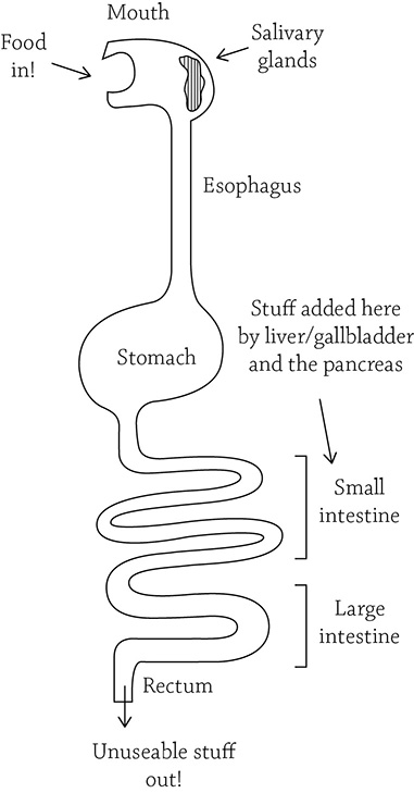
Simplified drawing of mammalian alimentary canal
■ Step 1 Mouth
Here it begins, that spoonful of cereal you just guided into your mouth will now meet the mechanical destruction of your teeth, grinding up big pieces into smaller pieces. At the same time, salivary glands secrete an enzyme called amylase that begins the chemical breakdown of starch and glycogen (both polymers of glucose). The saliva also contains a buffer to help neutralize acids (protects your tooth enamel) and lysozyme, an antimicrobial compound (because, I hate to tell you this, but your food may have a fair amount of bacteria contaminating it).
Meanwhile, your tongue is helping to shape this saliva-softened glob of food into a bolus, the official term for a saliva-laden glob of chewed up food. When you swallow, the food is guided into the pharynx (back of the throat), where it will pass down the esophagus, into the stomach. To make sure the food bolus goes down the correct esophageal path, there is a flap of cartilage that kindly covers your trachea, preventing food from entering your lungs.
■ Step 2 Stomach
After the food bolus is pushed down the esophagus by peristaltic waves, it passes through a sphincter and into the stomach. The sphincter is the connection point between the food tube and the stomach, and it helps keep gastric juice from bubbling up and burning the tender esophagus (acid reflux, or “heartburn”). The stomach is responsible for both mechanical (churning) digestion and chemical (HCl and pepsin) digestion.
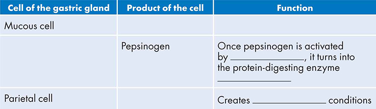
Once food passes through the sphincter, the stomach revs up and begins muscular contractions that mix the stomach soup (technically called chyme) once every 20 seconds. Digestive fluids are dumped into the mix. The gastric juice contains hydrochloric acid (HCl) and pepsin, an enzyme that digests proteins, along with mucus to help protect the lining of the stomach. The parts of the stomach responsible for making the gastric juice are called gastric glands, and they are found all along the stomach lining. Interestingly, each gastric gland is composed of three types of cells:
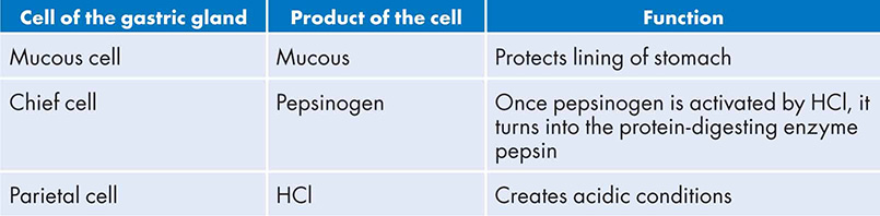
All of these cells secrete their products to create the gastric juices. The mucous protects the lining of the stomach from self-digestion. In fact, conditions are so harsh, the stomach replaces the eroded epithelial layer once every 3 days.
![]()
IRL
Some unfortunate folks may develop an ulcer, an open sore in the lining of the stomach. For a long time, people thought ulcers were caused by stress, or eating spicy food, or maybe drinking too much coffee. We now know that ulcers are caused by these acidic-loving bacteria called Helicobacter pylori. Happily, we now know to treat an ulcer with antibiotics, not antacids and bland foods.
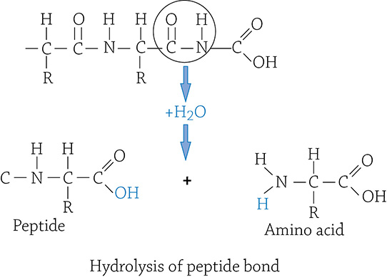
The hydrochloric acid is provided by parietal cells, and it drops the pH of the stomach to a 2. This is helpful because it kills most bacteria (unfortunately not those nasty H. pylori) and denatures proteins, exposing the peptide bonds for pepsin to attack.
In a clever attempt to limit self-digestion, the chief cells secrete an inactive form of the protein-digesting enzyme, called pepsinogen. Once pepsinogen hits the acidic stomach soup, however, it is converted into the active form pepsin. Not only does it begin to cleave apart proteins, the pepsin also activates more pepsinogen by converting it into pepsin. Yes, one of those relatively rare positive feedback loops!
Anywhere between 2 and 6 hours later, the chyme is slowly released through the lower sphincter that guards the passage from the stomach into the small intestine.
■ Step 3 Small intestine
Most of the digestion occurs here, in the approximately 20 feet of small intestine currently snuggled up in your abdominal cavity. Most of the digestion is through enzymatic hydrolysis, though there is a bit of mechanical digestion, compliments of bile. Bile qualifies as mechanical digestion because it breaks up big blobs of fat into smaller blobs of fat (emulsification). It is not chemical digestion because no covalent bonds are enzymatically broken.
The small intestine is made up of three sections: the first part is the duodenum, where most of the digestion is completed. The second and third parts are the jejunum and ileum, the major sites of nutrient absorption. The chyme from the stomach is slowly squirted into the duodenum, where it is mixed with digestive juices from the pancreas, the liver, and the gallbladder. Although we know the pancreas as the master of glucose metabolism, it also plays a large role in digestion. The pancreas helps neutralize the scalding acidity of the chyme by adding bicarbonate solution. It also secretes trypsin and chymotrypsin, a protein-digesting protease pair. The liver makes bile that is piped into the gallbladder for storage. Bile emulsifies fats, meaning it breaks up large fat globules into smaller ones.
Digesting the food is only half the battle. In order for the nutrients to do any good, they must get into the circulatory system. As the digested food travels down the inner space of the digestive tract (the lumen), the nutrients will pass through the walls of the small intestine and into the capillaries. This process is facilitated by the highly folded inner lining of the small intestine. There are large folds, each covered by smaller folds called villi. On top of that, each villus is covered with epithelial cells that have their own tiny hair-like projections called microvilli (also called the brush border).
![]()
A highly folded tissue is an excellent example of form fits function. If nutrients need to pass through the intestinal wall, natural selection would favor a high surface area. A highly folded structure provides a high surface area. When you flatten out the folds—all the villi and microvilli—you get a surface area the size of a tennis court!
■ Step 4 Large intestine
This is the final path along our digestion journey. Once all the useable nutrients are absorbed by the small intestine, the stuff that’s left over moves into the large intestine. The large intestine includes the colon, cecum, and the rectum. The cecum is a dead-end pouch where the large intestine connects with the small intestine, and it is the happy home for bacteria that ferment cellulose. It makes sense, therefore, that the more cellulose and plant matter an animal eats, the larger the cecum. Once again, natural selection has sculpted a form that best fits its function.
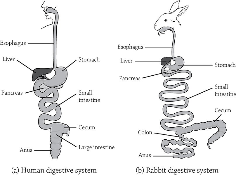
The relative lengths of the cecum in carnivore versus herbivore
Authors: Charles Molnar and Jane Gair. https://opentextbc.ca/biology/wp-content/uploads/sites/96/2015/03/Figure_34_01_05ab.jpg
The major role of the large intestine is to reclaim water before it passes out of the body. About 90% of the water that enters the digestive tract is reabsorbed by the small and large intestines! Problems occur if the feces move along too quickly and not enough water is absorbed (diarrhea) or they move too slowly and too much water is removed (constipation). Once it’s all said and done, feces will pass out of the body through the rectum. Fecal matter consists of undigested material, such as cellulose fiber. About a third of poop is composed of the harmless bacteria that live inside your colon. The brown color is due to the bile that is dumped into the mix earlier in the small intestine. Bile contains a pigment called bilirubin that is produced when the hemoglobin in red blood cells is broken down. As bilirubin is digested, it produces the brown color of your feces.
REVIEW QUESTIONS
1. The first stage of digestion is _______________, when you take a bite of food. That food is broken up during the process of _______________, when large molecules are broken up into smaller components. The small components are more easily taken up into the bloodstream through the process of _______________. Finally, any indigestible material is passed out of the body through _______________.
2. What type of enzymatic reaction is responsible for chemical digestion of food?
3. What structural detail of the small intestine helps increase its ability to absorb nutrients?
4. Fill in the following table about gastric cell function:
5. Explain the difference between mechanical digestion and chemical digestion.
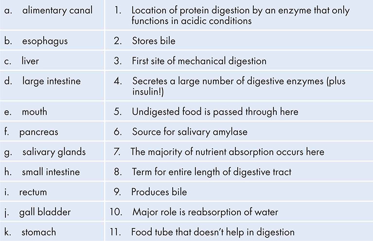
6. Match the following terms with their correct definition:
7. If you compared the lengths of the ceca (plural of cecum) of a tiger versus a rabbit, what difference would you expect and why?
8. Interesting poop trivia! About a third of your poop is composed of _______________, and poop is brown because of pigments produced from the breakdown of _______________.