Must Know High School Biology - Kellie Ploeger Cox 2019
PART TWO Cells
Eukaryotic Cells and Their Organelles
MUST ![]() KNOW
KNOW
![]() Organelles create internal compartments that allow cells to increase in complexity.
Organelles create internal compartments that allow cells to increase in complexity.
When a cell grows too big, it runs into a problem—its surface-area-to-volume ratio decreases. This means that there isn’t enough membrane (the surface area) to deal with the large amount of cytoplasm (the volume). The cell membrane is the interface between the inside of the cell and the surrounding environment, so it’s very important that there’s enough membrane to deal with all the goings-on inside the cell. Keeping this in mind, it makes sense that by compartmentalizing the cell (our must know for this chapter), there is an increase in “surface” to deal with all that the cell needs to do. These compartments are the organelles of the eukaryotic cell. Organelles create smaller spaces for “microenvironments,” meaning an organelle can create conditions within it that are different from other areas of the cell. Organelles can store things, they can have a pH other than the peaceful 7 (neutral) of most cells’ interiors, and they can stockpile enzymes for specific functions. This compartmentalization allowed cells to grow in complexity because it now has these different little “rooms” in which separate events could occur.
Organelles
Each organelle performs a very specific function in the eukaryotic cell, and is only able to do so because it is a smaller compartment that can create special conditions within it. The nucleus, for example, is fondly called the brain of the cell because it houses the DNA (and thus controls the cell’s functions). The nuclear membrane surrounds the nucleus and, unlike the outer cell membrane, it is riddled with holes called nuclear pores. These pores allow movement of “messages” (mRNA, which you will learn about in Chapter 11) between the DNA and the protein-producing ribosomes waiting in the cytoplasm. The number of ribosomes in a cell correlate with the amount of proteins the cell needs to make. Ribosomes can be floating around freely in the cytoplasm, waiting for instructions to make proteins that the cell intends to keep and use. Ribosomes that are stuck onto the endoplasmic reticulum, however, make proteins that are destined for export out of the cell.
The endoplasmic reticulum (ER) is an extensive network of membranes surrounding the nucleus. There is a ton of ER; almost half of a eukaryotic cell’s membranes compose the ER! Two types of ER differ in both structure and function: the smooth ER and the rough ER. The rough ER (rER) looks rough because it has a constant supply of ribosomes stuck onto it. These ribosomes are constantly grabbing on to the rough ER and letting go; the ribosomes that latch onto the rough ER do so because they have instructions to make a protein destined for export out of the cell. The rough ER has the special role to help create proteins for export; the ribosomes thread their protein into the inner cavity (the lumen) of the rough ER.
The rough ER will then bleb off a tiny vesicle that contains this protein, and send it on the beginning of its journey. A cell that specializes in exporting massive amounts of proteins tends to have a large amount of rER to help it do its job. The pancreas is an organ that makes many important types of hormones and digestive enzymes, including insulin. The pancreas’s beta cells make and export insulin; beta cells, therefore, have a high amount of rough ER to help expedite the process.
The rough ER has an organelle cousin called the smooth ER. The smooth ER (sER) lacks ribosomes, so it does not play a role in protein production. The sER, however, still has a number of important purposes, depending on the cell type in which it resides. Primarily, the sER makes more membranes for the cell (a worthy task, considering that organelles are made of membranes). The sER also plays a role in detoxification. The sER creates a space in which many catabolic enzymes can be concentrated. Alcohol and other drugs can stimulate the synthesis of more sER, and thus leads to an increased tolerance to these drugs, since they are broken down quicker. This highly active sER can also, unfortunately, wipe out and remove some types of antibiotics and other helpful medicines.
![]()
The term lumen refers to a cavity of some sort. We will use the term again when we talk about the lumen of the small intestine and capillaries!
![]()
IRL
Think about this. If the smooth ER is used in detoxification, can you hypothesize what cell types would tend to contain a larger amount of smooth ER? To figure this out, think of the human body as a whole. What organs specialize in removing nasty things from circulation? The skin, the lungs, the kidneys … and the liver! The liver, specifically, carefully checks the blood and looks for poisons and other toxins. Liver cells, therefore, have a high amount of smooth ER to help them do their jobs. This is also why there is no need to go on a “detox diet.” Your body is the result of millions of years of evolution; it can detox your blood better than any lemon-and-honey smoothie.
Remember those proteins created by ribosomes on the rough ER? These are the proteins destined for places other than the cell in which they were created. Considering that this protein has some specific target and purpose, it needs directions; these directions are provided by the Golgi apparatus (or Golgi body). The Golgi apparatus is responsible for taking freshly made proteins, modifying them (such as adding tiny sugar molecules or phospholipids), and then sending them to their final destination. Cells that specialize in secretion (such as our pancreatic beta cells) have a high number of Golgi bodies hanging around.
Lysosomes are organelles that act as the recycling centers of the cell. A lysosome can also be considered the “stomach” of the cell, since it’s filled with digestive enzymes that break up large macromolecules into their smaller monomers. The lysosome is a perfect example of the importance of compartmentalization in a eukaryotic cell. A lysosome is a membranous bag full of digestive enzymes. When an old, nonfunctioning organelle needs to be recycled, a lysosome will engulf it and the enzymes will chew up the organelle into its macromolecule components. It would be disastrous if these digestive chemicals leaked out of the lysosome and into the cytoplasm of the cell … imagine the havoc! The cell would digest itself! Cleverly enough, the microenvironment inside the lysosome creates a type of fail-safe system. These digestive enzymes only work in acidic conditions, and the inside of the lysosome is, indeed, acidic. If the lysosome broke open and released a flood of enzymes into the cytosol, they would not work—the pH of the cytosol is neutral!
Endosymbiotic theory, the mysterious history behind mitochondria and chloroplasts
As mentioned earlier, the first cells to evolve were prokaryotic cells. There is a theory, the endosymbiotic theory, that hundreds of millions of years ago, a smaller prokaryotic cell was engulfed by a larger prokaryotic cell. Instead of the smaller cell being digested and destroyed, it stayed put and served a very specific (and helpful) purpose: it provided energy for the larger cell. The larger cell, in turn, gave the smaller cell a nice, safe place to live. This mutualistic relationship, the endosymbiotic theory suggests, is how the energy-producing organelles first came into being.
If you can guess, these two organelles are the mitochondria (found in all eukaryotic cells) and the chloroplasts (found in plant cells and photosynthetic protists). Both of these organelles have some unique and special traits: they can reproduce autonomously within the cell, they contain their own ribosomes, and strangely enough, they have their own DNA! How do these characteristics support the endosymbiotic theory? Well, if chloroplasts and mitochondria originated from free-living bacterial ancestors, it makes sense that they maintain some remnants of their independent lives.
How cool is it that those old bacterial ancestors reside in every one of your own cells? You may have also heard of mitochondrial DNA (mtDNA) ancestry. Your mitochondria were a gift from your mom, because all of the organelles in a newly formed zygote are provided by the fertilized egg. Therefore, mitochondrial DNA is passed down through the mother’s lineage.
![]()
IRL
There has been recent research suggesting we actually inherit mitochondrial DNA from both our parents … keep an eye out for further findings about your father’s mtDNA.
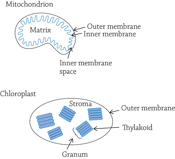
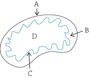
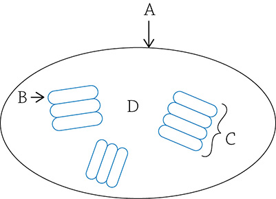
The mitochondrion is, yes, the powerhouse of the cell. What does that mean, specifically? The mitochondria supply the cell with a chemical energy source called ATP. It is able to create ATP because it creates a compartment within itself that serves as a reservoir for hydrogen ions (H+), and this high concentration of H+ is used to make ATP. The mitochondria also house many enzymes specialized in the methodical breakdown of the sugar glucose; these are in the innermost space called the matrix. We will cover all of this is more detail when we discuss cellular respiration in a bit. Similarly, the chloroplast also has a compartment within itself that holds a high concentration of H+ for the same purpose (to create ATP). The thylakoids are the hollow sacs that hold H+; the surface of the thylakoids is embedded with special pigment molecules that grab the sun’s energy. The thylakoids—a stack of which is called a granum—are floating in a liquidy goo called the stroma. The stroma is the location of a bunch of enzymes used to create glucose from carbon dioxide. We will also discuss the process of photosynthesis later.
Plant versus Animal Cells
Both plant and animal cells are eukaryotic, meaning they are filled with organelles and are larger than their prokaryotic cousins. The differences between the plant and animal cells, however, are significant: plant cells have chloroplasts and are photosynthetic, whereas animals are defined as not having the ability to photosynthesize. Plants do not have skeletons, so they rely on the structure provided for them by their individual cells, a phenomenon called turgor pressure. A plant cell has an organelle called a central vacuole that fills with water and applies supportive pressure on the cell wall. Animal cells have neither a cell wall nor a central vacuole. One thing to keep in mind: when I ask my students to describe the differences between plant and animal cells, a common mistake is to think that mitochondria are only found in animal cells (since plant cells have their chloroplasts). This is wrong! Plant cells most certainly do have mitochondria and undergo cellular respiration, just like animal cells do. The difference is, the glucose needed for cellular respiration in a plant cell is created from sun energy in the chloroplasts, whereas animals acquire their glucose by eating it (heterotrophs).
REVIEW QUESTIONS
1. How are organelles helpful to a eukaryotic cell?
2. A cell with a high amount of rough endoplasmic reticulum would be specialized to do what?
3. Match the organelle with its correct function:
4. What are two interesting traits of chloroplasts and mitochondria that suggest they were once autonomous (free-living) prokaryotic cells?
5. Label the mitochondrion with the following terms: inner membrane space, inner membrane, matrix, outer membrane.
6. The glucose needed for cellular respiration in an animal cell comes from________________, whereas the glucose needed for cellular respiration in a plant cell comes from________________.
7. What three things does a plant cell have that an animal cell does not?
8. How is the lysosome a perfect example of creating a microenvironment within a cell?
9. Fill in the blanks with the correct organelles: When a protein is made for export, it is created by________________ stuck onto the surface of the ________________.
10. True or False: Only plant cells have chloroplasts, and only animal cells have mitochondria.
11. Label the chloroplast with the following terms: granum, outer membrane, thylakoid, stroma.
12. How is turgor pressure formed in a plant cell?