Biology of Humans
19. Chromosomes and Cell Division
In the previous few chapters, we considered reproduction and development. In this chapter, we examine the role of two types of cell division, mitosis and meiosis, in the human life cycle. We consider the physical basis of heredity—the chromosomes—and we consider how the chromosomes are parceled out during mitosis and meiosis. We finish the chapter by examining why it is important for each cell to have the correct number of chromosomes.
Two Types of Cell Division
We begin life as a single cell called a zygote, formed by the union of an egg and a sperm. By adulthood, our bodies consist of trillions of cells. What happened in the intervening years? How did we go from a single cell to the multitude of cells that make up the tissues of a fully functional adult? The answer is cell division, which happened over and over again as we grew. Even in adults, many cells continue to divide for growth and repair of body tissues. With very few exceptions, each of those cells carries the same genetic information as its ancestors. The type of nuclear division that results in identical body cells is called mitosis.
In Chapter 17 you learned that males and females produce specialized reproductive cells called gametes (eggs or sperm). You'll recall that meiosis is a special type of nuclear division that gives rise to gametes. In females, meiosis occurs in the ovaries and produces eggs. In males, meiosis occurs in the testes and produces sperm. Meiosis is important because through it the gametes end up with half the amount of genetic information (half the number of chromosomes) in the original cell. When the nuclei of an egg and sperm unite (fertilization), the chromosome number is restored to that of the original cell. As a result, the number of chromosomes in body cells remains constant from one generation to the next.
· Down syndrome, which results from an error in cell division, is the most frequent inherited cause of mild to moderate retardation.
The roles of mitosis (which produces new body cells) and meiosis (which forms gametes) are summarized in the diagram of the human life cycle in Figure 19.1. You will learn more about both mitosis and meiosis later in this chapter.
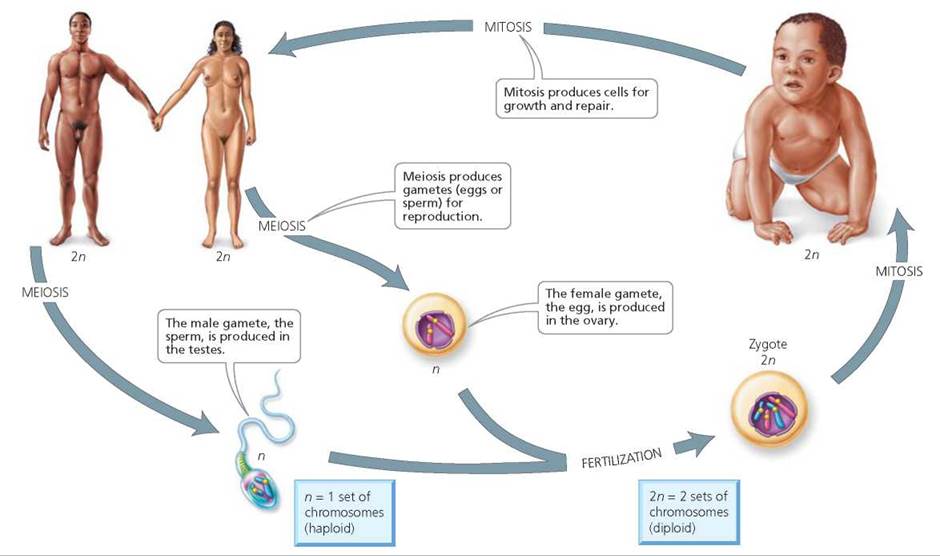
FIGURE 19.1. The human life cycle
Form of Chromosomes
A chromosome is a tightly coiled combination of a DNA molecule (which contains genetic information for the organism) and specialized proteins called histones. Chromosomes are found in the cell nucleus. The information contained in the DNA molecules in chromosomes directs the development and maintenance of the body. The histones combined with the DNA are for support and control of gene activity. A gene is a specific segment of the DNA that directs the synthesis of a protein, which in turn plays a structural or functional role within the cell. By coding for a specific protein, a gene determines the expression of a particular characteristic, or trait. Each chromosome in a human cell contains a specific assortment of genes. Like beads on a string, genes are arranged in a fixed sequence along the length of specific chromosomes.
In the human body, somatic cells—that is, all cells except for eggs or sperm—have 46 chromosomes. Those 46 chromosomes are actually 23 pairs of chromosomes. One member of each pair came from the mother's egg, and another member of each pair came from the father's sperm. Thus, each cell contains 23 homologous chromosome pairs, a pair being two chromosomes (one from the mother and one from the father) with genes for the same traits. Homologous pairs are called homologues for short. Any cell with two of each kind of chromosome is described as being diploid (annotated as 2n, with n representing the number of each kind of chromosome). In diploid cells, then, genes also occur in pairs. The members of each gene pair are located at the same position on homologous chromosomes.
One of the 23 pairs of chromosomes consists of the sex chromosomes that determine whether a person is male or female. There are two types of sex chromosomes, X and Y. A person who has two X chromosomes is described as XX and is genetically female; a person who has an X and a Y chromosome is described as XY and is genetically male. The other 22 pairs of chromosomes are called the autosomes. The autosomes determine the expression of most of a person's inherited characteristics.
The Cell Cycle
In mitosis, one nucleus divides into two daughter nuclei containing the same number and kinds of chromosomes. But mitosis is only one phase during the life of a dividing cell. The entire sequence of events that a cell goes through from its origin in the division of its parent cell through its own division into two daughter cells is called the cell cycle (Figure 19.2). The cell cycle consists of two major phases: interphase and cell division.
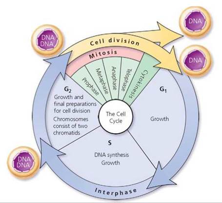
FIGURE 19.2. The cell cycle
Interphase
Interphase is the period of the cell cycle between cell divisions. It accounts for most of the time that elapses during a cell cycle. During active growth and divisions (depending on the type of cell), an entire cell cycle might take about 16 to 24 hours to complete, and only 1 to 2 hours are spent in division. Interphase is not a "resting period," as once thought. Instead, interphase is a time when the cell carries out its functions and grows. If the cell is going to divide, interphase is a time of intense preparation for cell division. During interphase, the DNA and organelles are duplicated. These preparations ensure that when the cell divides, each of its resulting cells, called daughter cells, will receive the essentials for survival.
Interphase consists of three parts: G1 (first "gap"), S (DNA synthesis), and G2 (second "gap"). All three parts of interphase are times of cell growth, characterized by the production of organelles and the synthesis of proteins and other macromolecules. There are, however, some events specific to certain parts of interphase:
• G1: A time of major growth before DNA synthesis begins.
• S: The time during which DNA is synthesized (replicated).
• G2: A time of growth after DNA is synthesized and before mitosis begins.
The details of DNA synthesis (replication) are described in Chapter 21. Our discussion in this chapter introduces some basic terminology pertaining to the cell cycle.
Throughout interphase, the genetic material is in the form of long, thin threads that are often called chromatin (Figure 19.3). They twist randomly around one another like tangled strands of yarn. In this state, DNA can be synthesized (replicated) and genes can be active. At the start of interphase, during G1, each chromosome consists of a DNA molecule and proteins. When the chromosomes are being replicated during the S phase, the chromosome copies remain attached. The two copies, each an exact replicate of the original chromosome, stay attached to one another at a region called the centromere. As long as the replicate copies remain attached, each copy is called a chromatid. The two attached chromatids are genetically identical and are called sister chromatids.
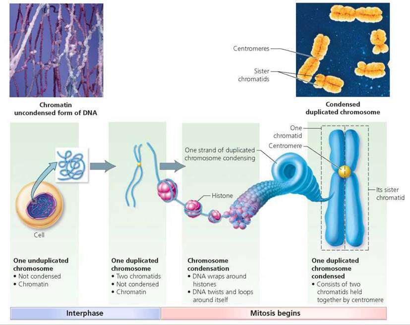
FIGURE 19.3. Changes in chromosome structure because of DNA replication during interphase and preparation for nuclear division in mitosis
Describe the difference in the structure of a chromosome between the start of interphase and at the end of interphase.
At the start of interphase, a chromosome is a single strand of DNA. At the end of interphase, a chromosome consists of two sister chromatids that are replicate copies of the original strand of DNA.
Division of the Nucleus and the Cytoplasm
Body cells divide continually in the developing embryo and fetus. Such division also plays an important role in the growth and repair of body tissues in children. In the adult, specialized cells, such as most nerve cells, lose their ability to divide. Late in G1 of interphase, these cells enter what is called the G0 stage; they are carrying out their normal cellular activities but do not divide. Other adult cells, such as liver cells, stop dividing but retain the ability to undergo cell division should the need for tissue repair and replacement arise. Still other cells actively divide throughout life. For example, the ongoing cell division in skin cells in adults serves to replace the enormous numbers of cells worn off each day.
We see, then, that the cell cycle requires precise timing and accuracy. Proteins monitor the environment within the cell to ensure that it is appropriate for cell division and that the DNA has been accurately replicated. Healthy cells will not divide unless these two conditions are met. However, as we will see in Chapter 21a, cancer cells escape this regulation and divide uncontrollably.
The division of body cells (after interphase) consists of two processes that overlap somewhat in time. The first process, division of the nucleus, is called mitosis. The second process is cytokinesis, which is the division of the cytoplasm that occurs toward the end of mitosis (Figure 19.4).
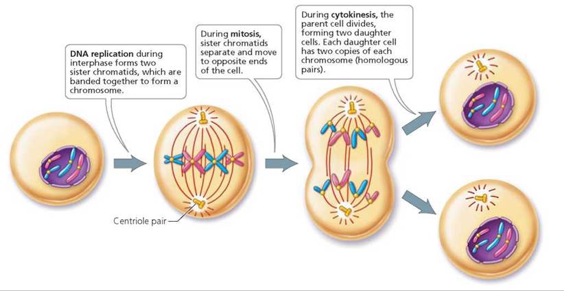
FIGURE 19.4. An overview of mitosis
Mitosis: Creation of Genetically Identical Diploid Body Cells
For the purpose of discussion, mitosis is usually divided into four stages: prophase, metaphase, anaphase, and telophase. The major events of each stage are depicted in Figure 19.5 (pp. 396-397).
• Prophase Mitosis begins with prophase, a time when changes occur in the nucleus as well as the cytoplasm. In the nucleus, the chromatin condenses and forms chromosomes as DNA wraps around histones. The DNA then loops and twists to form a tightly compacted structure (see Figure 19.3). When DNA is in this condensed state, it cannot be replicated, and gene activity is shut down. In this condensed state, the sister chromatids are easier to separate without breaking. At about this time, the nuclear membrane also begins to break down.
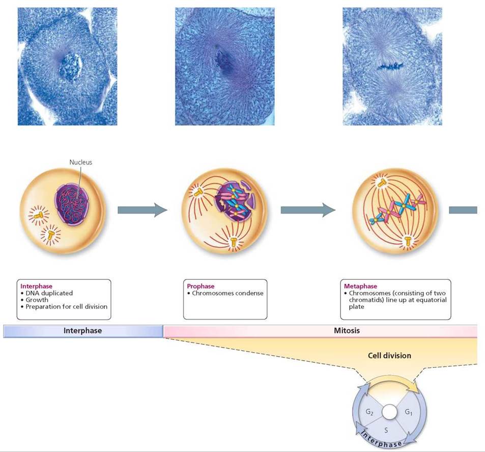
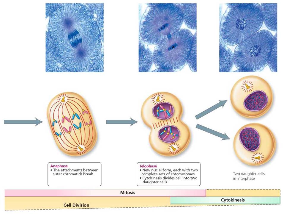
FIGURE 19.5. The stages of cell division (mitosis and cytokinesis) captured In light micrographs and depicted in schematic drawings
Outside the nucleus, in the cytoplasm, the mitotic spindle forms. The mitotic spindle is made of microtubules associated with the centrioles (see Chapter 3). During prophase, the centrioles, duplicated during interphase, move away from each other toward opposite ends of the cell.
• Metaphase During the next stage of mitosis, metaphase, the chromosomes attach to the mitotic spindles, forming a line at what is called the equator (center) of the mitotic spindles. This alignment ensures each daughter cell receives one chromatid from each of the 46 chromosomes when the chromosomes separate at the centromere. Thus each daughter cell is a diploid cell that is genetically identical to the parent cell.
• Anaphase Anaphase begins when the sister chromatids of each chromosome begin to separate, splitting at the centromere. Now separate entities, the sister chromatids are considered chromosomes in their own right. The spindle fibers pull the chromosomes toward opposite poles of the cell. By the end of anaphase, equivalent collections of chromosomes are located at the two poles of the cell.
• Telophase During telophase, a nuclear envelope forms around each group of chromosomes at each pole, and the mitotic spindle disassembles. The chromosomes also become more threadlike in appearance.
Stop and think
Cancer cells divide rapidly and without end. One type of drug used in cancer chemotherapy inhibits the formation of spindle fibers. Why can this be an effective anticancer treatment?
Cytokinesis
Cytokinesis—division of the cytoplasm—begins toward the end of mitosis, sometime during telophase. During this period, a band of microfilaments in the area where the chromosomes originally aligned contracts and forms a furrow, as shown in Figure 19.6. The furrow deepens, eventually pinching the cell in two.
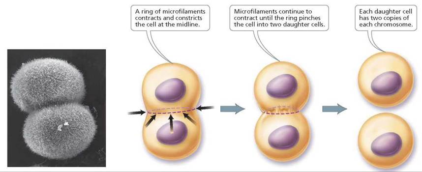
FIGURE 19.6. Cytokinesis is the division of the cytoplasm to form two daughter cells.
Stop and think
What would happen if a cell completed mitosis but did not complete cytokinesis?
Karyotypes
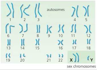
As we have seen, a major feature of cell division is the shortening and thickening of the chromosomes. In this state, the chromosomes are visible with a light microscope and can be used for diagnostic purposes, such as when potential parents want to check their own chromosomal makeup for defects. One often-used method takes white blood cells from a blood sample and grows them for a while in a nourishing medium. The culture then is treated with a drug that destroys the mitotic spindle, thus preventing separation of the chromosomes and halting cell division at metaphase. Next the cells are fixed, stained, and photographed so that the images of the chromosomes can be arranged in pairs based on physical characteristics such as location of the centromere and overall length. The chromosomes are numbered from largest to smallest, in an arrangement called a karyotype (Figure 19.7). Karyotypes can be checked for irregularities in number or structure of chromosomes.
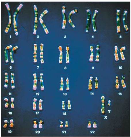
FIGURE 19.7. Chromosomes in dividing cells can be examined for defects in number or structure. A karyotype is constructed by arranging the chromosomes from photographs based on size and centromere location.
Meiosis: Creation of Haploid Gametes
We have seen that the somatic cells contain a homologous pair of each type of chromosome, one member of each pair from the father and one member of each pair from the mother. Recall that a cell with homologous pairs of chromosomes is described as being diploid, 2n. The gametes—eggs or sperm—differ from somatic cells in that they are haploid, indicated by n, meaning that they have only one member of each homologous pair of chromosomes. As you read earlier in the chapter, gametes are produced by a type of cell division called meiosis, which is actually two divisions that result in up to four haploid daughter cells. When a sperm fertilizes an egg, a new cell—the zygote—is created. Because the egg and sperm both contribute a set of chromosomes to the zygote, it is diploid. After many mitotic cell divisions, a zygote can eventually develop into a new individual.
Functions of Meiosis
Meiosis serves two important functions in sexual reproduction:
• Meiosis keeps the number of chromosomes in a body cell constant from generation to generation.
• Meiosis increases genetic variability in the population.
Meiosis keeps the number of chromosomes in a body cell constant over generations because it creates haploid gametes (sperm and eggs) with only one member of each homologous pair of chromosomes. If gametes were produced by mitosis, they would be diploid; each sperm and egg would contain 46 chromosomes instead of 23. Then, if a sperm containing 46 chromosomes fertilized an egg with 46 chromosomes, the zygote would have 92 chromosomes. The zygote of the next generation would have 184 chromosomes, having been formed by an egg and sperm each containing 92 chromosomes. The next generation would have 368 chromosomes in each cell, and the next one 736—and so on. You can see that the chromosome number would quickly become unwieldy and, what is more important, alter the amount of genetic information in each cell. As we will see toward the chapter's end, even one extra copy of a single chromosome usually causes an embryo to die.
Meiosis also increases genetic variability in the population. Later in this chapter we consider the mechanisms by which it accomplishes this increase. Genetic variability is important because it provides the raw material through which natural selection can act, leading to the changes described collectively as evolution. The relationship between genetic variability and evolution is discussed in Chapter 22.
Two Meiotic Cell Divisions: Preparation for Sexual Reproduction
First, let's consider how meiosis keeps the chromosome number constant. The stages in meiosis are summarized in Figure 19.8. Meiosis and mitosis begin the same way. Both are preceded by the same event—the replication of chromosomes. Unlike mitosis, however, meiosis involves two divisions. In the first division, the chromosome number is reduced, because the two homologues of each pair of chromosomes (each replicated into two chromatids attached by a centromere) are separated into two cells so that each cell has one member of each homologous pair of chromosomes. In the second division, the replicated chromatids of each chromosome are separated. We see, then, that meiosis begins with one diploid cell and, two divisions later, produces four haploid cells. The orderly movements of chromosomes during meiosis ensure that each haploid gamete produced contains one member of each homologous pair of chromosomes. Although not shown in the summary figure, each of the two meiotic divisions has four stages similar to those in mitosis: prophase, metaphase, anaphase, and telophase.
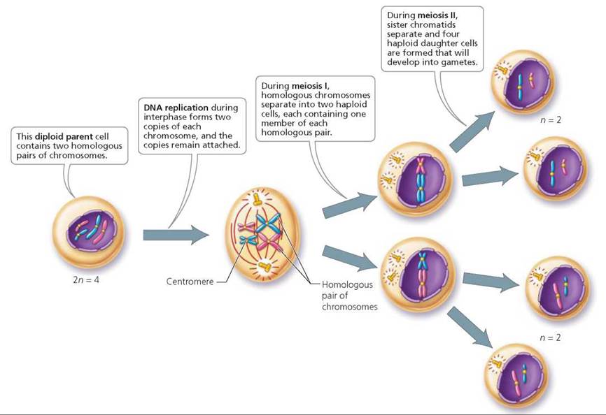
FIGURE 19.8. Overview of meiosis. Meiosis reduces the chromosome number from the diploid number to the haploid number. Meiosis involves two cell divisions.
Meiosis I. The first meiotic division—meiosis I—produces two cells, each with 23 chromosomes. Note that the daughter cells do not contain a random assortment of any 23 chromosomes. Instead, each daughter cell contains one member of each homologous pair, with each chromosome consisting of two sister chromatids.
It is important that each daughter cell receive one of each kind of chromosome during meiosis I. If one of the daughter cells had two of chromosome 3 and no chromosome 6, it would not survive. Although there would still be 23 chromosomes present, part of the instructions for the structure and function of the body (chromosome 6) would be missing. The separation of homologous chromosomes occurs reliably during meiosis I because, during prophase I (the I indicates this phase takes place during meiosis I), members of homologous pairs line up next to one another by a phenomenon called synapsis ("bringing together"). For example, the chromosome 1 that was originally from your father would line up with the chromosome 1 originally from your mother. Paternal chromosome 2 would pair with maternal chromosome 2, and so on. During metaphase I, matched homologous pairs become positioned at the midline of the cell and attach to spindle fibers. The pairing of homologous chromosomes helps ensure that the daughter cells will receive one member of each homologous pair. Consider the following analogy. By pairing your socks before putting them in a drawer, you are more likely to put matching socks on your feet than if you randomly pulled out two socks.
Next, during anaphase I, the members of each homologous pair of chromosomes separate, and each homologue moves to opposite ends of the cell. During telophase I, cytokinesis begins, resulting in two daughter cells, each with one member of each chromosome pair. Each chromosome in each daughter cell still consists of two replicated sister chromatids. Telophase I is followed by interkinesis, a brief interphase-like period. Interkinesis differs from mitotic interphase in that there is no replication of DNA during interkinesis.
Meiosis II During the second meiotic division—meiosis II— each chromosome lines up in the center of the cell independently (as occurs in mitosis), and the sister chromatids (attached replicates) making up each chromosome separate. Separation of the sister chromatids occurs in both daughter cells that were produced in meiosis I. This event results in four cells, each containing one of each kind of chromosome. The events of meiosis II are similar to those of mitosis, except that only 23 chromosomes are lining up independently in meiosis II compared with the 46 chromosomes aligning independently in mitosis. Figure 19.9 depicts the events of meiosis. Table 19.1 and Figure 19.10 compare mitosis and meiosis.
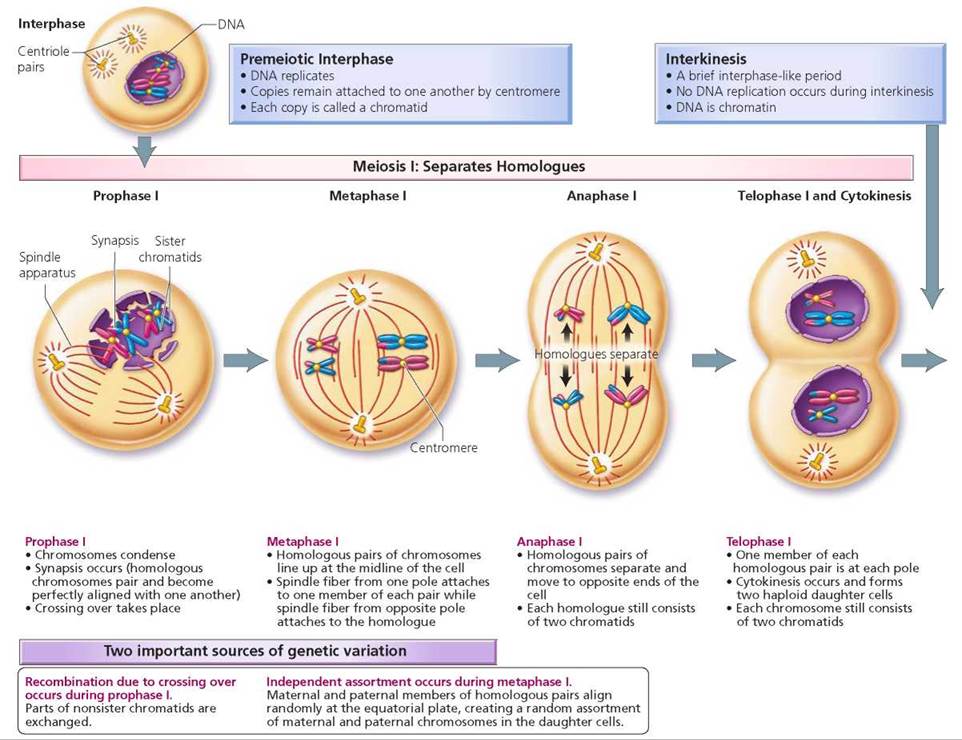
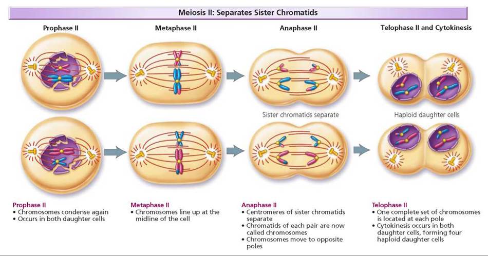
FIGURE 19.9. Stages of meiosis
TABLE 19.1. Mitosis and Meiosis Compared
|
Mitosis |
Meiosis |
|
Involves one cell division |
Involves two cell divisions |
|
Produces two diploid cells |
Produces up to four haploid cells |
|
Occurs in somatic cells |
Occurs only in ovaries and testes during the formation of gametes (egg and sperm) |
|
Results in growth and repair |
Results in gamete (egg and sperm) production |
|
No exchange of genetic material |
Parts of chromosomes are exchanged in crossing over |
|
Daughter cells are genetically similar |
Daughter cells are genetically dissimilar |
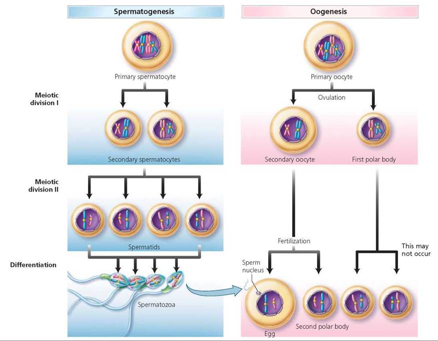
FIGURE 19.11. Comparison of spermatogenesis and oogenesis. Meiosis results in haploid cells that differentiate into mature gametes. Spermatogenesis produces four sperm cells that are specialized to transport the male’s genetic information to the egg. Oogenesis produces up to three polar bodies and one ovum that is packed with nutrients to nourish the early embryo.
Genetic Variability: Crossing Over and Independent Assortment
At the moment of fertilization, when the nuclei of an egg and a sperm fuse, a new, unique individual is formed. Although certain family characteristics may be passed along, each child bears its own assortment of genetic characteristics (Figure 19.12).
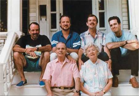
FIGURE 19.12. Each child inherits a unique combination of maternal and paternal genetic characteristics due to the shuffling of chromosomes that occurs during meiosis. This photograph shows Eric and Mary Goodenough with their four sons: Derick, Stephen, David, and John.
Genetic variation arises largely because of the shuffling of maternal and paternal forms of genes during meiosis. One way this mixing occurs is through a process called crossing over, in which corresponding pieces of chromatids of maternal and paternal homologues (nonsister chromatids) are exchanged during synapsis when the homologues are aligned side by side. After crossing over, the affected chromatids have a mixture of DNA from the two parents. Because the homologues align gene by gene during synapsis, the exchanged segments contain genetic information for the same traits. However, because the genes of the mother and those of the father may direct different expressions of the trait—attached or unattached earlobes, for instance—the chromatids affected by crossing over have a new, novel combination of genes. Thus, crossing over increases the genetic variability of gametes (Figure 19.13).
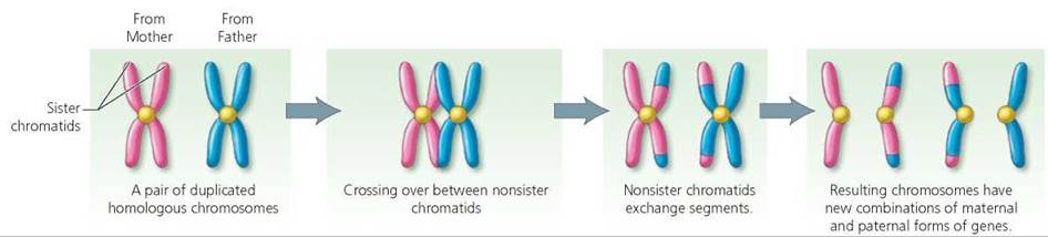
FIGURE 19.13. Crossing over. During synapsis, when the homologous chromosomes of the mother and the father are closely aligned, corresponding segments of nonsister chromatids are exchanged. Each of the affected chromatids has a mixture of maternal and paternal genetic information.
Independent assortment is a second way that meiosis provides for the shuffling of genes between generations (Figure 19.14). Recall that the homologous pairs of chromosomes line up at the equator (midpoint) of the mitotic spindles during metaphase I. However, the orientation of the members of the pair is random with respect to which member is closer to which pole. Thus, like the odds that a flipped coin will come up heads, there is a fifty-fifty chance that a given daughter cell will receive the maternal chromosome from a particular pair. Each of the 23 pairs of chromosomes orients independently during metaphase I. The orientations of all 23 pairs will determine the assortments of maternal and paternal chromosomes in the daughter cells. Thus, each child (other than identical siblings) of the same parents has a unique genetic makeup.
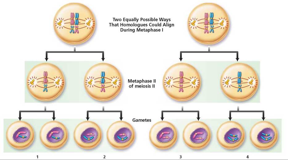
FIGURE 19.14. Independent assortment. The relative positioning of homologous maternal and paternal chromosomes with respect to the poles of the cell is random. The members of each homologous pair orient independently of the other pairs. Notice that with only two homologous pairs, there are four possible combinations of chromosomes in the resulting gametes.
Extra or Missing Chromosomes
Most of the time, meiosis is a precise process that results in the chromosomes being distributed evenly to gametes. But meiosis is not foolproof. A pair of chromosomes or sister chromatids may adhere so tightly to one another that they do not separate during anaphase. As a result, both go to the same daughter cell, and the other daughter cell receives none of this type of chromosome (Figure 19.15). The failure of homologous chromosomes to separate during meiosis I or of sister chromatids to separate during meiosis II is called nondisjunction.
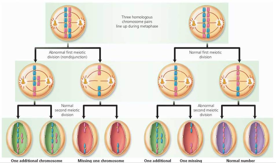
FIGURE 19.15. Nondisjunction is a mistake that occurs during cell division in which homologous chromosomes or sister chromatids fail to separate during anaphase. One of the resulting daughter cells will have three of one type of chromosome, and the other daughter cell will be missing that type of chromosome.
Ethical Issue
Trisomy 21
One in every 700 infants is born with three copies of chromosome 21 (trisomy 21), a condition known as Down syndrome. Symptoms of Down syndrome include moderate to severe mental retardation, short stature or shortened body parts due to poor skeletal growth, and characteristic facial features (Figure 19.A). Individuals with Down syndrome typically have a flattened nose, a forward-protruding tongue that forces the mouth open, upward-slanting eyes, and a fold of skin at the inner corner of each eye. Approximately 50% of all infants with Down syndrome have heart defects, and many of them die as a result of this defect. Blockage in the digestive system, especially in the esophagus or small intestine, is also common and may require surgery shortly after birth.
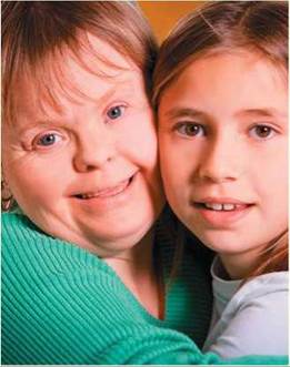
FIGURE 19.A. A person with Down syndrome is moderately to severely mentally retarded and has a characteristic appearance.
The risk of having a baby with Down syndrome increases with the mother's age. A 30-year-old woman is twice as likely to give birth to a child with Down syndrome as is a 20-year-old woman. After age 30, the risk rises dramatically. At age 45, a mother is 45 times as likely to give birth to a Down syndrome infant as is a 20-year-old woman.
Today, people with Down syndrome live longer and with a higher quality life than they did in the past. These improvements are due to better healthcare, more effective teaching approaches, and a greater range of opportunities. Life expectancy is now approaching 60 years in many countries.
Prenatal screening for Down syndrome is common and usually recommended for pregnant women aged over 30 years. Approximately 95% of the “positive” screening tests are wrong. Nonetheless, allwomen who initially test positive for carrying a fetus with Down syndrome are encouraged to undergo more invasive tests and 1% to 2% of the pregnancies tested by these procedures result in miscarriage. As a result, prenatal screening for Down syndrome poses a risk to 700,000 pregnancies each year.
Questions to Consider
Down Syndrome International is encouraging reviews of screening policies and public debate about the acceptance of genetic screening for mental and physical disabilities.
• If you or a loved one were pregnant, would you advocate for prenatal screening for Down syndrome? Why or why not?
• Who should pay for prenatal screening? The person? Health insurer? The government?
• Do you agree that genetic screening for mental and physical disabilities should be recommended?
What happens if nondisjunction creates a gamete with an extra or a missing chromosome and that gamete is then united with a normal gamete during fertilization? The resulting zygote will have an excess or deficit of chromosomes. For instance, if the abnormal gamete has an extra chromosome, the resulting zygote will have three of one type of chromosome and two of the rest. This condition, in which there are three representatives of one chromosome, is called trisomy. If, on the other hand, a gamete that is missing a representative of one type of chromosome joins with a normal gamete during fertilization, the resulting zygote will have only one of that type of chromosome, rather than the normal two chromosomes. The condition in which there is only one representative of a particular chromosome in a cell is called monosomy. The imbalance of chromosome numbers usually causes abnormalities in development. Most of the time, the resulting malformations are severe enough to cause the death of the fetus, which will result in a miscarriage. Indeed, in about 70% of miscarriages, the fetus has an abnormal number of chromosomes.
When a fetus inherits an abnormal number of certain chromosomes—for instance, chromosome 21 or the sex chromosomes—the resulting condition is usually not fatal (see Ethical Issue essay, Trisomy 21). The upset in chromosome balance does, however, cause a specific syndrome. (A syndrome is a group of symptoms that generally occur together.)
Like autosomes, sex chromosomes may fail to separate during anaphase. This error can occur during either egg or sperm formation. A male is chromosomally XY, so when the X and Y separate during anaphase, equal numbers of X-bearing and Y-bearing sperm are produced. However, if nondisjunction of the sex chromosomes occurs during sperm formation, half of the resulting sperm will carry both X and Y chromosomes, whereas the other resulting sperm will not contain any sex chromosome. A female is chromosomally XX, so each of the eggs she produces should contain a single X chromosome. When nondisjunction of sex chromosomes occurs, however, an egg may contain two X chromosomes or none at all. When a gamete with an abnormal number of sex chromosomes is joined with a normal gamete during fertilization, the resulting zygote has an abnormal number of sex chromosomes (Figure 19.16).
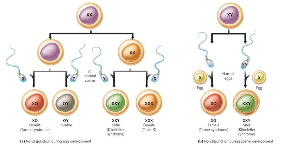
FIGURE 19.16. The sex chromosomes may fail to separate during formation of a gamete. Here an egg with an abnormal number of sex chromosomes joins a normal sperm in fertilization; the resulting zygote has an abnormal number of sex chromosomes. Imbalances of sex chromosomes upset normal development of reproductive structures.
Turner syndrome occurs in individuals who have only a single X chromosome (XO). Approximately 1 in 5000 female infants is born with Turner syndrome, but this represents only a small percentage of the XO zygotes that are formed. Most of these XO zygotes are lost as miscarriages. A person with Turner syndrome has the external appearance of a female. The only hint of Turner syndrome may be a thick fold of skin on the neck. As she ages, however, she generally is noticeably shorter than her peers. Her chest is wide, and her breasts underdeveloped. In 90% of the women with Turner syndrome, the ovaries are also poorly developed, leading to infertility. Pregnancy may be possible through in vitro fertilization (see Chapter 18), in which a fertilized egg from a donor is implanted in her uterus.
Klinefelter syndrome is observed in males who are XXY. Although the extra X chromosome can be inherited as a result of nondisjunction during either egg or sperm formation, it is twice as likely to come from the egg. Increased maternal age may increase the risk slightly.
Klinefelter syndrome is fairly common. Approximately 1 in 500 to 1 in 1000 of all newborn males is XXY. However, not all XXY males display the symptoms of having an extra X chromosome. In fact, some of them live their lives without ever suspecting that they are XXY. When there are signs that a male has Klinefelter syndrome, they do not usually show up until puberty. During the teenage years, the testes of an XY male gradually increase in size. In contrast, the testes of many XXY males remain small and do not produce an adequate amount of the male sex hormone, testosterone. As a result of the testosterone insufficiency, these males may grow taller than average but remain less muscular. Secondary sex characteristics, such as facial and body hair, may fail to develop fully. The breasts may also develop slightly. The penis is usually of normal size, but the testes may not produce sperm; so men with Klinefelter syndrome may be sterile.
Nondisjunction can also result in a female with three X chromosomes (XXX, triple-X syndrome) or a male with two Y chromosomes (XYY, Jacob syndrome, produced when the chromatids of a replicated Y chromosome fail to separate). Most women with triple-X syndrome (XXX) have normal sexual development and are able to conceive children. Some triple-X females have learning disabilities and delayed language skills. Males with two Y chromosomes (XYY) are often taller than normal, and some have slightly lower than normal intelligence.
What would you do?
If you had a son with Klinefelter syndrome, would you want him to have testosterone treatments after puberty?
Looking ahead
In this chapter we considered cell division: mitosis, which gives rise to new body cells for growth and repair, and meiosis, which gives rise to the gametes (eggs and sperm). In the next chapter, we consider mitosis further and explore stem cells, which are unspecialized cells that can divide continuously and develop into different tissue types.
Highlighting the Concepts
Two Types of Cell Division (p. 392)
• The human life cycle requires two types of nuclear division— mitosis and meiosis. Mitosis creates cells that are exact copies of the original cell. Mitosis occurs in growth and repair. Meiosis creates cells with half the number of chromosomes as were in the original cell. Gamete production requires meiosis.
Form of Chromosomes (p. 393)
• A chromosome contains DNA and proteins called histones. A gene is a segment of DNA that codes for a protein that plays a structural or functional role in the cell. Genes are arranged along a chromosome in a specific order. Each of the 23 different kinds of chromosomes in human cells contains a specific sequence of genes.
• Somatic cells (all cells except for eggs and sperm) are diploid; that is, they contain pairs of chromosomes, one member of each pair from each parent. Homologous chromosomes carry genes for the same traits. In humans, the diploid number of chromosomes is 46—or 23 homologous pairs. One pair of chromosomes, the sex chromosomes, determines gender. Males are XY, and females are XX. The other 22 pairs of chromosomes are called autosomes. Eggs and sperm are haploid; they contain only one set of chromosomes.
The Cell Cycle (p. 394)
• The cell cycle consists of two major phases: interphase and cell division. Interphase is the period between cell divisions.
• During interphase, DNA and organelles become replicated in preparation for the cell to divide and produce two identical daughter cells. Somatic cell division consists of mitosis (division of the nucleus) and cytokinesis (division of the cytoplasm).
Mitosis: Creation of Genetically Identical Diploid Body Cells (pp. 394-398)
• In mitosis, the original cell, having replicated its genetic material, distributes it equally between its two daughter cells. There are four stages of mitosis: prophase, metaphase, anaphase, and telophase.
• Cytokinesis, division of the cytoplasm, usually begins sometime during telophase. A band of microfilaments at the midline of the cell contracts and forms a furrow. The furrow deepens and eventually pinches the cell in two.
Karyotypes (p. 398)
• A karyotype is an arrangement of chromosomes based on their physical characteristics, such as length and position of the centromere.
Meiosis: Creation of Haploid Gametes (pp. 398-407)
• Meiosis, a special type of nuclear division that occurs in the ovaries or testes, begins with a diploid cell and produces four haploid cells that will become gametes (eggs or sperm).
• Meiosis is important because it halves the number of chromosomes in gametes, thereby keeping the chromosome number constant between generations. When a sperm fertilizes an egg, a diploid cell called a zygote is created. After many successful mitotic divisions, the zygote may develop into a new individual.
• Before meiosis begins, the chromosomes are replicated, and the copies remain attached to one another by centromeres. The attached replicated copies are called sister chromatids.
• There are two cell divisions in meiosis. During the first meiotic division (meiosis I), members of homologous pairs are separated. Thus, the daughter cells contain only one member of each homologous pair (although each chromosome still consists of two replicated sister chromatids). During the second meiotic division (meiosis II), the sister chromatids are separated.
• Genetic recombination during meiosis results in variation among offspring from the same two parents. One cause of genetic recombination is crossing over, in which corresponding segments of DNA are exchanged between maternal and paternal homologues, creating new combinations of genes in the resulting chromatids.
• A second cause of genetic recombination is the independent assortment of maternal and paternal homologues into daughter cells during meiosis I. The orientation of the members of the pair relative to the poles of the cell determines whether a daughter cell will receive the maternal or the paternal chromosome from a given pair. Each pair aligns independently of the others.
• Nondisjunction is the failure of homologous chromosomes or sister chromatids to separate during cell division. It results in an abnormal number of chromosomes in the resulting gametes, and in zygotes created by fertilization involving these gametes, which generally results in death of the fetus. Nondisjunction of chromosome 21 can result in Down syndrome.
Reviewing the Concepts
1. Explain the relationship between genes and a chromosome. р. 393
2. Define mitosis and cytokinesis. pp. 394-398
3. Why is meiosis important? p. 398
4. Describe the alignment of chromosomes at the midline during meiosis I and meiosis II. Explain the importance of these alignments in creating haploid gametes from diploid cells. pp. 400-403
5. Explain how crossing over and independent assortment result in genetic recombination that causes variability among offspring (aside from identical twins) from the same two parents. pp. 403-404
6. Define nondisjunction. Explain how nondisjunction can result in abnormal numbers of chromosomes in a person. p. 405
7. What causes Down syndrome? What are the usual characteristics of the condition? p. 405
8. The process of mitosis results in
a. two haploid cells.
b. two diploid cells.
с. four haploid cells.
d. four diploid cells.
9. DNA is synthesized (replicated) during
a. interphase.
b. prophase.
c. metaphase.
d. anaphase.
10. Crossing over occurs during which stage of meiosis?
a. Prophase I
b. Metaphase I
c. Prophase II
d. Metaphase II
11. During meiosis, the processes of _____ and _____ increase genetic diversity.
12. _____ chromosomes carry genes for the same traits.
13. _____ is the pairing of chromosomes during meiosis.
14. The stage of mitosis during which sister chromatids separate is _____.
15. The stage of meiosis during which sister chromatids separate is _____.
Applying the Concepts
1. A cell biologist is studying the cell cycle. She is growing the cells in culture, and they are actively dividing mitotically. One particular cell has half as much DNA as most of the other cells. Which stage of mitosis is this cell in? How do you know?
2. What would happen if the spindle fibers failed to form during mitosis?
3. What condition is indicated by the following karyotype?
Becoming Information Literate
Several genetic disorders are caused by too many or too few chromosomes. Use at least three reliable sources (books, journals, websites) to describe at least one such disorder other than Down syndrome, Turner syndrome, and Klinefelter syndrome. Indicate which chromosomes are extra or missing in the disorder, and note the symptoms of the disorder. List each source you considered, and explain why you chose the three sources you used.