THE LIVING WORLD
Unit Six. Animal Life
22. The Animal Body and How It Moves
22.7. Types of Skeletons
With muscles alone, the animal body could not move—it would simply pulsate as its muscles contracted and relaxed in futile cycles. For a muscle to produce movement, it must direct its force against another object. Animals are able to move because the opposite ends of their muscles are attached to a rigid scaffold, or skeleton, so that the muscles have something to pull against. There are three types of skeletal systems in the animal kingdom: hydraulic skeletons, exoskeletons, and endoskeletons.
Hydraulic skeletons are found in soft-bodied invertebrates such as earthworms and jellyfish. In this case, a fluid- filled cavity is encircled by muscle fibers that raise the pressure of the fluid when they contract. The earthworm in figure 22.8 moves forward by a wave of contractions of circular muscles that begins anteriorly and compresses the body, so that the fluid pressure pushes it forward. Contractions of longitudinal muscles then pull the rest of the body along.
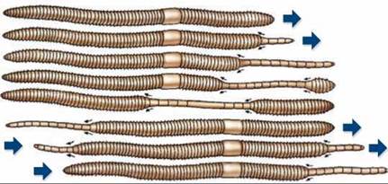
Figure 22.8. Earthworms have a hydraulic skeleton.
When an earthworm's circular muscles contract, the internal fluid presses on the longitudinal muscles, which then stretch to elongate segments of the earthworm. A wave of contractions down the body of the earthworm produces forward movement.
Exoskeletons surround the body as a rigid hard case to which muscles attach internally. When a muscle contracts, it moves the section of exoskeleton to which it is attached. Arthropods, such as crustaceans (figure 22.9) and insects, have exoskeletons made of the polysaccharide chitin. An animal with an exoskeleton cannot get too large because its exoskeleton would have to become thicker and heavier to prevent collapse. If an insect were the size of an elephant, its exoskeleton would have to be so thick and heavy it would hardly be able to move.
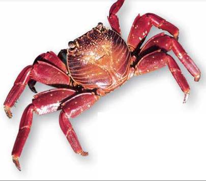
Figure 22.9 Crustaceans have an exoskeleton.
The exoskeleton of this rock crab is bright orange.
Endoskeletons, found in vertebrates and echinoderms, are rigid internal skeletons to which muscles are attached. Vertebrates have a soft, flexible exterior that stretches to accommodate the movements of their skeleton. The endoskeleton of most vertebrates is composed of bone (figure 22.10). Unlike chitin, bone is a cellular, living tissue capable of growth, selfrepair, and remodeling in response to physical stresses.
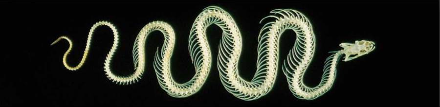
Figure 22.10. Snakes have an endoskeleton.
The endoskeleton of most vertebrates is made of bone. A snake's skeleton is specialized for quick lateral movement.
A Vertebrate Endoskeleton: The Human Skeleton
The human skeleton is made up of 206 individual bones. If you saw them as a pile of bones jumbled together, it would be hard to make any sense of them. To understand the skeleton, it is necessary to group the 206 bones according to their function and position in the body. The 80 bones of the axial skeleton, the purple-colored bones in figure 22.11, support the main body axis, while the remaining 126 bones of the appendicular skeleton, the tan-colored bones, support the arms and legs. These two skeletons function more or less independently—that is, the muscles controlling the axial skeleton (postural muscles) are managed by the brain separately from those controlling the appendages (manipulatory muscles).
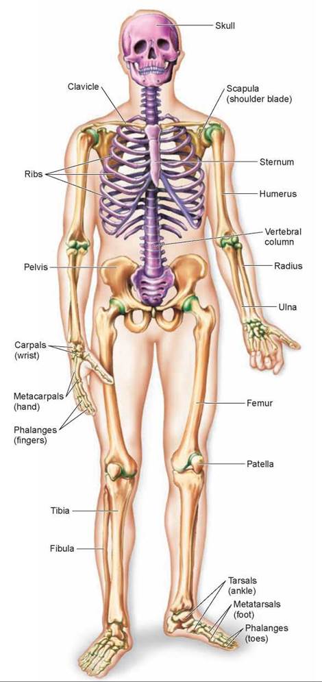
Figure 22.11. Axial and appendicular skeletons.
The axial skeleton is shown in purple, and the appendicular skeleton is shown in tan. Some of the joints are shown in green.
The Axial Skeleton
The axial skeleton is made up of the skull, backbone, and rib cage. Of the skull’s 28 bones, only 8 form the cranium, which encases the brain; the rest are facial bones and middle ear bones.
The skull is attached to the upper end of the backbone, which is also called the spine, or vertebral column. The spine is made up of 26 vertebrae, stacked one on top of the other to provide a flexible column surrounding and protecting the spinal cord. Curving forward from the vertebrae are 12 pairs of ribs, attached at the front to the breastbone, or sternum, and forming a protective cage around the heart and lungs.
The Appendicular Skeleton
The 126 bones of the appendicular skeleton are attached to the axial skeleton at the shoulders and hips. The shoulder, or pectoral girdle, is composed of two large, flat shoulder blades, each connected to the top of the sternum by a slender, curved collarbone (clavicle). The arms are attached to the pectoral girdle; each arm and hand contains 30 bones. The clavicle is the most frequently broken bone of the body. Can you guess why? Because if you fall on an outstretched arm, a large component of the force is transmitted to the clavicle.
The pelvic girdle forms a bowl that provides strong connections for the legs, which must bear the weight of the body. Each leg and foot contains a total of 30 bones.
Joints, points where two bones come together confer flexibility to the rigid endoskeleton, allowing a range of motion determined by the type of joint. There are three main classes of joints based on mobility. Immovable joints, such as the sutures of the skull, are capable of little to no movement. Slightly movable joints, such as the joints between vertebrae in the spine, allow the bones some movement. And freely movable joints allow a range of motion and are seen in the joints of the limbs (for example, the shoulder, elbow, hip, knee), the jaw, and fingers and toes. Depending on the type of joint, bones are held together at the joint by cartilage, fibrous connective tissue, or a fibrous capsule filled with a lubricating fluid.
Key Learning Outcome 22.7. The animal skeletal system provides a framework against which the body's muscles can pull. Many soft-bodied invertebrates employ a hydraulic skeleton, whereas arthropods have a rigid, hard exoskeleton surrounding their body. Echinoderms and vertebrates have an internal endoskeleton to which muscles attach.