THE LIVING WORLD
Unit Six. Animal Life
23.6. Amphibian and Reptile Circulation
The advent of lungs involved a major change in the pattern of circulation. After blood is pumped by the heart to the lungs, it does not go directly to the tissues of the body but instead returns to the heart. This results in two circulations: one that goes between the heart and the lungs, called the pulmonary circulation, and one that goes between the heart and the rest of the body, called the systemic circulation.
If no changes had occurred in the structure of the heart, the oxygenated blood from the lungs would be mixed in the heart with the deoxygenated blood returning from the rest of the body. Consequently, the heart would pump a mixture of oxygenated and deoxygenated blood rather than fully oxygenated blood. The amphibian heart has several structural features that help reduce this mixing. First, the atrium is divided by a septum, or dividing wall, into two chambers: The right atrium (the blue-colored area in figure 23.12a) receives deoxygenated blood from the systemic circulation, and the left atrium (colored red) receives oxygenated blood from the lungs. The septum prevents the two stores of blood from mixing in the atria, but some mixing might be expected when the contents of each atrium enter the single, common ventricle (the purple-colored area). Surprisingly, however, little mixing actually occurs. Two factors contribute to the separation of the two blood supplies. First, the ventricle in some amphibians is lined with folds that help direct the flow of blood from the atria. Second, the conus arteriosus (the branched structure in the foreground) is partially separated by another septum that directs deoxygenated blood into the pulmonary arteries toward the lungs, and oxygenated blood into the aorta, the major artery of the systemic circulation to the body. To trace the blood flow through the heart, follow the deoxygenated blood, the blue arrows, from the body to the lungs and then the oxygenated blood, the red arrows, from the lungs out to the body.
Amphibians in water supplement the oxygenation of the blood by obtaining additional oxygen by diffusion through their skin. This process is called cutaneous respiration.
Among reptiles, additional modifications have reduced the mixing of blood in the heart still further. In addition to having two separate atria, reptiles have a septum that partially subdivides the ventricle, with red and blue halves of the ventricle in figure 23.12b. This results in an even greater separation of oxygenated and deoxygenated blood within the heart. The separation is complete in one order of reptiles, the crocodiles, which have two separate ventricles divided by a complete septum. Crocodiles therefore have a completely divided pulmonary and systemic circulation. Another change in the circulation of reptiles is that the conus arteriosus has become incorporated into the trunks of the large arteries leaving the heart.
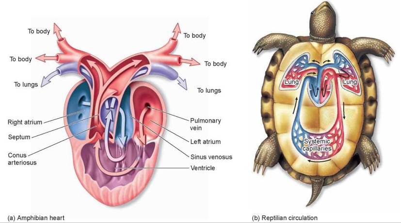
Figure 23.12. The amphibian heart and reptilian circulation.
(a) The frog heart has two atria but only one ventricle, which pumps blood both to the lungs and to the body. Despite the potential for mixing, the oxygenated and deoxygenated bloods (red and blue, respectively) mix very little as they are pumped to the body and lungs. (b) In reptiles, not only are there two separate atria, but the ventricle is also partially divided.
Key Learning Outcome 23.6. Amphibians and reptiles have two circulations, pulmonary and systemic, that deliver blood to the lungs and to the rest of the body, respectively.