THE LIVING WORLD
Unit Six. Animal Life
25. The Path of Food Through the Animal Body
25.3. Vertebrate Digestive Systems
In humans and other vertebrates, the digestive system consists of a tubular gastrointestinal tract and accessory digestive organs (figure 25.5). Working through the figure from the top down, the initial components of the gastrointestinal tract are the mouth and the pharynx, which is the common passage of the oral and nasal cavities. The pharynx leads to the esophagus, a muscular tube that delivers food to the stomach, where some preliminary digestion occurs. From the stomach, food passes to the first part of the small intestine, where a battery of digestive enzymes continues the digestive process. The products of digestion then pass across the wall of the small intestine into the bloodstream. The small intestine empties what remains into the large intestine, where water and minerals are absorbed. In most vertebrates other than mammals, the waste products emerge from the large intestine into a cavity called the cloaca (see the salamander in figure 25.4), which also receives the products of the urinary and reproductive systems. In mammals, the urogenital products are separated from the fecal material in the large intestine, also called the colon; the fecal material enters the rectum and is expelled through the anus.
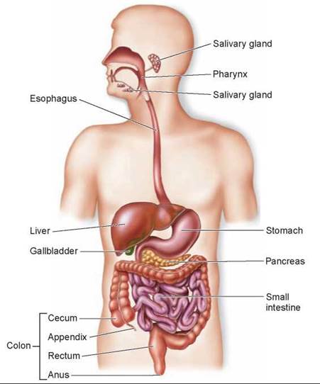
Figure 25.5. The human digestive system.
The tubular gastrointestinal tract and accessory digestive organs are shown. The colon extends from the cecum to the anus.
In general, carnivores have shorter intestines for their size than do herbivores. A short intestine is adequate for a carnivore, but herbivores ingest a large amount of plant cellulose, which resists digestion. These animals have a long, convoluted small intestine. In addition, mammals called ruminants (such as cows) that consume grass and other vegetation have stomachs with multiple chambers, where bacteria aid in the digestion of cellulose. Other herbivores, including rabbits and horses, digest cellulose (with the aid of bacteria) in a blind pouch called the cecum located at the beginning of the large intestine (these will be discussed in more detail in section 25.7). Accessory digestive organs described later in this chapter include the liver, the gallbladder, and the pancreas.
The tubular gastrointestinal tract of a vertebrate, as you can see in figure 25.6, has a characteristic layered structure. Working from the inside (the lumen) outward, the innermost layer (light pink) is the mucosa, an epithelium that lines the lumen. The next major tissue layer, composed of connective tissue, is called the submucosa (darker pink). Just outside the submucosa is the muscularis, which consists of a double layer of smooth muscles. The muscles in the inner layer have a circular orientation, and those in the outer layer are arranged longitudinally. An outer connective tissue layer, the serosa, covers the external surface of the tract. Nerves, intertwined in regions called plexuses, are located in the submucosa and help regulate the gastrointestinal activities.
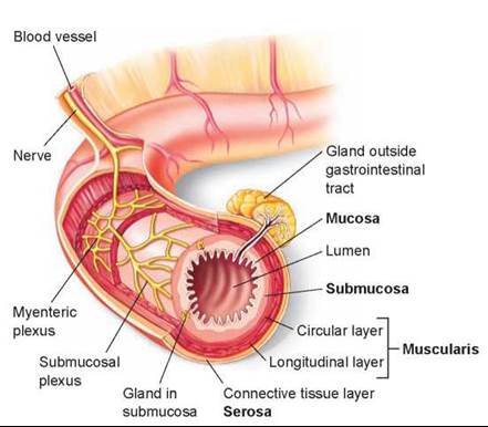
Figure 25.6. The layers of the gastrointestinal tract.
The mucosa contains a lining epithelium, the submucosa is composed of connective tissue (as is the outer serosa layer), and the muscularis consists of smooth muscles.
Key Learning Outcome 25.3. The vertebrate digestive system consists of a tubular gastrointestinal tract, which is modified in different animals, composed of a series of tissue layers.