THE LIVING WORLD
Unit Six. Animal Life
27. How the Animal Body Defends Itself
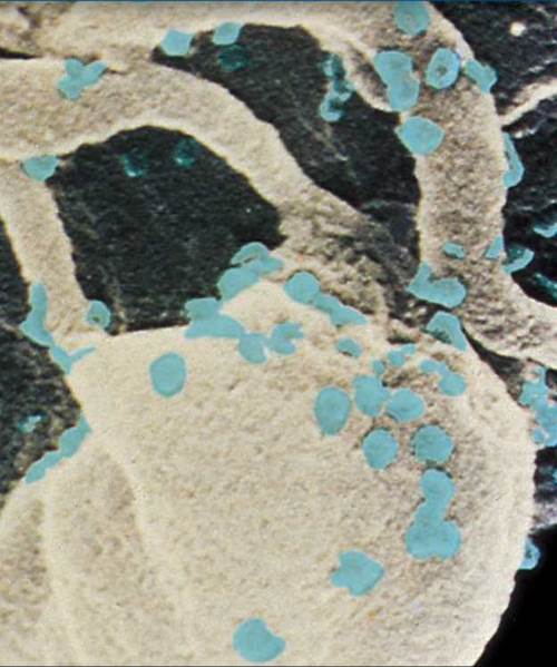
All animals are constantly at war with bacteria and viruses that attempt to use the rich resources of the cellular environment to fuel their own reproduction. Another war is fought on a very different front by animals against their own cells when cells become cancerous and begin to grow without restraint. Both of these wars, against invading microbes and against cancer, are fought with the same defensive weapons: the immune system. This chapter focuses on the vertebrate immune system and how it defends the body in the face of these onslaughts. Sometimes the immune system itself is the target of infection, leading to a loss of the body’s ability to defend itself. AIDS results from just this sort of infection by a virus called HIV (human immunodeficiency virus). The cell you see here is a human immune system cell called a macrophage that has been infected by HIV. Progeny HIV viruses are being released from the infected cell, budding out from its surface. These viruses soon spread to neighboring lymphocytes, infecting and killing them. Eventually a large majority of lymphocytes become infected, and the immune defense is destroyed. The individual viruses, colored blue in this scanning electron micrograph, are extremely small; over 200 million would fit on the period at the end of this sentence.
27.1. Skin: The First Line of Defense
Multicellular bodies offer a feast of nutrients for tiny, single-celled creatures, as well as a warm, sheltered environment in which they can grow and reproduce. We live in a world awash with microbes, and no animal can long withstand their onslaught unprotected. Animals survive because they have a variety of very effective defenses against this constant attack.
Overview of the Three Lines of Defense
The vertebrate body is defended from infection the same way knights defended medieval cities. “Walls and moats” make entry difficult; “roaming patrols” attack strangers; and “guards” challenge anyone wandering about and signal for an attack if a proper “ID” is not presented.
1. Walls and moats. The outermost layer of the vertebrate body, the skin, is the first barrier to penetration by microbes. Mucous membranes in the respiratory and digestive tracts are also important barriers that protect the body from invasion.
2. Roaming patrols. If the first line of defense is penetrated, the response of the body is to mount a cellular counterattack, using a battery of cells and chemicals that kill microbes. These defenses act very rapidly after the onset of infection.
3. Guards. Lastly, the body is also policed by cells that circulate in the bloodstream and scan the surfaces of every cell they encounter. They are part of the specific immune response. One kind of immune cell aggressively attacks and kills any cell identified as foreign, whereas the other type marks the foreign cell or virus for elimination by the roaming patrols.
The Skin
Skin, like the thick, tough skin of the elephants in figure 27.1, is the outermost layer of the vertebrate body and provides the first defense against invasion by microbes. Skin is our largest organ, comprising some 15% of our total weight. One square centimeter of skin from your forearm (about the size of a dime) contains 200 nerve endings, 10 hairs and muscles, 100 sweat glands, 15 oil glands, 3 blood vessels, 12 heat-sensing organs, 2 cold-sensing organs, and 25 pressure-sensing organs. The section of skin you see in figure 27.2 has two distinct layers: an outer epidermis and a lower dermis. A subcutaneous layer lies underneath the dermis. Cells of the outer epidermis are continually being worn away and replaced by cells moving up from below—in one hour your body loses and replaces approximately 1.5 million skin cells!
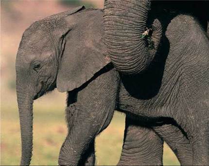
Figure 27.1. Skin is the body's first line of defense.
This young elephant has tough, leathery skin thicker than a belt, allowing it to follow the herd through dense thickets without injury.
The epidermis of skin is from 10 to 30 cells thick, about as thick as this page. The outer layer, called the stratum corneum, is the one you see when you look at your arm or face. Cells from this layer are continuously subjected to damage. They are abraded, injured, and worn by friction and stress during the body’s many activities. They also lose moisture and dry out. The body deals with this damage not by repairing cells but by replacing them. Cells from the stratum corneum are shed continuously, replaced by new cells produced deep within the epidermis (the dark layer of cells at the border of the epidermis and dermis). The cells of this inner basal layer are among the most actively dividing cells of the vertebrate body. New cells formed there migrate upward, and as they move they manufacture keratin protein, which makes them tough. Each cell eventually arrives at the outer surface and takes its turn in the stratum corneum, residing there for about a month before it is shed and replaced by a newer cell. Persistent dandruff (psoriasis) is a chronic skin disorder in which new cells reach the epidermal surface every three or four days, about eight times faster than normal.
The dermis of the skin is from 15 to 40 times thicker than the epidermis. It provides structural support for the epidermis, as well as a matrix for many specialized cells residing within the skin. The wrinkling that occurs as we grow older occurs here. The leather used to manufacture belts and shoes is derived from thick animal dermis. The layer of subcutaneous tissue below the dermis is composed of fat-rich cells that act as shock absorbers and provide insulation, which conserves body heat.
The skin not only defends the body by providing a nearly impermeable barrier, but it also reinforces this defense with chemical weapons. For example, the oil glands that occur along the shaft of the hair and sweat glands, which look like yellow coiled spaghetti in figure 27.2, make the skin’s surface very acidic (pH of between 4 and 5.5), which inhibits the growth of many microbes. Sweat also contains the enzyme lysozyme, which attacks and digests the cell walls of many bacteria.
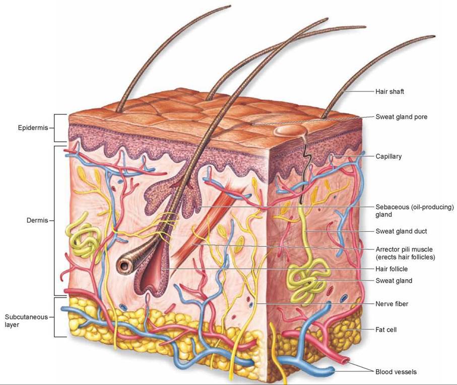
Figure 27.2. A section of human skin.
The skin defends the body by providing a barrier with sweat and oil glands, whose secretions make the skin's surface acidic enough to inhibit the growth of microorganisms.
Other External Surfaces
In addition to the skin, other surfaces, such as the eyes, are exposed to the outside. Like sweat, the tears that bathe the eyes contain lysozyme to fight bacterial infections. Two other potential routes of entry by viruses and microorganisms must be guarded: the digestive tract and the respiratory tract. Microbes are present in food, but many are killed by saliva (which also contains lysozyme), by the very acidic environment of the stomach (pH of 2), and by digestive enzymes in the intestine. Microorganisms are also present in inhaled air. The cells lining the smaller bronchi and bronchioles secrete a layer of sticky mucus that traps most microorganisms before they can reach the warm, moist lungs, which would provide ideal breeding grounds for them. Other cells lining these passages have cilia that continually sweep the mucus up toward the glottis in the throat, like an escalator. There it can be coughed out or swallowed, carrying potential invaders out of the lungs.
The surface defenses are very effective, but they are occasionally breached. Through breathing, eating, or cuts and nicks, bacteria and viruses now and then enter our bodies. When these invaders reach deeper tissue, a second line of defense comes into play, a cellular counterattack.
Key Learning Outcome 27.1. Skin and the mucous membranes lining the digestive and respiratory tracts are the body's first defenses.