THE LIVING WORLD
Unit Six. Animal Life
28.2. Neurons Generate Nerve Impulses
Neurons
The basic structural unit of the nervous system, whether central, motor, or sensory, is the nerve cell, or neuron. All neurons have the same basic structure as you can see by comparing the three cell types in figure 28.1 and the generalized cell in figure 28.3a. The cell body in figure 28.3a is the flat region of the neuron containing the nucleus. Short, slender branches called dendrites extend from one end of a neuron’s cell body. Dendrites are input channels. Nerve impulses travel inward along them toward the cell body. Motor and association neurons possess a profusion of highly branched dendrites, enabling those cells to receive information from many different sources simultaneously. Projecting out from the other end of the cell body is a single, long, tubelike extension called an axon. Axons are output channels. Nerve impulses travel outward along them, away from the cell body, toward other neurons or to muscles or glands.
Most neurons are unable to survive alone for long; they require the nutritional support provided by companion neuroglial cells. More than half the volume of the human nervous system is composed of supporting neuroglial cells. Two of the most important kinds of neuroglial cells are Schwann cells and oligodendrocytes, which envelop the axons of many neurons with a sheath of fatty material called myelin that acts as an electrical insulator. Schwann cells produce myelin in the PNS, whereas oligodendrocytes produce myelin in the CNS. During development, these cells associate with the axon, as shown at the top in figure 28.3b, and begin to wrap themselves around the axon several times to form a myelin sheath, an insulating covering consisting of multiple layers of membrane. Axons that have myelin sheaths are said to be myelinated, and those that don’t are unmyelinated. The myelin sheath is interrupted at intervals, leaving uninsulated gaps called nodes of Ranvier (the nodes are where the yellow underlying axon can be seen). At the node regions, the axon is in direct contact with the surrounding fluid. The nerve impulse jumps from node to node, speeding its travel down the axon. Multiple sclerosis (see chapter 27) and Tay-Sachs (see chapter 10) are debilitating clinical disorders, and, in the case of Tay-Sachs, fatal. They result from the degeneration of the myelin sheath.
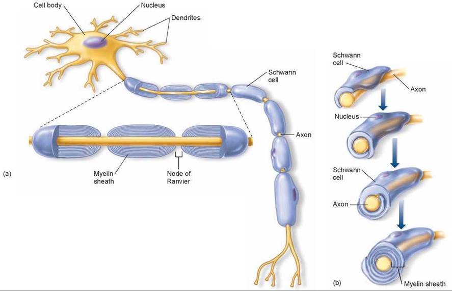
Figure 28.3. Structure of a typical neuron and formation of the myelin sheath.
(a) Extending from the cell body are many dendrites, which receive information and carry it to the cell body. A single axon transmits impulses away from the cell body. Many axons are encased by a myelin sheath, whose multiple membrane layers facilitate a more rapid conduction of impulses. The sheath is interrupted at regular intervals by small gaps called nodes of Ranvier. In the peripheral nervous system, myelin sheaths are formed by supporting Schwann cells. (b) The myelin sheath is formed by successive wrappings of Schwann cell membranes around the axon.
The Nerve Impulse
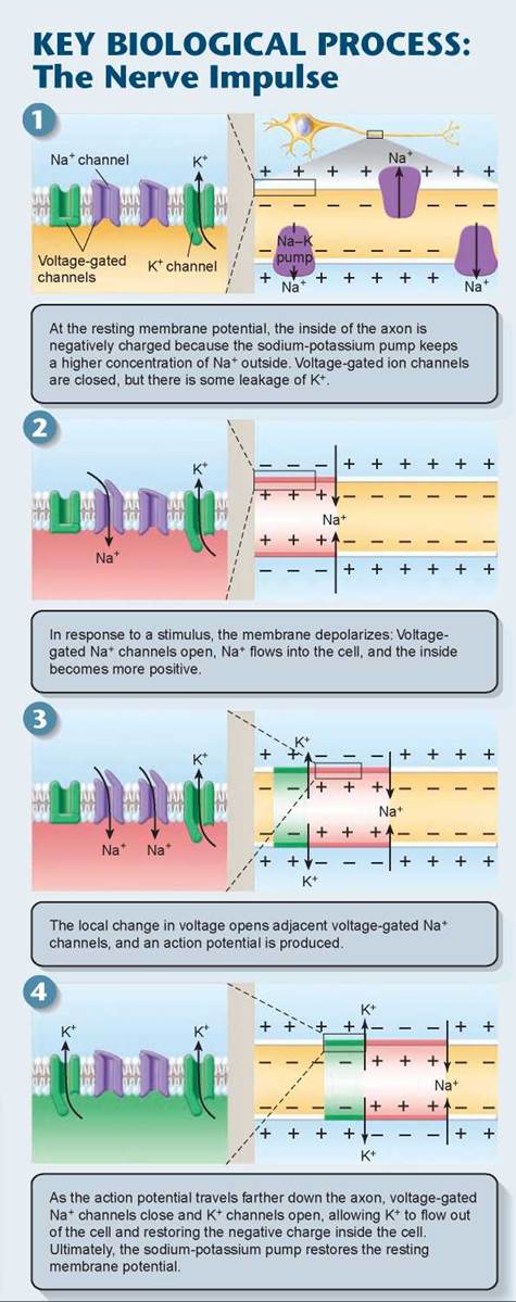
When a neuron is “at rest,” not carrying an impulse, active transport channels in the neuron’s plasma membrane transport sodium ions (Na+) out of the cell and potassium ions (K+) in. This sodium- potassium pump was described in chapter 4. Sodium ions cannot easily move back into the cell once they are pumped out, so the concentration of sodium ions builds up outside the cell. Similarly, potassium ions accumulate inside the cell, although they are not as highly concentrated because many potassium ions are able to diffuse out through open channels. This resting phase is indicated by the yellow coloring in panel 1 of the Key Biological Process illustration on the facing page. The result is to make the outside of the neuron more positive than the inside, a condition called the resting membrane potential. The resting plasma membrane is said to be “polarized.”
Neurons are constantly expending energy to pump sodium ions out of the cell, in order to maintain the resting membrane potential. The net negative charge of most proteins within the cell also adds to this charge difference. Using sophisticated instruments, scientists have been able to measure the voltage difference between the neuron interior and exterior as -70 millivolts (thousandth of a volt). The resting membrane potential is the starting point for a nerve impulse (panel 1).
A nerve impulse travels along the axon and dendrites as an electrical current caused by ions moving in and out of the neuron through voltage-gated channels (that is, protein channels in the neuron membrane that open and close in response to an electrical voltage). The impulse starts when pressure or other sensory inputs disturb a neuron’s plasma membrane, causing sodium channels on a dendrite to open (the purple channels in panel 2). As a result, sodium ions flood into the neuron from outside, down their concentration gradient, and for a brief moment a localized area inside of the membrane is “depolarized,” becoming more positive than the outside in that immediate area of the axon (indicated by the pink coloring in panel 2).
The sodium channels in the small patch of depolarized membrane remain open for only about a half a millisecond. However, if the change in voltage is large enough, it causes nearby voltage-gated sodium and potassium channels to open (panel 3). The sodium channels open first, which starts a wave of depolarization moving down the neuron. The opening of the gated channels causes nearby voltage-gated channels to open, like a chain of falling dominoes. This local reversal of voltage moving along the axon is called an action potential. An action potential follows an all-or-none law: A large enough depolarization produces either a full action potential or none at all because the voltage-gated Na+ channels open completely or not at all. Once they open, an action potential occurs. After a slight delay, potassium voltage-gated channels open and K+ flows out of the cells down its concentration gradient, making the inside of the cell more negative. The increasingly negative membrane potential (colored green in panel 4) causes the voltage-gated sodium channels to snap closed again. This period of time after the action potential has passed and before the resting membrane potential is restored is called the refractory period. A second action potential cannot fire during the refractory period, not until the resting potential is restored by the actions of the sodium-potassium pump.
The depolarization and restoration of the resting membrane potential takes only about 5 milliseconds. Fully 100 such cycles could occur, one after another, in the time it takes to say the word nerve.
Key Learning Outcome 28.2. Neurons are cells specialized to conduct impulses. Signals typically arrive along any of numerous dendrites, pass over the cell body's surface, and travel outward on a single long axon. Nerve impulses result from ion movements across the neuron plasma membrane through special protein channels that open and close in response to chemical or electrical stimulation.