THE LIVING WORLD
Unit Six. Animal Life
A nerve impulse travels along a neuron until it reaches the end of the axon, usually positioned very close to another neuron, a muscle cell, or gland. Axons, however, do not actually make direct contact with other cells. Instead, a narrow gap, 10 to 20 nanometers across, called the synaptic cleft, separates the axon tip and the target cell. This junction of an axon with another cell is called a synapse. A synapse is shown in figure 28.4. The membrane on the axon side of the synapse (on the left here) belongs to the presynaptic cell; the cell on the receiving side of the synapse (on the right) is called the postsynaptic cell.
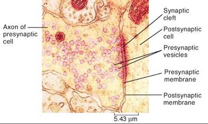
Figure 28.4. A synapse between two neurons.
This micrograph clearly shows the space between the presynaptic and postsynaptic membranes, which is called the synaptic cleft.
Neurotransmitters
When a nerve impulse reaches the end of an axon, its message must cross the synapse if it is to continue. Messages do not “jump” across synapses. Instead, they are carried across by chemical messengers called neurotransmitters. These chemicals are packaged in tiny sacs, or vesicles, at the tip of the axon. When a nerve impulse arrives at the tip, it causes the sacs to release their contents into the synapse, as shown in figure 28.5a. The neurotransmitters diffuse across the synaptic cleft and bind to receptors (the purple structures) in the postsynaptic membrane. The signal passes to the postsynaptic cell when the binding of the neurotransmitter opens special ion channels, allowing ions to enter the postsynaptic cell and cause a change in electrical charge across its membrane. The enlarged view of figure 28.5a shows how the channel opens and the ion (the yellow ball) enters the cell. Because these channels open when stimulated by a chemical, they are said to be chemically gated.
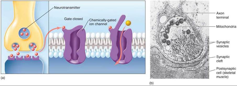
Figure 28.5 Events at the synapse.
(a) When a nerve impulse reaches the end of an axon, it releases a neurotransmitter into the synaptic cleft. The neurotransmitter molecules diffuse across the synapse and bind to receptors on the postsynaptic cell, opening ion channels. (b) A transmission electron micrograph of the tip of an axon filled with synaptic vesicles.
Why go to all this trouble? Why not just wire the neurons directly together? For the same reason that the wires of your house are not all connected but instead are separated by a host of switches. When you turn on one light switch, you don’t want every light in the house to go on, the toaster to start heating, and the television to come on! If every neuron in your body were connected to every other neuron, it would be impossible to move your hand without moving every other part of your body at the same time. Synapses are the control switches of the nervous system. However, the control switch must be turned off at some point by getting rid of the neurotransmitter, or the postsynaptic cell would keep firing action potentials. In some cases, the neurotransmitter molecules diffuse away from the synapse. In other cases, the neurotransmitter molecules are either reabsorbed by the presynaptic cell, or are degraded in the synaptic cleft.
Kinds of Synapses
The vertebrate nervous system uses dozens of different kinds of neurotransmitters, each recognized by specific receptors on receiving cells. They fall into two classes, depending on whether they excite or inhibit the postsynaptic cell.
In an excitatory synapse, the receptor protein is usually a chemically gated sodium channel, meaning that a sodium channel through the membrane is opened by the neurotransmitter. On binding with a neurotransmitter whose shape fits it, the sodium channel opens, allowing sodium ions to flood inward. If enough sodium ion channels are opened by neurotransmitters, an action potential begins.
In an inhibitory synapse, the receptor protein is a chemically gated potassium or chloride channel. Binding with its neurotransmitter opens these channels, leading to the exit of positively charged potassium ions or the influx of negatively charged chloride ions, resulting in a more negative interior in the receiving cell. This inhibits the start of an action potential, because the negative voltage change inside means that even more sodium ion channels must be opened to get a domino effect started among voltage-gated sodium channels, and so start an action potential.
An individual nerve cell, like the neuron in figure 28.6, can possess both kinds of synaptic connections to other nerve cells. In the drawing, the excitatory synapses are colored red and the inhibitory synapses are colored blue. When signals from both excitatory and inhibitory synapses reach the cell body of a neuron, the excitatory effects (which cause less internal negative charge) and the inhibitory effects (which cause more internal negative charge) interact with one another. The result is a process of integration in which the various excitatory and inhibitory electrical effects tend to cancel or reinforce one another. An area at the base of the axon, called the axon hillock, is the site of this integration process. If the result of the integration is a large enough depolarization (that is, the inside of the cell becomes more positive), an action potential will fire. Neurons often receive many inputs. A single motor neuron in the spinal cord may have as many as 50,000 synapses on it!
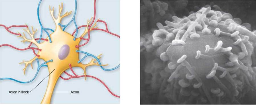
Figure 28.6. Integration.
Many different axons synapse with the cell body and dendrites of the postsynaptic neuron illustrated here. Excitatory synapses are shown in red and inhibitory synapses are shown in blue. The summed influence of their input at the axon hillock determines whether or not a nerve impulse will be sent down the axon extending below. The electron micrograph shows a neuronal cell body with numerous synapses.
Acetylcholine (ACh) is the neurotransmitter released at the neuromuscular junction, the synapse that forms between a neuron and a muscle fiber. ACh forms an excitatory synapse with skeletal muscle but has the opposite effect on cardiac muscle, causing an inhibitory synapse.
Glycine and GABA are inhibitory neurotransmitters. This inhibitory effect is very important for neural control of body movements and other brain functions. Interestingly, the drug diazepam (Valium) causes its sedative and other effects by enhancing the binding of GABA to its receptors.
Biogenic amines are a group of neurotransmitters that include dopamine, norepinephrine, serotonin, and the hormone epinephrine. These neurotransmitters have various effects on the body: Dopamine is important in controlling body movements; norepinephrine and epinephrine are involved in the autonomic nervous system, which will be discussed later; and serotonin is involved in sleep regulation and other emotional states. The drug PCP (angel dust) elicits its actions by blocking the elimination of biogenic amines from the synapse. The symptoms vary depending on the dosage.
Key Learning Outcome 28.3. A synapse is the junction of an axon with another cell. The cells are separated by a gap across which neurotransmitters carry a signal that has either an excitatory or inhibitory effect, depending on which ion channels are opened.