THE LIVING WORLD
Unit Six. Animal Life
The Cerebrum Is the Control Center of the Brain
Although vertebrate brains differ in the relative importance of different components, the human brain is a good model of how vertebrate brains function. About 85% of the weight of the human brain is made up of the cerebrum, the tan, convoluted area in figure 28.11. The cerebrum is a large, rounded area of the brain divided by a groove into right and left halves called cerebral hemispheres. The sectioned brain in figure 28.11 is cut along the center groove, with the left hemisphere removed, showing the right hemisphere. The hemispheres are further divided into the frontal, parietal, occipital, and temporal lobes. The cerebrum functions in language, conscious thought, memory, personality development, vision, and a host of other activities we call “thinking and feeling.” Figure 28.12 shows general areas of the brain and the functions they control (the different lobes of the brain are color-coded: yellow for the frontal lobe, orange for the parietal lobe, light green for the occipital lobe, and light purple for the temporal lobe). The cerebrum, which looks like a wrinkled mushroom, is positioned over and surrounding the rest of the brain, like a hand holding a fist. Much of the neural activity of the cerebrum occurs within a thin, gray outer layer only a few millimeters thick called the cerebral cortex (cortex is Latin for “bark of a tree”). This layer is gray because it is densely packed with neuron cell bodies. The human cerebral cortex contains the cell bodies of more than 10 billion nerve cells, roughly 10% of all the neurons in the brain. The wrinkles in the surface of the cerebral cortex increase its surface area (and number of cell bodies) threefold. Underneath the cortex is a solid white region of myelinated nerve fibers that shuttle information between the cortex and the rest of the brain.
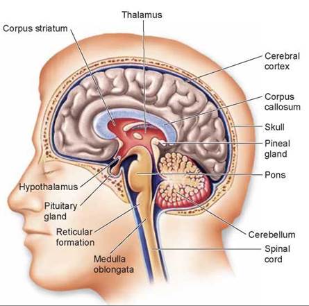
Figure 28.11. A section through the human brain.
The cerebrum occupies most of the brain. Only its outer layer, the cerebral cortex, is visible on the surface.
The right and left cerebral hemispheres are linked by bundles of neurons called tracts. These tracts serve as information highways, telling each half of the brain what the other half is doing. Because these tracts cross over, in the area of the brain called the corpus callosum (the blue-colored band in figure 28.11), each half of the brain controls muscles and glands on the opposite side of the body. Therefore, a touch on the right hand is relayed primarily to the left hemisphere, which may then initiate movement of the right hand in response to the touch.
Researchers have found that the two sides of the cerebrum can operate as two different brains. For instance, in some people the tract between the two hemispheres has been cut by accident or surgery. In laboratory experiments, one eye of an individual with such a “split brain” is covered and a stranger is introduced. If the other eye is then covered instead, the person does not recognize the stranger who was just introduced!
Sometimes blood vessels in the brain are blocked by blood clots, causing a disorder called a stroke. During a stroke, circulation to an area in the brain is blocked and the brain tissue dies. A severe stroke in one side of the cerebrum may cause paralysis of the other side of the body.
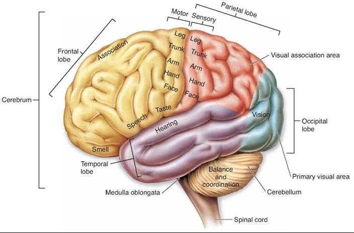
Figure 28.12. The major functional regions of the human brain.
Specific areas of the cerebral cortex are associated with different regions and functions of the body.
The Thalamus and Hypothalamus Process Information
Beneath the cerebrum are the thalamus and hypothalamus, important centers for information processing. The thalamus is the major site of sensory processing in the brain. Auditory (sound), visual, and other information from sensory receptors enter the thalamus and then are passed to the sensory areas of the cerebral cortex. The thalamus also controls balance. Information about posture, derived from the muscles, and information about orientation, derived from sensors within the ear, combine with information from the cerebellum and pass to the thalamus. The thalamus processes the information and channels it to the appropriate motor center on the cerebral cortex.
The hypothalamus integrates all the internal activities of the body. It controls centers in the brain stem that in turn regulate body temperature, blood pressure, respiration, and heartbeat. It also directs the secretions of the brain’s major hormone-producing gland, the pituitary gland. The hypothalamus is linked by an extensive network of neurons to some areas of the cerebral cortex. This network, along with parts of the hypothalamus and areas of the brain called the hippocampus and amygdala, make up the limbic system. The areas highlighted in green in figure 28.13 indicate the components of the limbic system. The operations of the limbic system are responsible for many of the most deep-seated drives and emotions of vertebrates, including pain, anger, sex, hunger, thirst, and pleasure, centered in the amygdala. You’ll recall on page 592 that the limbic system is the area of the brain affected by cocaine. It is also involved in memory, centered in the hippocampus.
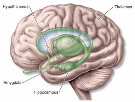
Figure 28.13. The limbic system.
The hippocampus and the amygdala are the major components of the limbic system, which controls our most deep-seated drives and emotions.
The Cerebellum Coordinates Muscle Movements
Extending back from the base of the brain is a structure known as the cerebellum. The cerebellum controls balance, posture, and muscular coordination. This small, cauliflowershaped structure, while well developed in humans and other mammals, is even better developed in birds. Birds perform more complicated feats of balance than we do, because they move through the air in three dimensions. Imagine the kind of balance and coordination needed for a bird to land on a branch, stopping at precisely the right moment without crashing into it.
The Brain Stem Controls Vital Body Processes
The brain stem, a term used to collectively refer to the midbrain, pons, and medulla oblongata, connects the rest of the brain to the spinal cord. This stalklike structure contains nerves that control your breathing, swallowing, and digestive processes, as well as the beating of your heart and the diameter of your blood vessels. A network of nerves called the reticular formation runs through the brain stem and connects to other parts of the brain. Their widespread connections make these nerves essential to consciousness, awareness, and sleep. One part of the reticular formation filters sensory input, enabling you to sleep through repetitive noises such as traffic yet awaken instantly when a telephone rings.
Language and Other Higher Functions
Although the two cerebral hemispheres seem structurally similar, they are responsible for different activities. The most thoroughly investigated example of this lateralization of function is language. The left hemisphere is the “dominant” hemisphere for language—the hemisphere in which most neural processing related to language is performed—in 90% of right-handed people and nearly two-thirds of left-handed people. There are two language areas in the dominant hemisphere: One is important for language comprehension and the formulation of thoughts into speech, and the other is responsible for the generation of motor output needed for language communication. Different language activities in figure 28.14 confirm that different parts of the brain are involved.
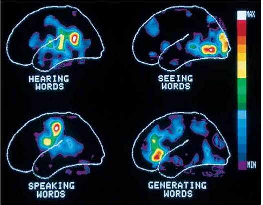
Figure 28.14. Different brain regions control various activities.
This illustration shows how the brain reacts in human subjects asked to listen to a spoken word, to read that same word silently, to repeat the word out loud, and then to speak a word related to the first. Regions of white, red, and yellow show the greatest activity. Compare this to figure 28.12 to see how regions of the brain are mapped.
While the dominant hemisphere for language is adept at sequential reasoning, like that needed to formulate a sentence, the nondominant hemisphere (the right hemisphere in most people) is adept at spatial reasoning, the type of reasoning needed to assemble a puzzle or draw a picture. It is also the hemisphere primarily involved in musical ability— a person with damage to the speech area in the left hemisphere may not be able to speak but may retain the ability to sing! Damage to the nondominant hemisphere may lead to an inability to appreciate spatial relationships and may impair musical activities such as singing. Reading, writing, and oral comprehension remain normal. The nondominant hemisphere is also important for the consolidation of memories of nonverbal experiences.
One of the great mysteries of the brain is the basis of memory and learning. There is no one part of the brain in which all aspects of a memory appear to reside. Although memory is impaired if portions of the brain, particularly the temporal lobes, are removed, it is not lost entirely. Many memories persist in spite of the damage, and the ability to access them gradually recovers with time. Therefore, investigators who have tried to probe the physical mechanisms underlying memory often have felt that they were grasping at a shadow. Although we still do not have complete understanding, we have learned a good deal about the basic processes in which memories are formed.
There appear to be fundamental differences between short-term and long-term memory. Short-term memory is transient, lasting only a few moments. Such memories can readily be erased by the application of an electrical shock, leaving previously stored long-term memories intact. This result suggests that short-term memories are stored electrically in the form of a transient neural excitation. Long-term memory, in contrast, appears to involve structural changes in certain neural connections within the brain. Two parts of the temporal lobes, the hippocampus and the amygdala, are involved in both short-term memory and its consolidation into long-term memory.
The Mechanism of Alzheimer Disease Still a Mystery
In the past, little was known about Alzheimer disease, a condition in which the memory and thought processes of the brain become dysfunctional. Scientists disagree about the biological nature of the disease and its cause. Two hypotheses have been proposed: One suggests that nerve cells in the brain are killed from the outside in, the other from the inside out.
In the first hypothesis, external proteins called b-amyloid peptides kill nerve cells. A mistake in protein processing produces an abnormal form of the peptide, which then forms aggregates, or plaques. The plaques begin to fill in the brain, which damages and kills nerve cells. However, recent clinical trails of a drug that inhibits b-amyloid synthesis worsened rather than cured Alzheimer disease.
The second hypothesis maintains that the nerve cells are killed by an abnormal form of an internal protein. This protein, called tau (t), normally functions to maintain protein transport microtubules. Abnormal forms of t assemble into helical segments that form tangles, which interfere with the normal functioning of the nerve cells. Researchers continue to study whether tangles and plaques are causes or effects of Alzheimer disease.
Key Learning Outcome 28.6. The associative activity of the brain is centered in the cerebral cortex, which lies over the cerebrum. Beneath, the thalamus and hypothalamus process information and integrate body activities. The cerebellum coordinates muscle movements.