THE LIVING WORLD
Unit Six. Animal Life
28.8. Voluntary and Autonomic Nervous Systems
As you learned in the opening discussion in section 28.1, the nervous system is divided into two main parts: the central nervous system (the pink boxes in figure 28.17, which include the brain and spinal cord) and the peripheral nervous system (the blue boxes, which include the motor and sensory pathways). The motor pathways of the peripheral nervous system of a vertebrate can be further subdivided into the somatic (voluntary) nervous system, which relays commands to skeletal muscles, and the autonomic (involuntary) nervous system, which stimulates glands and relays commands to the smooth muscles of the body and to cardiac muscle. The voluntary nervous system can be controlled by conscious thought. You can, for example, command your hand to move. The autonomic nervous system, by contrast, cannot be controlled by conscious thought. You cannot, for example, tell the smooth muscles in your digestive tract to speed up their action. The central nervous system issues commands over both voluntary and autonomic systems, but you are conscious of only the voluntary commands.
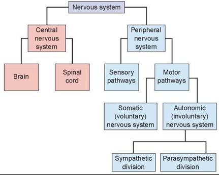
Figure 28.17. The divisions of the vertebrate nervous system.
The motor pathways of the peripheral nervous system are the somatic (voluntary) and autonomic nervous systems.
Voluntary Nervous System
Motor neurons of the voluntary nervous system stimulate skeletal muscles to contract in two ways. First, motor neurons may stimulate the skeletal muscles of the body to contract in response to conscious commands. For example, if you want to bounce a basketball, your CNS sends messages through motor neurons to the muscles in your arms and hands. However, skeletal muscle can also be stimulated as part of reflexes that do not require conscious control.
Reflexes Enable Quick Action. The motor neurons of the body have been wired to enable the body to act particularly quickly in time of danger—even before the animal is consciously aware of the threat. These sudden, involuntary movements are called reflexes. A reflex produces a rapid motor response to a stimulus because the sensory neuron bringing information about the threat passes the information directly to a motor neuron. The escape reaction of a fly about to be swatted is a reflex. One of the most frequently used reflexes in your body is blinking, a reflex that protects your eyes. If anything approaches your eye, such as an insect or a cloud of dust, the eyelid blinks closed even before you realize what has happened. The reflex occurs before the cerebrum is aware the eye is in danger.
Because they involve passing information between few neurons, reflexes are very fast. Many reflexes never reach the brain. The “danger” nerve impulse travels only as far as the spinal cord and then comes right back as a motor response. Most reflexes involve a single connecting interneuron between the sensory neuron and the motor neuron. A few, like the knee-jerk reflex in figure 28.18, are monosynaptic reflex arcs. You see in the figure that the sensory neuron, a stretch receptor embedded in a muscle, “senses” the stretching of the muscle when the tendon is tapped. This stretching could harm the muscle and so a nerve impulse is sent to the spinal cord where it synapses directly with a motor neuron—there is no interneuron between them. Similarly, if you step on something sharp, your leg jerks away from the danger. The prick causes nerve impulses in sensory neurons, which pass to the spinal cord and then to motor neurons, which cause your leg muscles to contract, jerking your leg up.
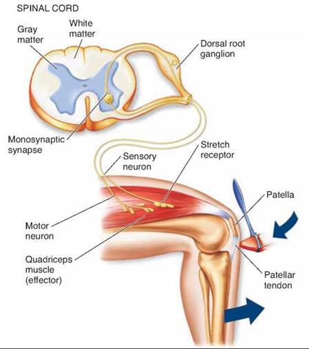
Figure 28.18. The knee-jerk reflex.
The most famous involuntary response, the knee jerk, is produced by activating stretch receptors in the quadriceps muscle. When a rubber mallet taps the patellar tendon, the muscle and stretch receptors in the muscle are stretched. A signal travels up a sensory neuron to the spinal cord, where the sensory neuron stimulates a motor neuron, which sends a signal to the quadriceps muscle to contract.
Some motor neurons are active all the time, even during sleep. These neurons carry messages from the CNS that keep the body going even when it is not active. These neurons make up the autonomic nervous system. The word autonomic means involuntary. The autonomic nervous system carries messages to muscles and glands that work without the animal noticing.
The autonomic nervous system is the command network used by the CNS to maintain the body’s homeostasis. Using it, the CNS regulates heartbeat and controls muscle contractions in the walls of the blood vessels. It directs the muscles that control blood pressure, breathing, and the movement of food through the digestive system. It also carries messages that help stimulate glands to secrete tears, mucus, and digestive enzymes.
The autonomic nervous system is composed of two divisions that act in opposition to one another. One division, the sympathetic nervous system, dominates in times of stress.
It controls the “fight-or-flight” reaction, increasing blood pressure, heart rate, breathing rate, and blood flow to the muscles. The sympathetic nervous system is colored pink in figure 28.19 and consists of a network of short motor axons extending out from the spinal cord to clusters of neuron cell bodies, called ganglia, indicated by the darker-colored band, located just to the right of the spinal cord in the figure. You can also see this chain of ganglia in figure 28.16. Long motor neurons extend from the ganglia directly to each target organ. Another division, the parasympathetic nervous system, has the opposite effect. It conserves energy by slowing the heartbeat and breathing rate and by promoting digestion and elimination. The parasympathetic nervous system is colored in blue and consists of a network of long axons extending out from motor neurons within the upper and lower sections of the spinal cord; these axons extend to ganglia in the immediate vicinity of an organ. It also consists of short motor neurons extending from the ganglia to the nearby organ.
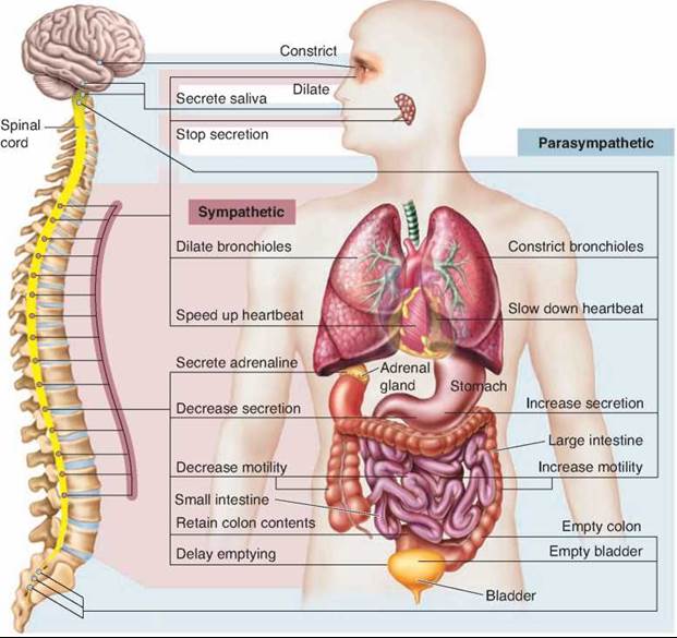
Figure 28.19. How the sympathetic and parasympathetic nervous systems interact.
A nerve path runs from both of the systems to every organ indicated except the adrenal gland, which is only innervated by the sympathetic nervous system.
Most glands, smooth muscles, and cardiac muscles get constant input from both the sympathetic and parasympathetic systems. The CNS controls activity by varying the ratio of the two signals to either stimulate or inhibit the organ.
Key Learning Outcome 28.8. The voluntary nervous system relays commands to skeletal muscles and can be controlled by conscious thought. The autonomic nervous system relays commands to muscles and glands that cannot be controlled by conscious thought.
Inquiry & Analysis
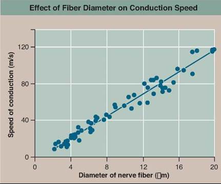
In this chapter, you learned that the axons of neurons can have an insulating covering called a myelin sheath, but many nerve axons do not have myelin covers. In addition, not all axons are the same. Some are thin, like fine wire, while others are much thicker. A motor axon to a human internal organ might have a diameter of 1 to 5 micrometers, while the giant motor axon to the mantle muscle of a squid is fully 500 micrometers in diameter.
Why the great difference in size? Physics tells us that there should be a relationship between the conduction velocity of a nerve axon and its diameter. To be precise, there should be a 10-fold increase in conduction speed for a 100-fold increase in fiber diameter. The squid giant axon is 100 times thicker than the human motor axons to internal organs—is it 10 times faster? Yes. The conduction velocity of the human axon is measured at 2 meters per second, while the conduction velocity of the squid giant axon is fully 25 meters per second—the very high velocity allows squids to contract their mantles fast enough to power their jet propulsion.
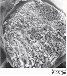
Most of the motor nerve axons in a vertebrate body like yours have insulating myelin covers, allowing them to transmit signals much faster, the electrical signal jumping down the axon over the insulated segments. The photo to the right shows a bundle of nerve axons, many of them myelinated and looking somewhat like doughnuts. axons also come in a wide range of sizes, from the 5 micrometer-diameter axons of skin temperature receptors, to the 20 micrometer-diameter fibers travelling to leg muscles. Would you expect fatter myelinated axons to transmit faster? Yes. Why? The transmission speed between axon node regions will depend upon how many ion channels open when an electrical impulse arrives at that node. If you think of the axon as a cylinder or pipe, then the number of ion channels exposed at a node of the axon will be proportional to the exposed surface area of the node, with the larger surface area of bigger axons exposing more ion channels and so transmitting the signal faster to the next node. The surface area considered at any one location of a node is simply the circumference of the axon at that point, which is the diameter times a constant—3.14159265 (called pi, symbolized n). Thus the velocity of a myelinated axon would be expected to be directly proportional to its diameter. Stated simply, doubling diameter should double speed. Is that the case?
With modern electrode technology, it is possible to directly measure the conduction velocity of axons within the body of a vertebrate, and so answer this question. The graph above shows the speed of conduction of myelinated axon fibers of a cat, plotting measured speed of conductance in meters per second against axon fiber diameter measured in micrometers.
1. Applying Concepts
a. What is the dependent variable?
b. Range. What is the range of nerve fiber diameters examined?
c. Frequency. Which were more frequently examined, narrow diameters (less than 8 micrometers), or thick diameters (more than 12 micrometers)?
2. Interpreting Data
a. For a nerve fiber diameter of 4 micrometers, what is the speed of conduction?
b. For a nerve fiber of twice that diameter, 8 micrometers, what is the speed of conduction?
c. For a nerve fiber twice that diameter, 16 micrometers, what is the speed of conduction?
3. Making Inferences
a. Is conduction velocity faster for larger diameter fibers?
b. When the nerve fiber diameter is doubled, what is the effect on speed of conductance?
4. Drawing Conclusions Do these data support the conclusion that conduction velocity is directly proportional to fiber diameter?
5. Further Analysis Do you imagine myelinated axons would transmit faster if the distance between nodes were shorter? If it were longer? How might you test this?
1. Complexity in animal nervous systems evolved with an increase in
a. animal body size.
b. animal body nutritive requirements.
c. the amount of associative neurons that eventually formed the “brain.”
d. types of animal behavior.
2. Which of the following is not found in the peripheral nervous system?
a. motor neurons
b. association neurons
c. sensory neurons
d. Schwann cells
3. An action potential is caused by a quick depolarization of the membrane in a nerve cell resulting from the
a. influx of sodium ions.
b. actions of the Na+/K+ pump.
c. influx of potassium ions.
d. All of the above.
4. The sodium-potassium pump
a. is used to propagate an action potential.
b. is important for maintenance of the resting membrane potential.
c. is important only at the synapse.
d. is a voltage-gated channel.
5. The junction between an axon and another cell is called a(n)
a. axon hillock.
b. axon terminal.
c. synapse.
d. action potential.
6. Excitatory neurotransmitters initiate an action potential in a postsynaptic neuron by opening
a. sodium ion gates in the postsynaptic cell.
b. potassium ion gates in the postsynaptic cell.
c. chloride ion gates in the postsynaptic cell.
d. calcium ion gates in the postsynaptic cell.
7. Most of the neural activity of the cerebrum occurs in the
a. cerebral cortex.
b. corpus callosum.
c. thalamus.
d. reticular formation.
8. The integration of the internal activities of the body is controlled by the
a. cerebrum.
b. cerebellum.
c. hypothalamus.
d. brain stem.
9. The purpose of the limbic system is to
a. coordinate olfactory information from the nose with visual information from the eyes.
b. process critical thought and learning.
c. coordinate optic information with the muscles.
d. coordinate emotions.
10. The purpose of the autonomic nervous system is to do all of the following except
a. stimulate glands.
b. relay messages to skeletal muscles.
c. relay messages to cardiac and smooth muscles.
d. regulate the body’s homeostasis.