THE LIVING WORLD
Unit Six. Animal Life
29. The Senses
29.4. Sensing Sounds: Hearing
When you hear a sound, you are detecting the air vibrating— waves of pressure in the air beating against your ear, pushing a membrane called the eardrum in and out. As you can see in figure 29.7, on the inner side of the eardrum are three small bones, called ossicles, that act as a lever system to increase the force of the vibration. They transfer the amplified vibration across a second membrane to fluid within the inner ear. The fluid-filled chamber of the inner ear is shaped like a tightly coiled snail shell and is called the cochlea, from the Latin name for “snail.” The middle ear, where the ossicles are located, is connected to the throat by the eustachian tube in such a way that there is no difference in air pressure between the middle ear and the outside. That is why your ears sometimes “pop” when you are landing in an airplane—the pressure is equalizing between the two sides of the eardrum. This equalized pressure is necessary for the eardrum to work.
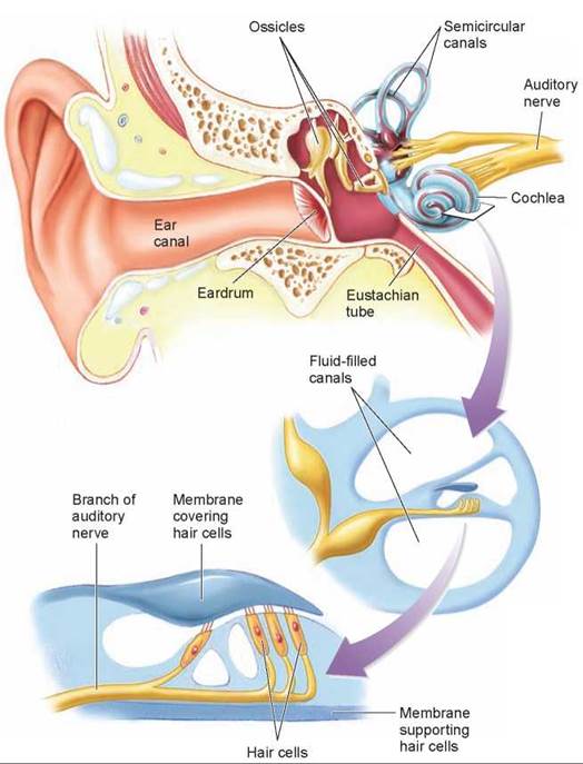
Figure 29.7. Structure and function of the human ear.
Sound waves passing through the ear canal beat on the eardrum, pushing a set of three small bones, or ossicles, against an inner membrane. This sets up a wave motion in the fluid filling the canals within the cochlea. The wave causes the membrane covering the hair cells to move back and forth against the hair cells, which causes associated neurons to fire impulses.
The sound receptors within the cochlea are hair cells that rest on a membrane that runs up and down the middle of the spiraling chamber, separating it into two halves, the upper and lower fluid-filled canals in the enlarged view. The hair cells do not project into the fluid-filled canals of the cochlea; instead, they are covered by a second membrane (the dark blue membrane in the figure). When a sound wave enters the cochlea, it causes the fluid in the chambers to move. The moving fluid causes this membrane “sandwich” to vibrate, bending the hairs pressed against the upper membrane and causing them to send nerve impulses to sensory neurons that travel to the brain.
Sounds of different frequencies cause different parts of the membrane to vibrate, and thus fire different sensory neurons—the identity of the sensory neuron being fired tells the CNS the frequency of the sound. Sound waves of higher frequencies, about 20,000 vibrations (or cycles) per second, also called hertz (Hz), move the membrane in the area closest to the middle ear. Medium-length frequencies, about 2,000 Hz, move the membrane in the area about midway down the length of the cochlea. The lowest-frequency sound waves, about 500 Hz, move the membrane near the tip of the cochlea.
The intensity of the sound is determined by how often the neurons fire. Our ability to hear depends upon the flexibility of the membranes within the cochlea. Humans cannot hear low-pitched sounds, below 20 vibrations (or cycles) per second, although some vertebrates can. As children, we can hear high-pitched sounds, up to 20,000 cycles per second, but this ability decreases as we get older. Other vertebrates can hear sounds at far higher frequencies. Dogs readily hear sounds of 40,000 cycles per second and so respond to a high-pitched dog whistle that seems silent to a human.
Frequent or prolonged exposure to loud noises can result in damage to the hair cells and membrane, especially in the high-frequency area to the cochlea. The loss of the ability to detect high-frequency sounds affects a person’s ability to hear certain sounds, especially in a noisy setting.
The Lateral Line System
A lateral line system supplements the fish’s sense of hearing. It is performed by a different sensory structure, and provides a sense of “distant touch.” A fish is able to sense objects that reflect pressure waves and low-frequency vibrations and thus can detect prey, for example, and swim in synchrony with the rest of its school. The lateral line system also enables a blind cave fish to sense its environment by monitoring changes in the patterns of water flow past the lateral line receptors. The same system is found in amphibian larvae, but it is lost at metamorphosis and is not present in any terrestrial vertebrate.
The lateral line system consists of sensory structures within a longitudinal canal in the fish’s skin, shown in figure 29.8. Canals extend along each side of the body, and there are several canals in the head. As you can see in the enlarged view, openings lead into the canal, which is lined with sensory structures known as hair cells because they have hairlike processes at their surface. The processes of the hair cells project into a gelatinous membrane called a cupula (Latin, “little cup”). The hair cells are innervated by sensory neurons that transmit impulses to the brain. Vibrations carried through the fish’s environment and down into the canal produce movements of the cupula, which cause the hairs to bend. When the hair cells bend, the associated sensory neurons are stimulated and generate a nerve impulse that is sent to the brain.
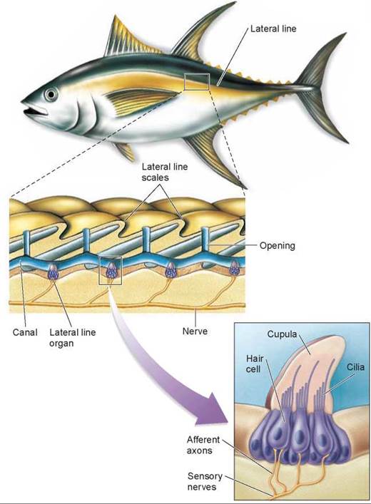
Figure 29.8. The lateral line system.
This system consists of canals running the length of the fish's body beneath the surface of the skin. Within these canals are sensory structures containing hair cells with cilia that project into a gelatinous cupula. Pressure waves traveling through the water in the canals deflect the cilia and depolarize the sensory neurons associated with the hair cells.
A few groups of mammals that live and obtain their food in dark environments have circumvented the limitations of darkness. A bat flying in a completely dark room easily avoids objects that are placed in its path—even a wire less than a millimeter in diameter. Shrews use a similar form of “lightless vision” beneath the ground, as do whales and dolphins beneath the sea. All of these mammals perceive distance by means of sonar. They emit sounds and then determine the time it takes these sounds to reach an object and return to the animal. This process is called echolocation. The bat in figure 29.9, for example, produces clicks that last 2 to 3 milliseconds and are repeated several hundred times per second. The three-dimensional imaging achieved with such an auditory sonar system can be quite sophisticated, and will help this bat find the moth.
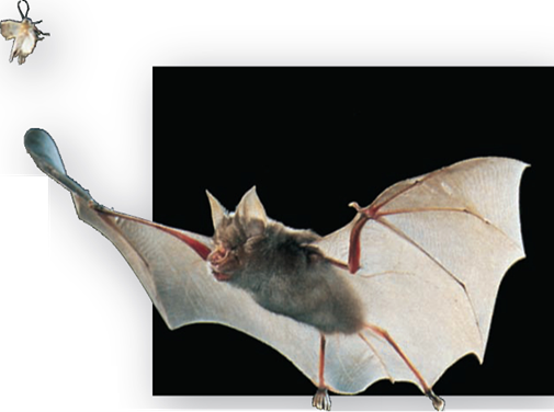
Figure 29.9. Using ultrasound to locate a moth.
This bat is emitting high-frequency "chirps" as it flies. It then listens for the sound's reflection against the moth. By timing how long it takes for a sound to return, the bat can "see" the moth even in total darkness.
Key Learning Outcome 29.4. Sound receptors detect air vibrations as waves of pressure pushing against the eardrum. Inside, these waves are amplified and press down hair cells that send signals to the brain. Fish sense pressure waves in water much as an ear senses sound. Many vertebrates sense distant objects by bouncing sounds off of them.