THE LIVING WORLD
Unit Seven. Plant Life
The organs of a plant—the roots, stem, leaves, and in some cases, flowers and fruits—are composed of different combinations of tissues, just as your legs are composed of bone, muscle, and connective tissue. A tissue is a group of similar cells—cells that are specialized in the same way—organized into a structural and functional unit. Most plants have three major tissue types: (1) ground tissue, in which the vascular tissue is embedded; (2) dermal tissue, the outer protective covering of the plant; and (3) vascular tissue, which conducts water and dissolved minerals up the plant and conducts the products of photosynthesis throughout.
Each major tissue type is composed of distinctive kinds of cells, whose structures are related to the functions of the tissues in which they occur. For example, vascular tissue is composed of xylem, which conducts water and dissolved minerals, and phloem, which conducts carbohydrates (mostly sugars) that the plant uses as food.
Ground Tissue
Parenchyma cells are the least specialized and the most common of all plant cell types; they form masses in leaves, stems, and roots. Parenchyma cells, unlike some other cell types, are characteristically alive at maturity, with fully functional cytoplasm and a nucleus. They are the cells that carry out the basic functions of living, including photosynthesis, cellular respiration, and food and water storage. The edible parts of most fruits and vegetables are composed of parenchyma cells. They are capable of cell division and are important in cell regeneration and wound healing. As you can see here, parenchyma cells have only thin cell walls, called primary cell walls, which are mostly cellulose that is laid down while the cells are still growing.
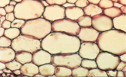
Parenchyma
Collenchyma cells, like parenchyma cells, are living at maturity and may live for many years. The cells, which are usually a little longer than they are wide, have walls that vary in thickness (as you can see in this photograph). Collenchyma cells provide support for plant organs. They are relatively flexible, allowing the organs to bend without breaking. They often form strands or continuous cylinders beneath the epidermis of stems or leaf stalks (petioles) and along veins in leaves. Strands of collenchyma provide much of the support for stems in which secondary growth has not taken place. The parts of celery that we eat (petioles) have “strings” that consist mainly of collenchyma and vascular bundles (conducting tissues).
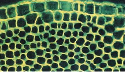
Collenchyma
Sclerenchyma cells, in contrast to parenchyma and collenchyma cells, have tough, thick cell walls called secondary cell walls; they usually do not contain living cytoplasm when mature. The secondary cell wall is laid down inside of the primary cell wall after the cell has stopped growing and expanding in size. The secondary cell wall provides cells with strength and rigidity. There are two types of sclerenchyma: fibers, which are long, slender cells that usually form strands, and sclereids, which are variable in shape but often branched. Sclereids, the reddish-colored cells in the photograph, are sometimes called “stone cells.” Clusters of sclereids form the gritty texture you feel in the flesh of pears. Both fibers and sclereids are thick-walled and strengthen the tissues in which they occur. Compare the thickness of the cell walls in the sclereid cells with the green-stained parenchyma cells that surround them.
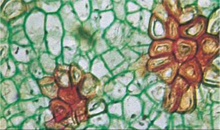
Sclerenchyma
Dermal Tissue
All parts of the outer layer of a primary plant body are covered by flattened epidermal cells. These are the most abundant cells in the plant epidermis, or outer covering. The epidermis is one cell layer thick and is a protective layer that provides an effective barrier against water loss. The epidermis is often covered with a thick, waxy layer called the cuticle, which protects against ultraviolet light damage and water loss. In some cases, the dermal tissue is more extensive and forms the bark of trees.
Trichomes are outgrowths of the epidermis that occur on the shoot, on the surfaces of stems and leaves. Trichomes vary greatly in form in different kinds of plants, from rounded-tip forms to globular- tipped ones. A “fuzzy” or “woolly” leaf is covered with trichomes, which when viewed under the microscope look like a thicket of fibers. Trichomes play an important role in regulating the heat and water balance of the leaf, much as the hairs of an animal’s coat provide insulation. Other trichomes are glandular, secreting sticky or toxic substances that may deter potential herbivores.
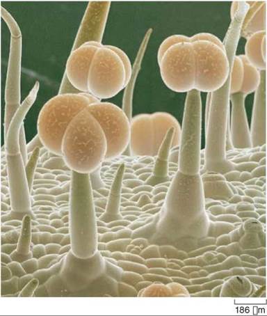
Trichomes
Guard cells are paired cells flanking a mouth-shaped epidermal opening that lies between them called a stoma (plural, stomata). Guard cells, unlike other epidermal cells, contain chloroplasts. Guard cells and stomata occur frequently in the epidermis of leaves and occasionally on other parts of the shoot, such as on stems or fruits. Oxygen, carbon dioxide, and water vapor pass across the epidermis almost exclusively through the stomata, which open and shut in response to external factors such as supply of moisture and light. There are from 1,000 to more than 1 million stomata per square centimeter of leaf surface. In many plants, stomata are more numerous on the lower epidermis of the leaf than on the upper epidermis, a design that minimizes water loss.
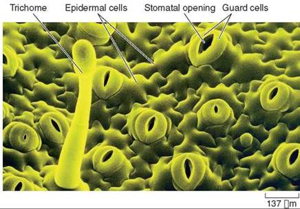
Stomata
Root hairs are tubular outgrowths of individual epidermal cells near the tips of young growing roots. Because a root hair is simply an extension of a cell’s cytoplasm and not a separate cell, there is no cross-wall isolating it from the epidermal cell. Root hairs keep the root in intimate contact with the surrounding particles of soil. Because root hairs greatly increase the surface area of the root, they profoundly increase the efficiency of absorption from the soil by the root. Indeed, most of the absorption of water and minerals occurs through root hairs, especially in herbaceous plants. As the root grows, root hairs at the older end slough off while new ones are produced behind the growing root tip.
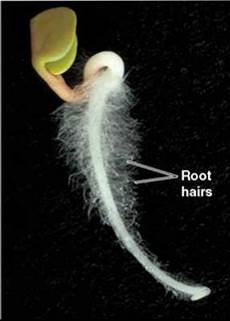
Root hairs
Vascular plants contain two kinds of conducting, or vascular, tissue: xylem and phloem.
Xylem is the plant’s principal water-conducting tissue, forming a continuous system that runs throughout the plant body. Within this system, water (and minerals dissolved in it) passes from the roots up through the shoot in an unbroken stream. When water reaches the leaves, much of it passes into the air as water vapor, through the stomata.
The two principal types of conducting cells in the xylem are tracheids and vessel elements, both of which have thick secondary walls that are laid down inside the primary cell wall. These cells are elongated, and they have no living cytoplasm (they are dead) at maturity. Tracheids are elongated cells that overlap at the ends. In conducting elements composed of tracheids, water flows from tracheid to tracheid through openings called pits in the secondary walls. In contrast, vessel elements are elongated cells that line up end-on-end.
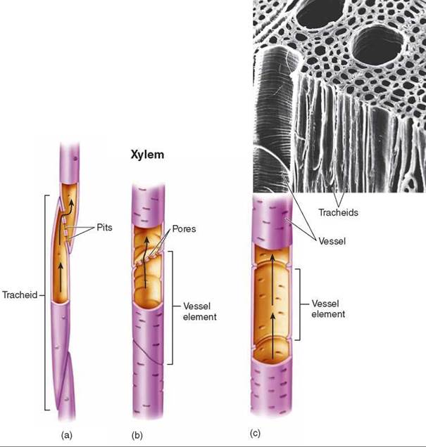
The end walls of vessel elements may be almost completely open or may have bars or strips of wall material perforated by pores through which water flows. A linked row of vessel elements forms a vessel (in the upper right, a scanning electron micrograph of the red maple Acer rubrum shows tracheids and vessels). Primitive angiosperms and other vascular plants have only tracheids, but the majority of angiosperms have vessels. Vessels conduct water much more efficiently than do strands of tracheids.
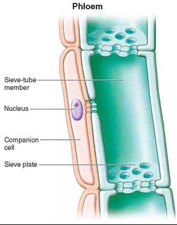
Phloem is the principal food-conducting tissue in vascular plants. Food conduction in phloem is carried out through two kinds of elongated cells: sieve cells and sieve-tube members. The cells differ in the extent of the perforations between the cells, with the sieve cells having smaller perforations between cells. Seedless vascular plants and gymnosperms have only sieve cells; most angiosperms have sieve-tube members. Clusters of pores known as sieve areas occur on both kinds of cells and connect the cytoplasms of adjoining sieve cells and sieve-tube members. Both cell types are living, but their nuclei are lost during maturation.
In sieve-tube members, some sieve areas have larger pores and are called sieve plates. Sieve-tube members occur end to end, as shown in the figure to the left, forming longitudinal series called sieve tubes. Specialized parenchyma cells known as companion cells occur regularly in association with sieve-tube members. The companion cells can be seen in the figure associated with the left side of the sieve-tube members. Companion cells apparently carry out some of the metabolic functions that are needed to maintain the associated sieve-tube member; their cytoplasms are connected to the sieve-tube members through openings called plasmodesmata.
In addition to conducting cells, xylem and phloem also contain fibers (sclerenchyma cells) and parenchyma cells. The parenchyma cells function in storage, and the fibers provide support and some storage. Xylem fibers are used in the production of paper.
Key Learning Outcome 33.2. Plants contain a variety of ground tissue, dermal tissue (outer coverings), and vascular tissues (conducting tissues).
We now consider the three kinds of vegetative organs that form the body of a plant: roots, stems, and leaves. While we will examine their basic structures, it is important to understand that these organs are often modified for different functions. For example, roots and stems can be modified for water and food storage, and leaves can be modified for defenses, such as cactus spines.